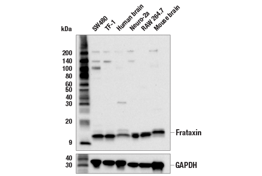
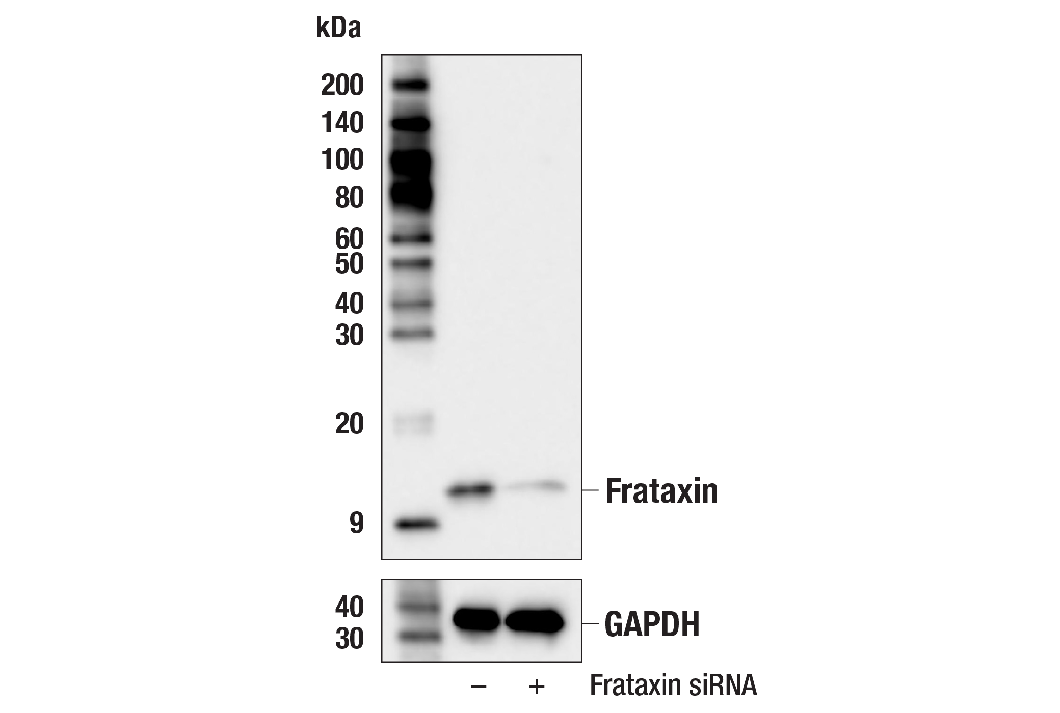
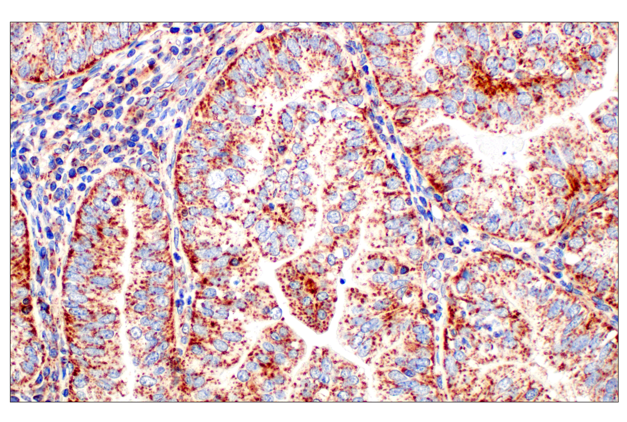
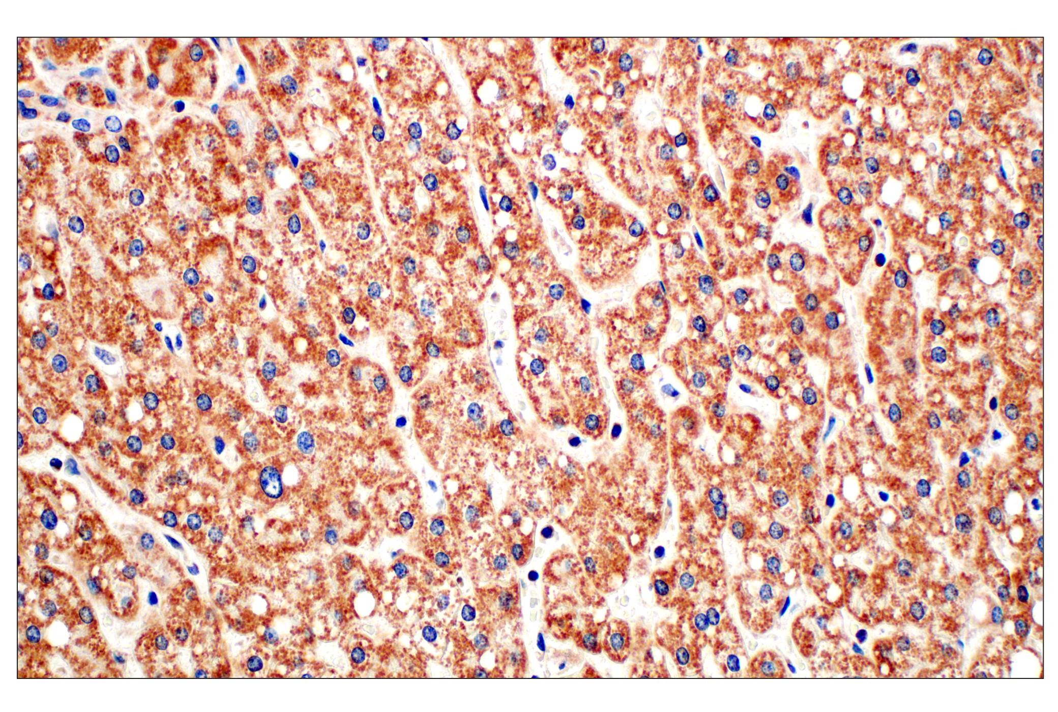
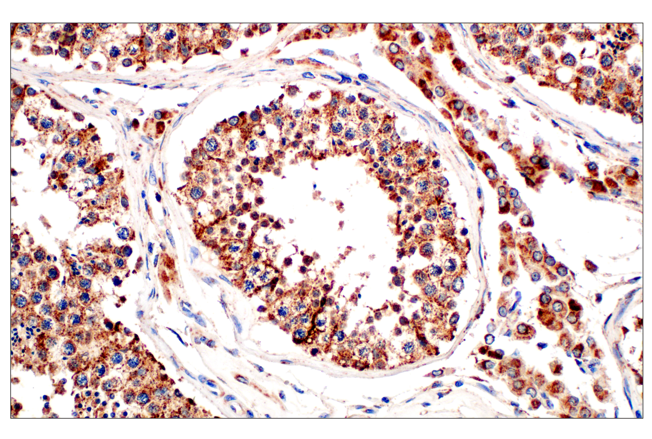
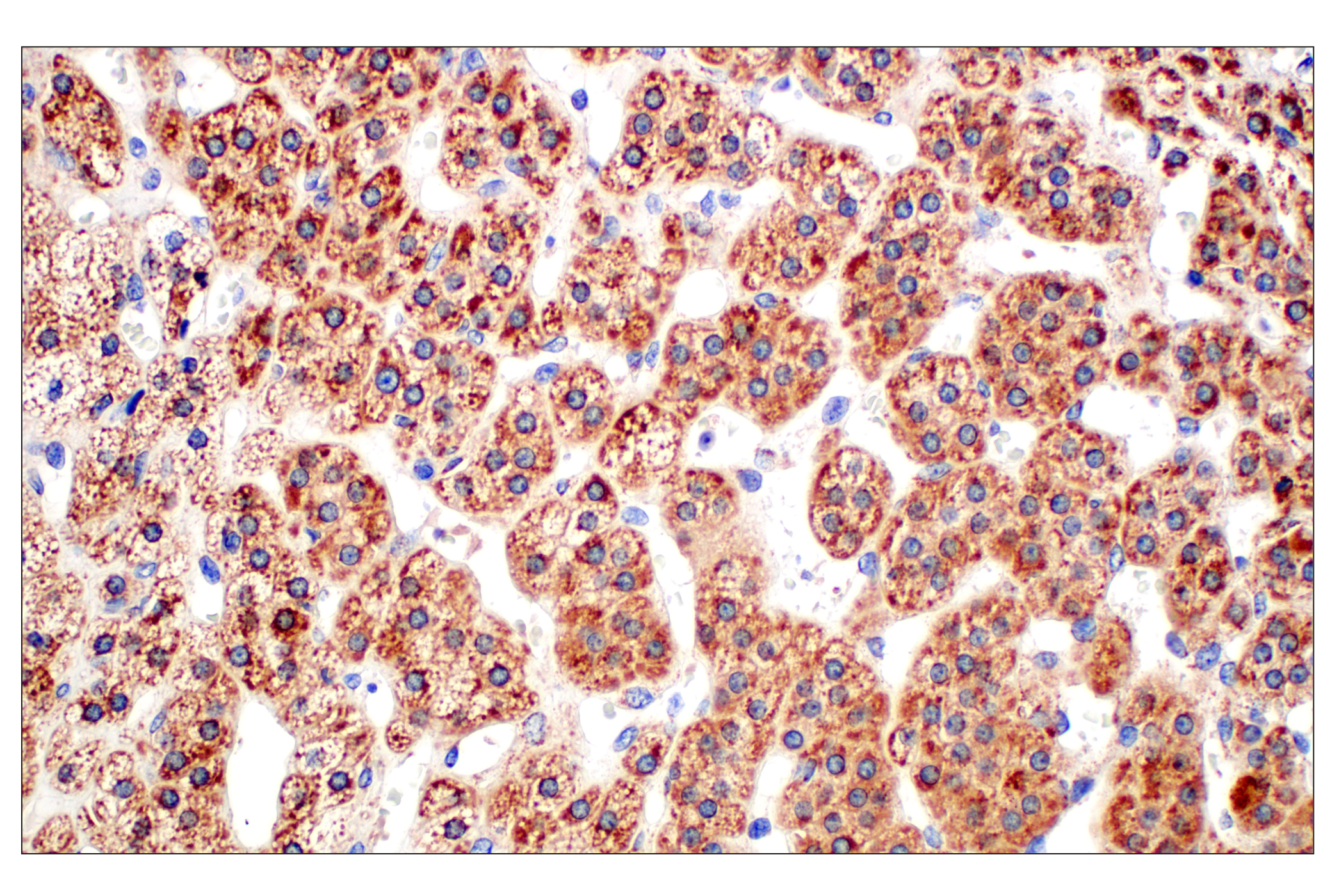
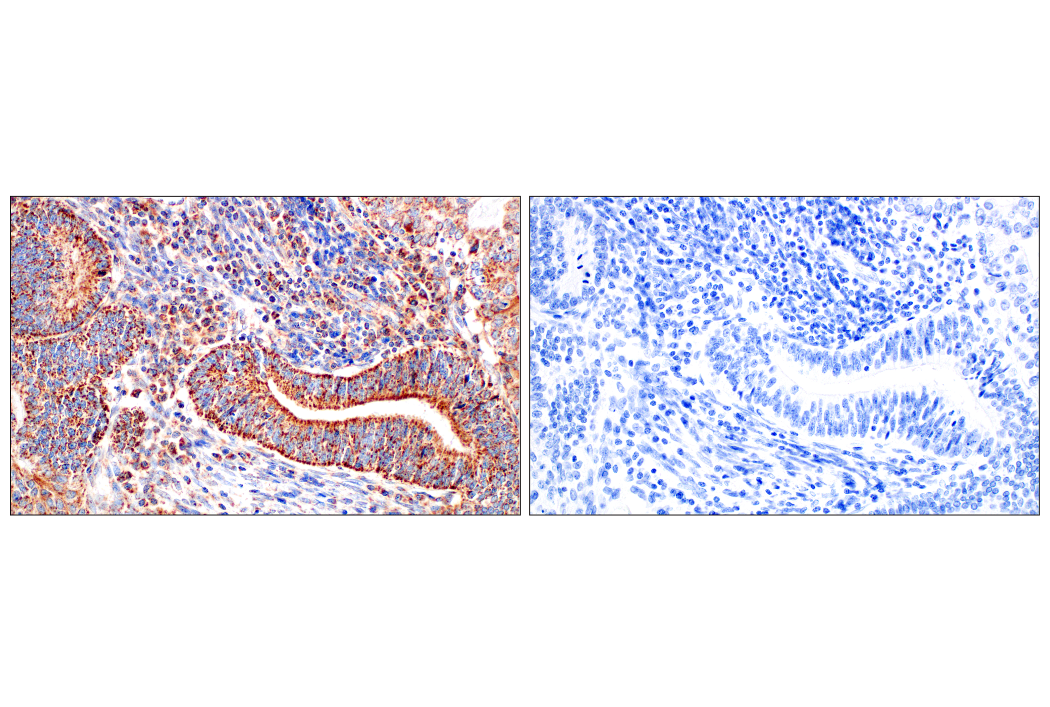
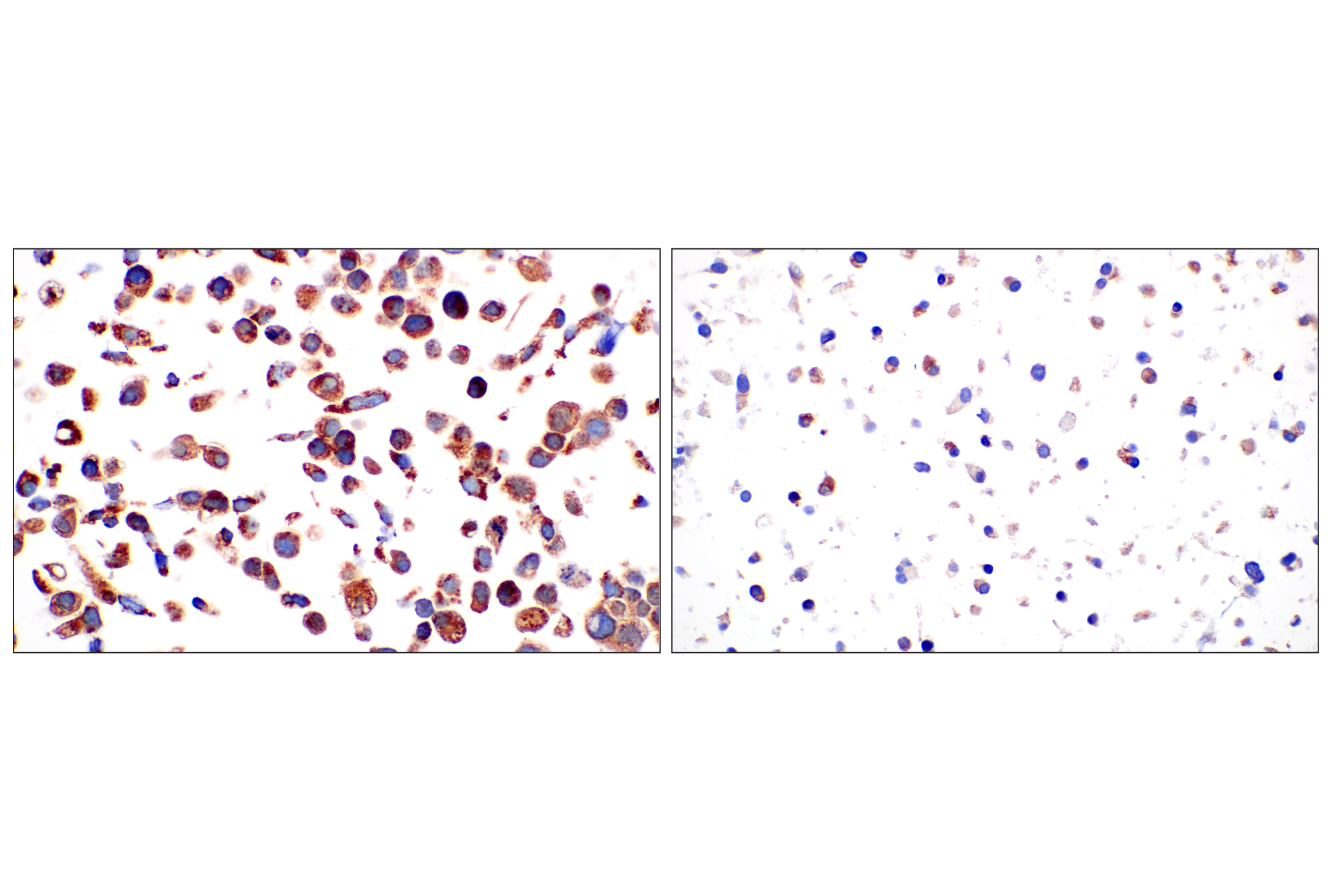
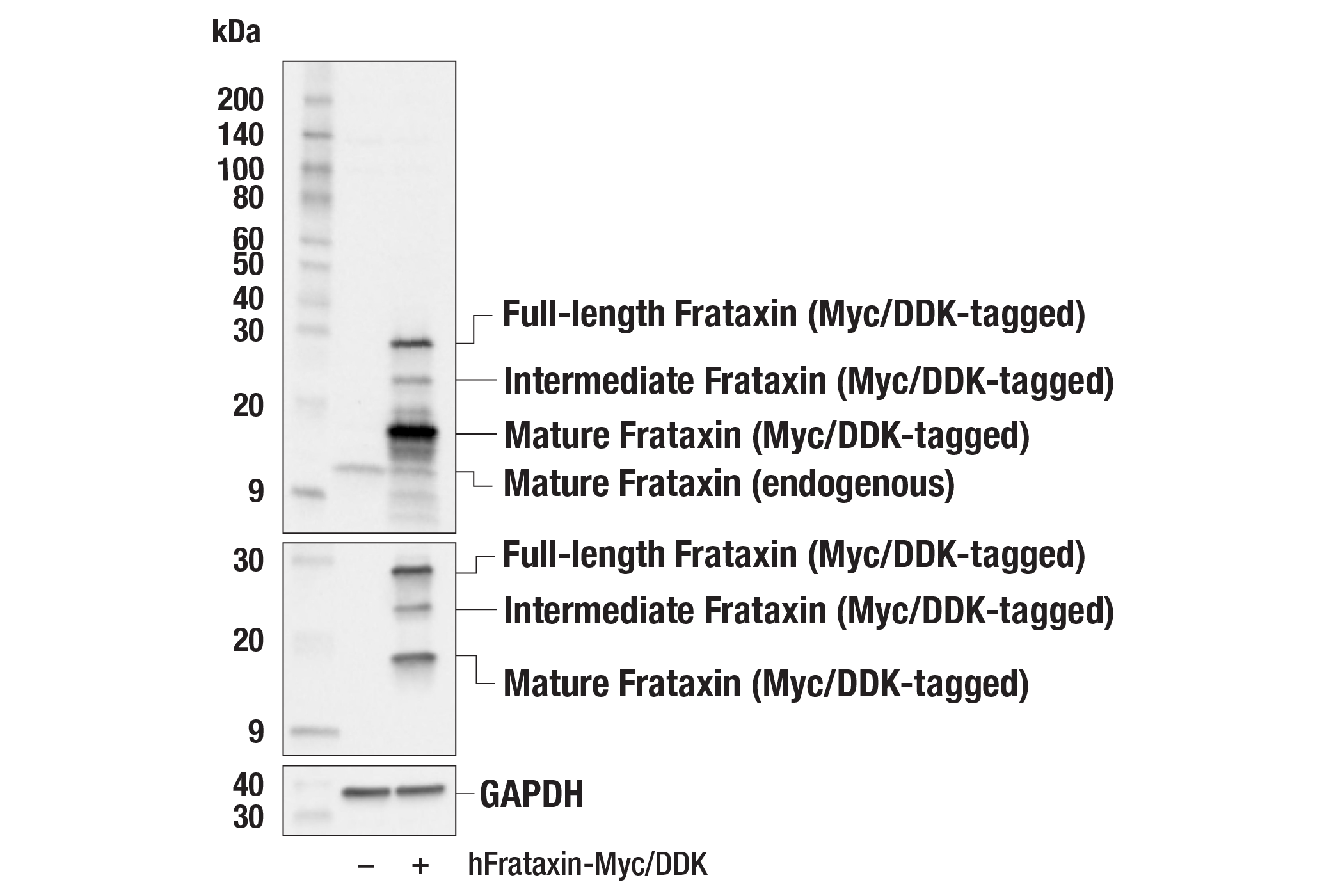
Revision 1

#64668Store at -20C

Cell Signaling Technology

Orders: 877-616-CELL (2355) [email protected]

Support: 877-678-TECH (8324)

Web: [email protected] cellsignal.com

3 Trask LaneDanversMassachusetts01923USA
For Research Use Only. Not for Use in Diagnostic Procedures.
Applications:

WB, IHC-P

REACTIVITY:

H M

SENSITIVITY:

Endogenous

MW (kDa):

Full-length 23 - Intermediate 17 - Mature 14

Source/Isotype:

Rabbit IgG

UniProt ID:

#Q16595

Entrez-Gene Id:

2395

Product Information

Product Usage Information

Application Dilution
Western Blotting 1:1000
Immunohistochemistry (Paraffin) 1:800 - 1:3200

Storage

Supplied in 10 mM sodium HEPES (pH 7.5), 150 mM NaCl, 100 µg/mL BSA, 50% glycerol, and less than 0.02% sodium azide. Store at –20°C. Do not aliquot the antibody.

Specificity / Sensitivity

Frataxin (F4V2S) Rabbit mAb recognizes endogenous levels of total frataxin protein. Species cross-reactivity for IHC-P is human only.

Species Reactivity:

Human, Mouse

Source / Purification

Monoclonal antibody is produced by immunizing animals with recombinant protein specific to the carboxy terminus of human frataxin protein.
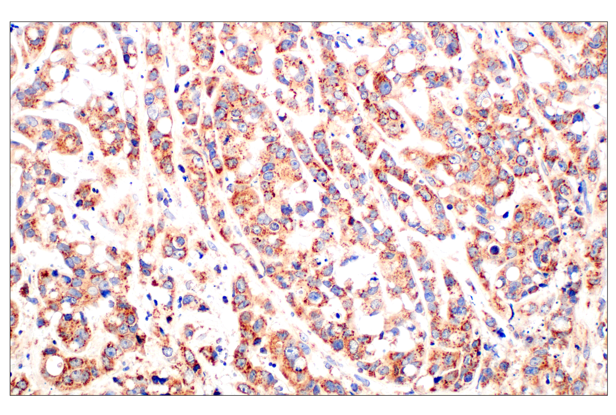
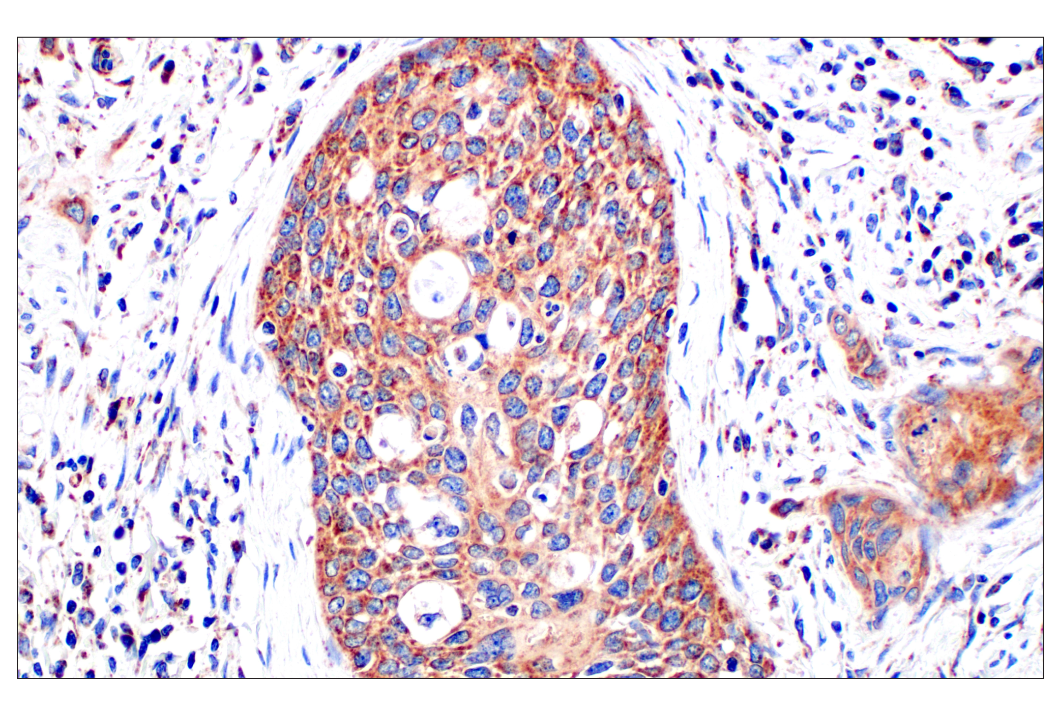
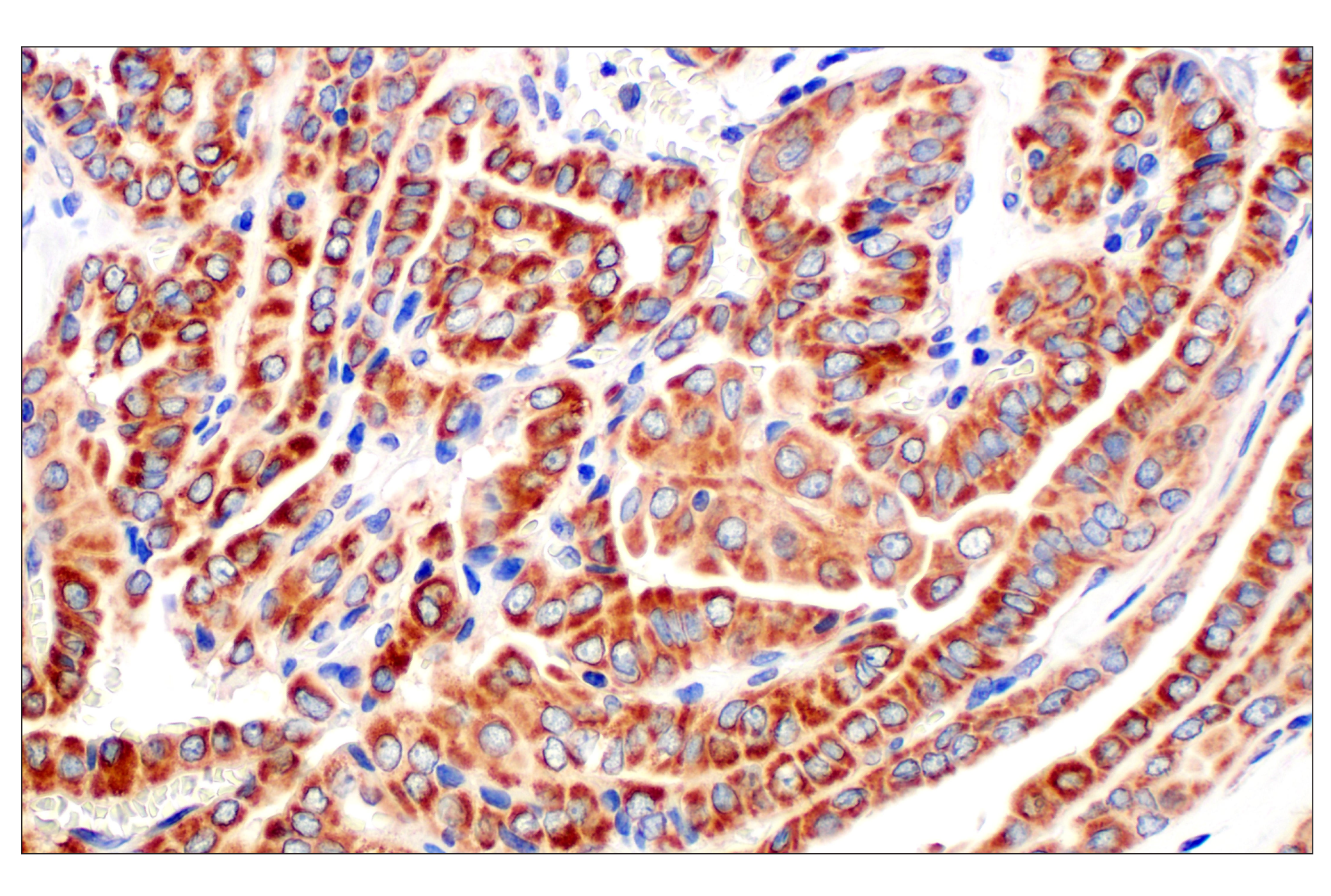
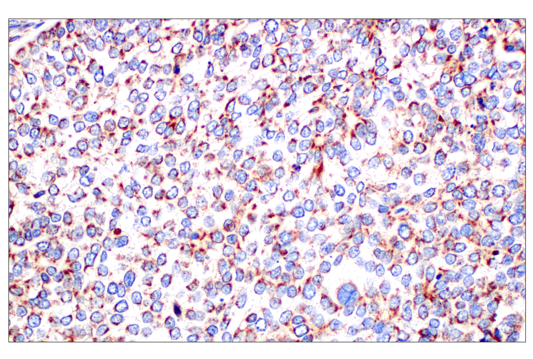
Background

Frataxin is a highly conserved, ubiquitously expressed mitochondrial protein implicated in iron-sulfide cluster (ISC) assembly and iron homeostasis. Synthesized as a cytosolic 210 amino acid precursor protein, frataxin undergoes a two-step proteolytic maturation before translocating to the mitochondria (1). The functions of frataxin in the mitochondria have yet to be fully elucidated, though it has been suggested to be an iron chaperone and activator of persulfide transfer from the cysteine desulfurase NFS1 to the scaffold protein ISCU, which occurs in the early stages of ISC assembly. Proper expression levels of frataxin are vital for cell survival, as is highlighted by the fact that complete loss of this protein is embryonically lethal in mice. In humans, the expansion of GAA repeats in the first intron of the frataxin gene (FXN) results in a reduction in its protein expression levels, leading to the development of  Friedreich’s ataxia (FRDA). FRDA is an early-onset neurodegenerative disorder and the most common inherited form of ataxia, affecting 1 in 50,000 people. The downregulation of frataxin in FRDA results in mitochondrial iron and reactive oxygen species (ROS) accumulation, mitochondrial dysfunction, and ferroptosis. Patients with FRDA present with a myriad of symptoms, including gait disturbances, cardiomyopathy, muscle weakness, and increased incidence of diabetes. Individuals with higher GAA repeats exhibit more severe and earlier onset of symptoms (2-4).

  1. Cherubini, F. et al. (2015) Hum Mol Genet 24, 4296-305.
  2. Ast, T. et al. (2019) Cell 177, 1507-1521.e16.
  3. Du, J. et al. (2020) Redox Biol 32, 101483.
  4. Ahmad, I. et al. (2024) Stem Cell Res 77, 103382.

Species Reactivity

Species reactivity is determined by testing in at least one approved application (e.g., western blot).

Western Blot Buffer

IMPORTANT: For western blots, incubate membrane with diluted primary antibody in 5% w/v nonfat dry milk, 1X TBS, 0.1% Tween® 20 at 4°C with gentle shaking, overnight.

Applications Key

WB: Western Blotting IHC-P: Immunohistochemistry (Paraffin)

Cross-Reactivity Key

H: human M: mouse R: rat Hm: hamster Mk: monkey Vir: virus Mi: mink C: chicken Dm: D. melanogaster X: Xenopus Z: zebrafish B: bovine Dg: dog Pg: pig Sc: S. cerevisiae Ce: C. elegans Hr: horse GP: Guinea Pig Rab: rabbit All: all species expected

Trademarks and Patents

Cell Signaling Technology is a trademark of Cell Signaling Technology, Inc.
All other trademarks are the property of their respective owners. Visit cellsignal.com/trademarks for more information.

使用に関する制限

法的な権限を与えられたCSTの担当者が署名した書面によって別途明示的に合意された場合を除き、 CST、その関連会社または代理店が提供する製品には以下の条件が適用されます。お客様が定める条件でここに定められた条件に含まれるものを超えるもの、 または、ここに定められた条件と異なるものは、法的な権限を与えられたCSTの担当者が別途書面にて受諾した場合を除き、拒絶され、 いかなる効力も効果も有しません。

研究専用 (For Research Use Only) またはこれに類似する表示がされた製品は、 いかなる目的についても FDA または外国もしくは国内のその他の規制機関により承認、認可または許可を受けていません。 お客様は製品を診断もしくは治療目的で使用してはならず、また、製品に表示された内容に違反する方法で使用してはなりません。 CST が販売または使用許諾する製品は、エンドユーザーであるお客様に対し、使途を研究および開発のみに限定して提供されるものです。 診断、予防もしくは治療目的で製品を使用することまたは製品を再販売 (単独であるか他の製品等の一部であるかを問いません) もしくはその他の商業的利用の目的で購入することについては、CST から別途許諾を得る必要があります。 お客様は以下の事項を遵守しなければなりません。(a) CST の製品 (単独であるか他の資材と一緒であるかを問いません) を販売、使用許諾、貸与、寄付もしくはその他の態様で第三者に譲渡したり使用させたりしてはなりません。また、商用の製品を製造するために CST の製品を使用してはなりません。(b) 複製、改変、リバースエンジニアリング、逆コンパイル、 分解または他の方法により製品の構造または技術を解明しようとしてはなりません。また、 CST の製品またはサービスと競合する製品またはサービスを開発する目的で CST の製品を使用してはなりません。(c) CST の製品の商標、商号、ロゴ、特許または著作権に関する通知または表示を除去したり改変したりしてはなりません。(d) CST の製品をCST 製品販売条件(CST’s Product Terms of Sale) および該当する書面のみに従って使用しなければなりません。(e) CST の製品に関連してお客様が使用する第三者の製品またはサービスに関する使用許諾条件、 サービス提供条件またはこれに類する合意事項を遵守しなければなりません。

Revision 1
#64668

Frataxin (F4V2S) Rabbit mAb

Western Blotting Image 1: Frataxin (F4V2S) Rabbit mAb Expand Image
Western blot analysis of extracts from various cell lines and tissues using Frataxin (F4V2S) Rabbit mAb (upper) or GAPDH (D16H11) XP® Rabbit mAb #5174 (lower).
Western Blotting Image 2: Frataxin (F4V2S) Rabbit mAb Expand Image
Western blot analysis of extracts from SW480 cells, transfected with control siRNA (-) or Frataxin siRNA (+), using Frataxin (F4V2S) Rabbit mAb (upper) or GAPDH (D16H11) XP® Rabbit mAb #5174 (lower).
Western Blotting Image 3: Frataxin (F4V2S) Rabbit mAb Expand Image
Western blot analysis of extracts from 293T cells, mock transfected (-) or transfected with a construct expressing Myc/DDK-tagged human frataxin protein (hFrataxin-Myc/DDK; +), using Frataxin (F4V2S) Rabbit mAb (upper), Myc-Tag (71D10) Rabbit mAb #2278 (middle), or GAPDH (D16H11) XP® Rabbit mAb #5174 (lower).
Immunohistochemistry Image 1: Frataxin (F4V2S) Rabbit mAb Expand Image
Immunohistochemical analysis of paraffin-embedded human endometrioid adenocarcinoma using Frataxin (F4V2S) Rabbit mAb.
Immunohistochemistry Image 2: Frataxin (F4V2S) Rabbit mAb Expand Image
Immunohistochemical analysis of paraffin-embedded human colon adenocarcinoma using Frataxin (F4V2S) Rabbit mAb.
Immunohistochemistry Image 3: Frataxin (F4V2S) Rabbit mAb Expand Image
Immunohistochemical analysis of paraffin-embedded human squamous cell carcinoma of the cervix using Frataxin (F4V2S) Rabbit mAb.
Immunohistochemistry Image 4: Frataxin (F4V2S) Rabbit mAb Expand Image
Immunohistochemical analysis of paraffin-embedded human papillary thyroid carcinoma using Frataxin (F4V2S) Rabbit mAb.
Immunohistochemistry Image 5: Frataxin (F4V2S) Rabbit mAb Expand Image
Immunohistochemical analysis of paraffin-embedded human squamous cell lung carcinoma using Frataxin (F4V2S) Rabbit mAb.
Immunohistochemistry Image 6: Frataxin (F4V2S) Rabbit mAb Expand Image
Immunohistochemical analysis of paraffin-embedded normal human liver using Frataxin (F4V2S) Rabbit mAb.
Immunohistochemistry Image 7: Frataxin (F4V2S) Rabbit mAb Expand Image
Immunohistochemical analysis of paraffin-embedded normal human testis using Frataxin (F4V2S) Rabbit mAb.
Immunohistochemistry Image 8: Frataxin (F4V2S) Rabbit mAb Expand Image
Immunohistochemical analysis of paraffin-embedded normal human adrenal gland using Frataxin (F4V2S) Rabbit mAb.
Immunohistochemistry Image 9: Frataxin (F4V2S) Rabbit mAb Expand Image
Immunohistochemical analysis of paraffin-embedded human endometrioid adenocarcinoma using Frataxin (F4V2S) Rabbit mAb (left) compared to concentration-matched Rabbit (DA1E) mAb IgG XP® Isotype Control #3900 (right). 
Immunohistochemistry Image 10: Frataxin (F4V2S) Rabbit mAb Expand Image
Immunohistochemical analysis of paraffin-embedded HepG2 cell pellet (left, high-expressing) or U-87MG cell pellet (right, low-expressing) using Frataxin (F4V2S) Rabbit mAb.