WB, IP, IF-IC
H M R Mk
Endogenous
60, 70
Rabbit
#Q9UN86
9908
Product Information
Product Usage Information
| Application | Dilution |
|---|---|
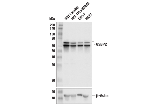
| Western Blotting | 1:1000 |
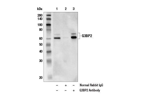
| Immunoprecipitation | 1:50 |
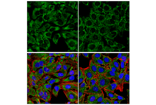
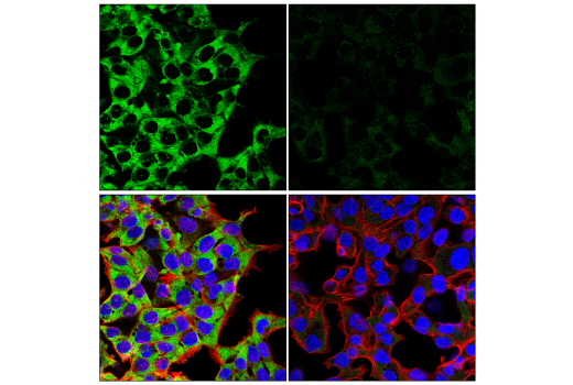
| Immunofluorescence (Immunocytochemistry) | 1:100 - 1:400 |
Storage
Specificity / Sensitivity
Species Reactivity:
Human, Mouse, Rat, Monkey
Source / Purification
Polyclonal antibodies are produced by immunizing animals with a synthetic peptide corresponding to residues near the carboxy terminus of human G3BP2 protein. Antibodies are purified by peptide affinity chromatography.
Background
Stress granules (SGs) are cytoplasmic mRNA-protein condensates formed in response to cellular stressors, such as oxidative stress, ultraviolet radiation, and viral infection (1). The Ras-GTPase-activating protein-binding proteins (G3BPs), consisting of G3BP1 and G3BP2, are key nucleating factors essential for SG formation (2). As their name suggests, G3BPs were originally identified as proteins that interact with the SH3 domain of Ras GTPase-activating protein (RasGAP) (3). However, a recent study argues that G3BP1 may not be a genuine RasGAP-binding partner, suggesting that further studies are necessary to validate this particular function (4). Rather, G3BPs are well known to be involved in mRNA decay and inhibition of translation initiation (5,6). The SARS-CoV-2 virus has also been reported to target G3BPs via interaction between the viral N protein and the N-terminal domain of G3BP. This binding prevents G3BPs from interacting with other SG-associated proteins, therefore suppressing SG assembly and promoting viral replication (7). G3BP2 in particular has also been reported to play a role in the initiation of breast tumor formation, as well as affect the expression of PD-L1 in breast and glioblastoma cells under stress conditions (8,9).
- Riggs, C.L. et al. (2020) J Cell Sci 133, jcs242487. doi: 10.1242/jcs.242487.
- Wang, F. et al. (2020) Pharmacol Res 161, 105143.
- Parker, F. et al. (1996) Mol Cell Biol 16, 2561-9.
- Annibaldi, A. et al. (2011) PLoS One 6, e29024.
- Fischer-Kešo, R. et al. (2014) Mol Cell Biol 34, 4244-56.
- Fischer, J.W. et al. (2020) Mol Cell 78, 70-84.e6.
- Luo, L. et al. (2021) Sci Bull (Beijing) 66, 1194-1204.
- Gupta, N. et al. (2017) Proc Natl Acad Sci U S A 114, 1033-1038.
- Zhang, Y. et al. (2021) Mol Oncol, doi: 10.1002/1878-0261.12915.
Species Reactivity
Species reactivity is determined by testing in at least one approved application (e.g., western blot).
Western Blot Buffer
IMPORTANT: For western blots, incubate membrane with diluted primary antibody in 5% w/v BSA, 1X TBS, 0.1% Tween® 20 at 4°C with gentle shaking, overnight.
Applications Key
WB: Western Blotting IP: Immunoprecipitation IF-IC: Immunofluorescence (Immunocytochemistry)
Cross-Reactivity Key
H: human M: mouse R: rat Hm: hamster Mk: monkey Vir: virus Mi: mink C: chicken Dm: D. melanogaster X: Xenopus Z: zebrafish B: bovine Dg: dog Pg: pig Sc: S. cerevisiae Ce: C. elegans Hr: horse GP: Guinea Pig Rab: rabbit All: all species expected
Trademarks and Patents
使用に関する制限
法的な権限を与えられたCSTの担当者が署名した書面によって別途明示的に合意された場合を除き、 CST、その関連会社または代理店が提供する製品には以下の条件が適用されます。お客様が定める条件でここに定められた条件に含まれるものを超えるもの、 または、ここに定められた条件と異なるものは、法的な権限を与えられたCSTの担当者が別途書面にて受諾した場合を除き、拒絶され、 いかなる効力も効果も有しません。
研究専用 (For Research Use Only) またはこれに類似する表示がされた製品は、 いかなる目的についても FDA または外国もしくは国内のその他の規制機関により承認、認可または許可を受けていません。 お客様は製品を診断もしくは治療目的で使用してはならず、また、製品に表示された内容に違反する方法で使用してはなりません。 CST が販売または使用許諾する製品は、エンドユーザーであるお客様に対し、使途を研究および開発のみに限定して提供されるものです。 診断、予防もしくは治療目的で製品を使用することまたは製品を再販売 (単独であるか他の製品等の一部であるかを問いません) もしくはその他の商業的利用の目的で購入することについては、CST から別途許諾を得る必要があります。 お客様は以下の事項を遵守しなければなりません。(a) CST の製品 (単独であるか他の資材と一緒であるかを問いません) を販売、使用許諾、貸与、寄付もしくはその他の態様で第三者に譲渡したり使用させたりしてはなりません。また、商用の製品を製造するために CST の製品を使用してはなりません。(b) 複製、改変、リバースエンジニアリング、逆コンパイル、 分解または他の方法により製品の構造または技術を解明しようとしてはなりません。また、 CST の製品またはサービスと競合する製品またはサービスを開発する目的で CST の製品を使用してはなりません。(c) CST の製品の商標、商号、ロゴ、特許または著作権に関する通知または表示を除去したり改変したりしてはなりません。(d) CST の製品をCST 製品販売条件(CST’s Product Terms of Sale) および該当する書面のみに従って使用しなければなりません。(e) CST の製品に関連してお客様が使用する第三者の製品またはサービスに関する使用許諾条件、 サービス提供条件またはこれに類する合意事項を遵守しなければなりません。