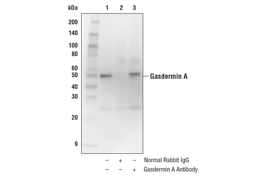
WB, IP
H
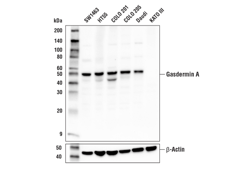
Endogenous
49
Rabbit
#Q96QA5
284110
Product Information
Product Usage Information
| Application | Dilution |
|---|---|
| Western Blotting | 1:1000 |
| Immunoprecipitation | 1:50 |
Storage
Specificity / Sensitivity
Species Reactivity:
Human
Source / Purification
Polyclonal antibodies are produced by immunizing animals with a synthetic peptide corresponding to residues surrounding Gly256 of human Gasdermin A protein. Antibodies are purified by protein A and peptide affinity chromatography.
Background
The gasdermin family, which includes GSDMA, GSDMB, GSDMC, GSDMD, and GSDME, has been shown to play a role in inflammation and cell death. Gasdermin D has been reported to have a critical role as a downstream effector of pyroptosis (1,2). Pyroptosis is a lytic type of cell death triggered by inflammasomes, multiprotein complexes assembled in response to pathogen-associated molecular patterns (PAMPs) or danger-associated molecular patterns (DAMPs) that result in the activation of caspase-1 and subsequent cleavage of pro-inflammatory cytokines IL-1β and IL-18 (3). Gasdermin D was identified by two independent groups as a substrate of inflammatory caspases, caspase-1 and caspase-11/4/5, producing two fragments: GSDMD-N and GSDMD-C. Cleavage results in release of an intramolecular inhibitory interaction between the N- and C-terminal domains, allowing the N-terminal fragment GSDMD-N to initiate pyroptosis through the formation of pores on the plasma membrane (4-7).
Gasdermin A (GSDMA) is preferentially expressed in the epithelium of the skin and gastrointestinal tract and is frequently suppressed in gastric cancer (8-10). Mice express three Gasdermin A genes termed GSDMA1-3 (8). The role of Gasdermin A has been associated with cellular differentiation, apoptosis, pyroptosis, and autophagy (10-15). Expression of an N-terminal fragment of GSDMA3, but not the full-length protein, induces pyroptosis, but the mechanisms of any cleavage of GSDMA3 are unknown (12). The most widely studied mouse form, GSDMA3, is expressed in mouse keratinocytes and is associated with skin differentiation and inflammation, and a dominant mutation has been found to play a causative role in alopecia (16,17).
- Kayagaki, N. et al. (2015) Nature 526, 666-71.
- Shi, J. et al. (2015) Nature 526, 660-5.
- Broz, P. and Dixit, V.M. (2016) Nat Rev Immunol 16, 407-20.
- Aglietti, R.A. et al. (2016) Proc Natl Acad Sci USA 113, 7858-63.
- Ding, J. et al. (2016) Nature 535, 111-6.
- Liu, X. et al. (2016) Nature 535, 153-8.
- Sborgi, L. et al. (2016) EMBO J 35, 1766-78.
- Tamura, M. et al. (2007) Genomics 89, 618-29.
- Saeki, N. et al. (2000) Mamm Genome 11, 718-24.
- Saeki, N. et al. (2007) Oncogene 26, 6488-98.
- Li, J. et al. (2010) Biochem Biophys Res Commun 403, 18-23.
- Shi, J. et al. (2015) Nature 526, 660-5.
- Shi, P. et al. (2015) Biochem J 468, 325-36.
- Lei, M. et al. (2011) Histochem Cell Biol 136, 335-43.
- Lei, M. et al. (2012) Histochem Cell Biol 138, 385-96.
- Runkel, F. et al. (2004) Genomics 84, 824-35.
- Zhou, Y. et al. (2012) Am J Pathol 180, 763-74.
Species Reactivity
Species reactivity is determined by testing in at least one approved application (e.g., western blot).
Western Blot Buffer
IMPORTANT: For western blots, incubate membrane with diluted primary antibody in 5% w/v BSA, 1X TBS, 0.1% Tween® 20 at 4°C with gentle shaking, overnight.
Applications Key
WB: Western Blotting IP: Immunoprecipitation
Cross-Reactivity Key
H: human M: mouse R: rat Hm: hamster Mk: monkey Vir: virus Mi: mink C: chicken Dm: D. melanogaster X: Xenopus Z: zebrafish B: bovine Dg: dog Pg: pig Sc: S. cerevisiae Ce: C. elegans Hr: horse GP: Guinea Pig Rab: rabbit All: all species expected
Trademarks and Patents
使用に関する制限
法的な権限を与えられたCSTの担当者が署名した書面によって別途明示的に合意された場合を除き、 CST、その関連会社または代理店が提供する製品には以下の条件が適用されます。お客様が定める条件でここに定められた条件に含まれるものを超えるもの、 または、ここに定められた条件と異なるものは、法的な権限を与えられたCSTの担当者が別途書面にて受諾した場合を除き、拒絶され、 いかなる効力も効果も有しません。
研究専用 (For Research Use Only) またはこれに類似する表示がされた製品は、 いかなる目的についても FDA または外国もしくは国内のその他の規制機関により承認、認可または許可を受けていません。 お客様は製品を診断もしくは治療目的で使用してはならず、また、製品に表示された内容に違反する方法で使用してはなりません。 CST が販売または使用許諾する製品は、エンドユーザーであるお客様に対し、使途を研究および開発のみに限定して提供されるものです。 診断、予防もしくは治療目的で製品を使用することまたは製品を再販売 (単独であるか他の製品等の一部であるかを問いません) もしくはその他の商業的利用の目的で購入することについては、CST から別途許諾を得る必要があります。 お客様は以下の事項を遵守しなければなりません。(a) CST の製品 (単独であるか他の資材と一緒であるかを問いません) を販売、使用許諾、貸与、寄付もしくはその他の態様で第三者に譲渡したり使用させたりしてはなりません。また、商用の製品を製造するために CST の製品を使用してはなりません。(b) 複製、改変、リバースエンジニアリング、逆コンパイル、 分解または他の方法により製品の構造または技術を解明しようとしてはなりません。また、 CST の製品またはサービスと競合する製品またはサービスを開発する目的で CST の製品を使用してはなりません。(c) CST の製品の商標、商号、ロゴ、特許または著作権に関する通知または表示を除去したり改変したりしてはなりません。(d) CST の製品をCST 製品販売条件(CST’s Product Terms of Sale) および該当する書面のみに従って使用しなければなりません。(e) CST の製品に関連してお客様が使用する第三者の製品またはサービスに関する使用許諾条件、 サービス提供条件またはこれに類する合意事項を遵守しなければなりません。