| Product Includes | Product # | Quantity | Mol. Wt | Isotype/Source |
|---|---|---|---|---|
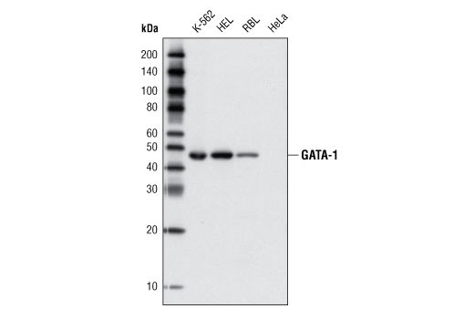
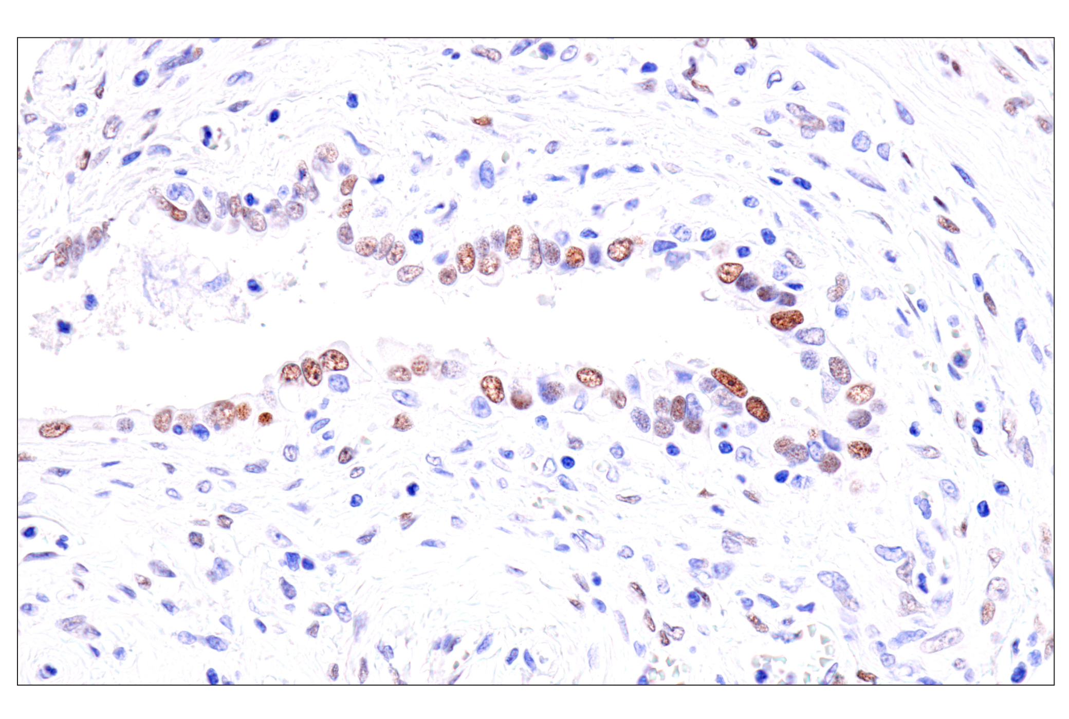
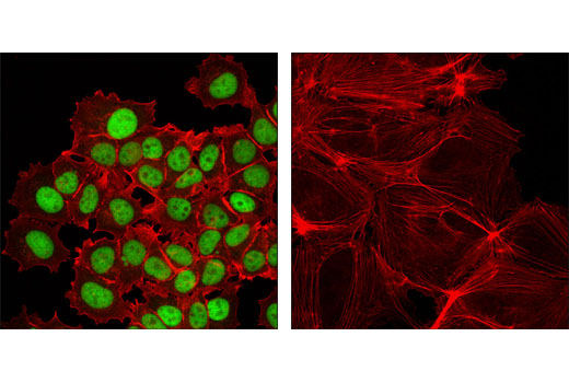
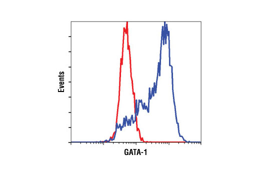
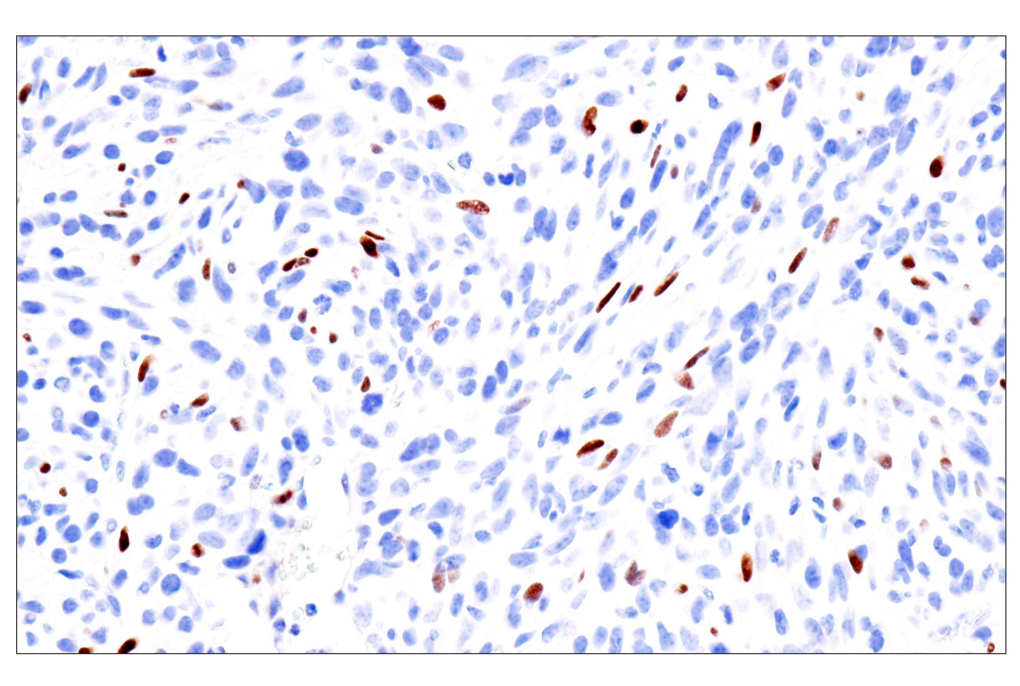
| GATA-1 (D52H6) XP® Rabbit mAb | 3535 | 20 µl | 43 kDa | Rabbit IgG |
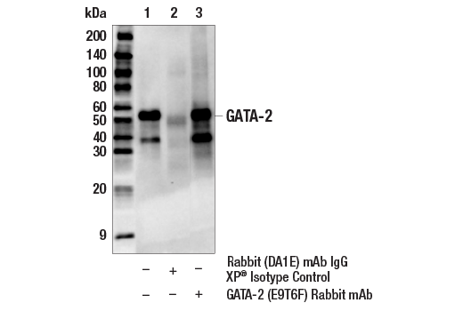
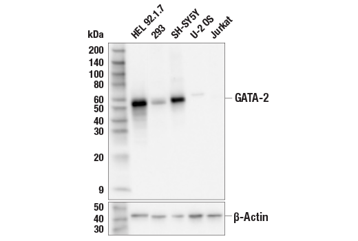
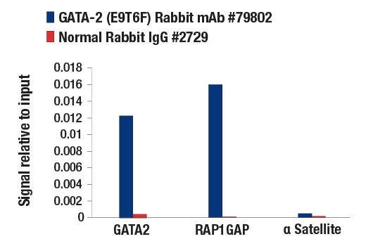
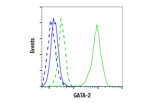
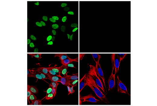
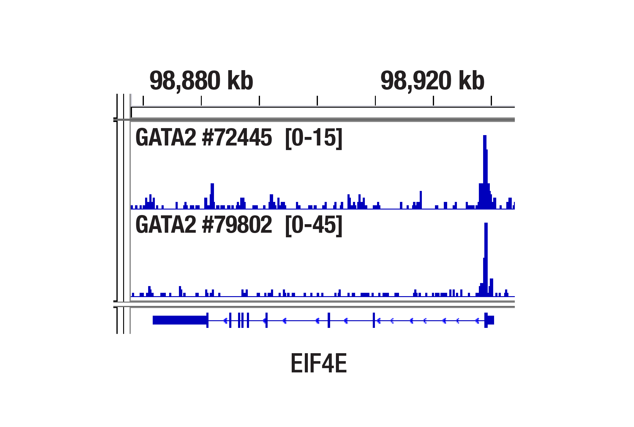
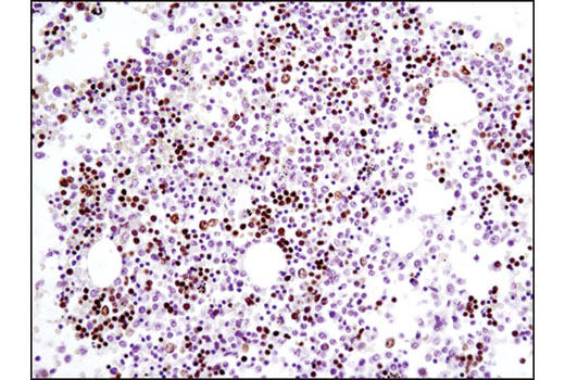
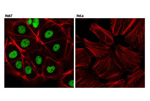
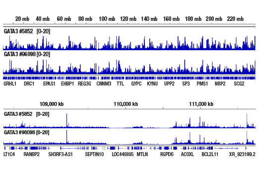
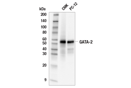
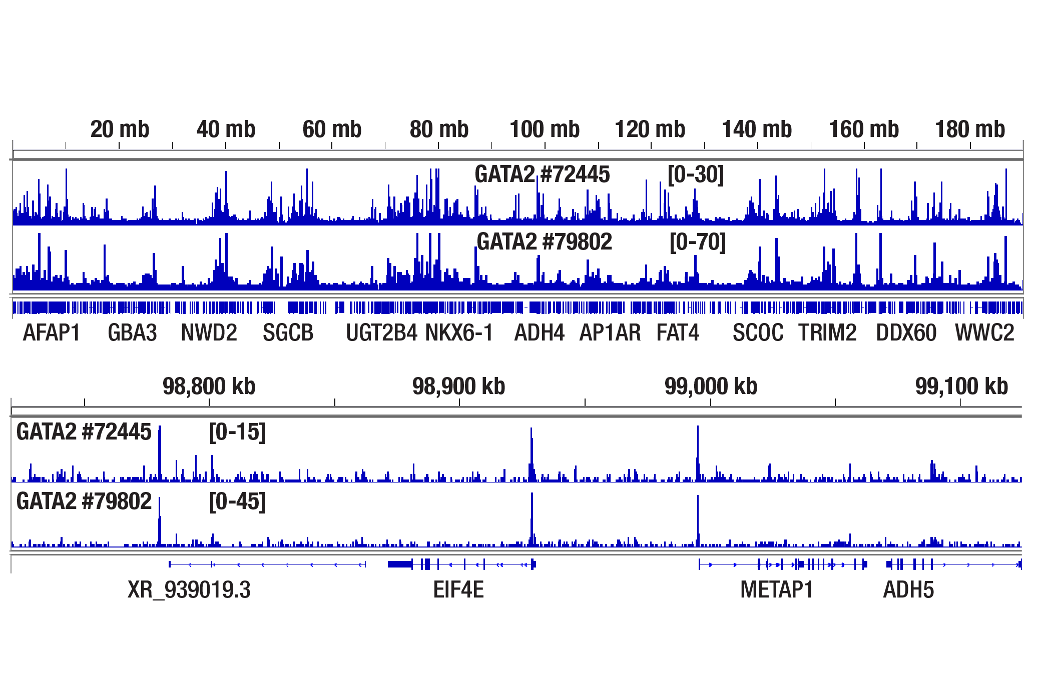
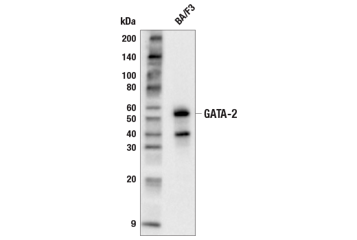
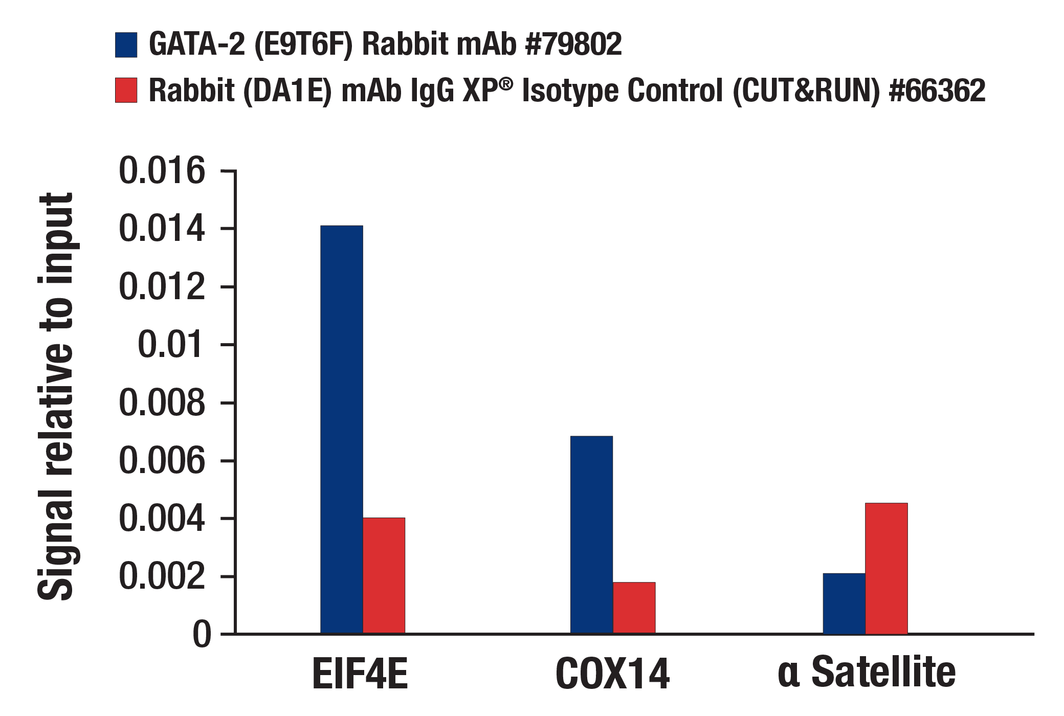
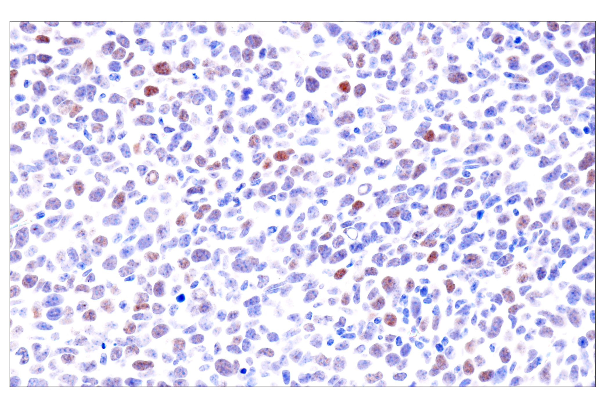
| GATA-2 (E9T6F) Rabbit mAb | 79802 | 20 µl | 51 kDa | Rabbit IgG |
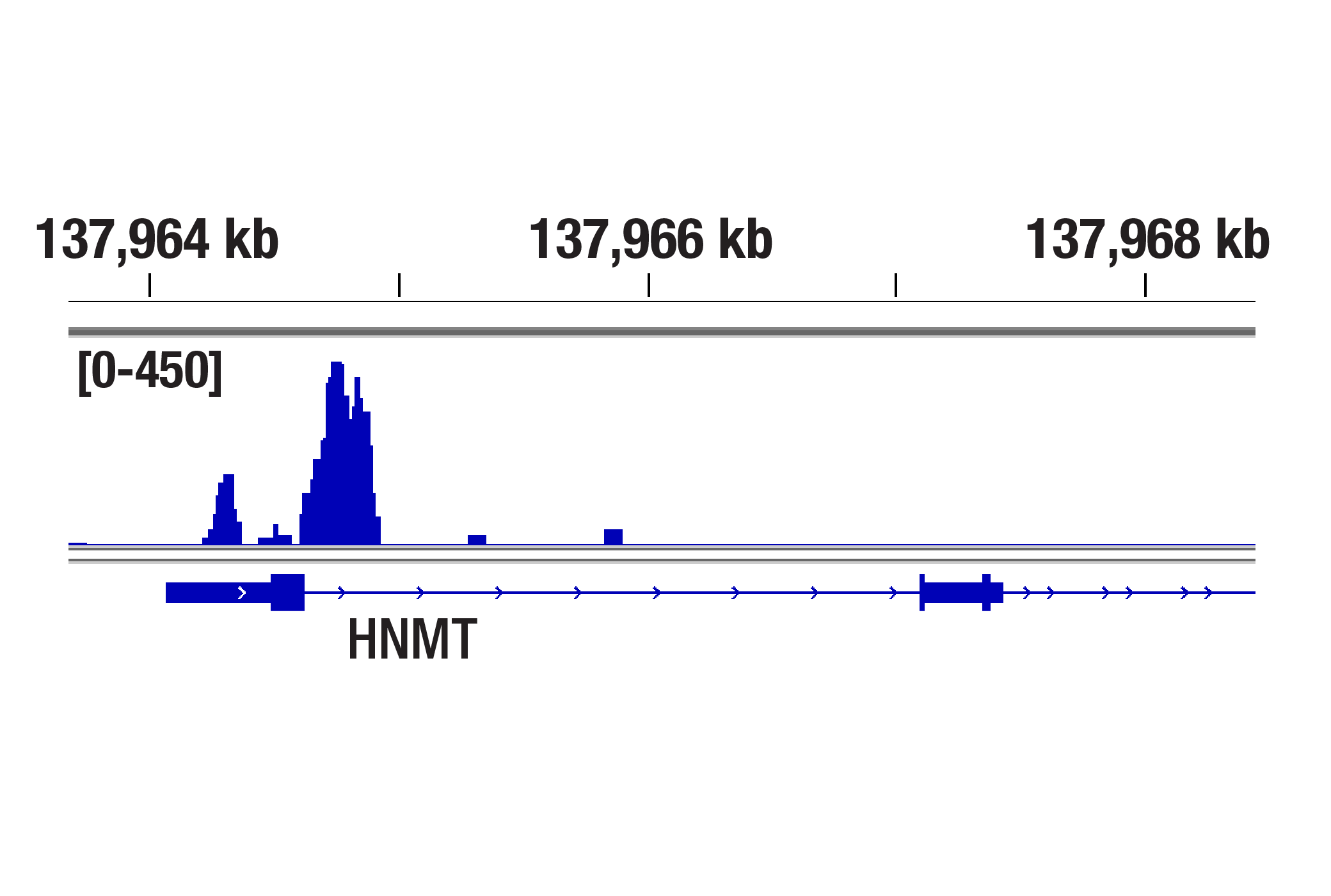
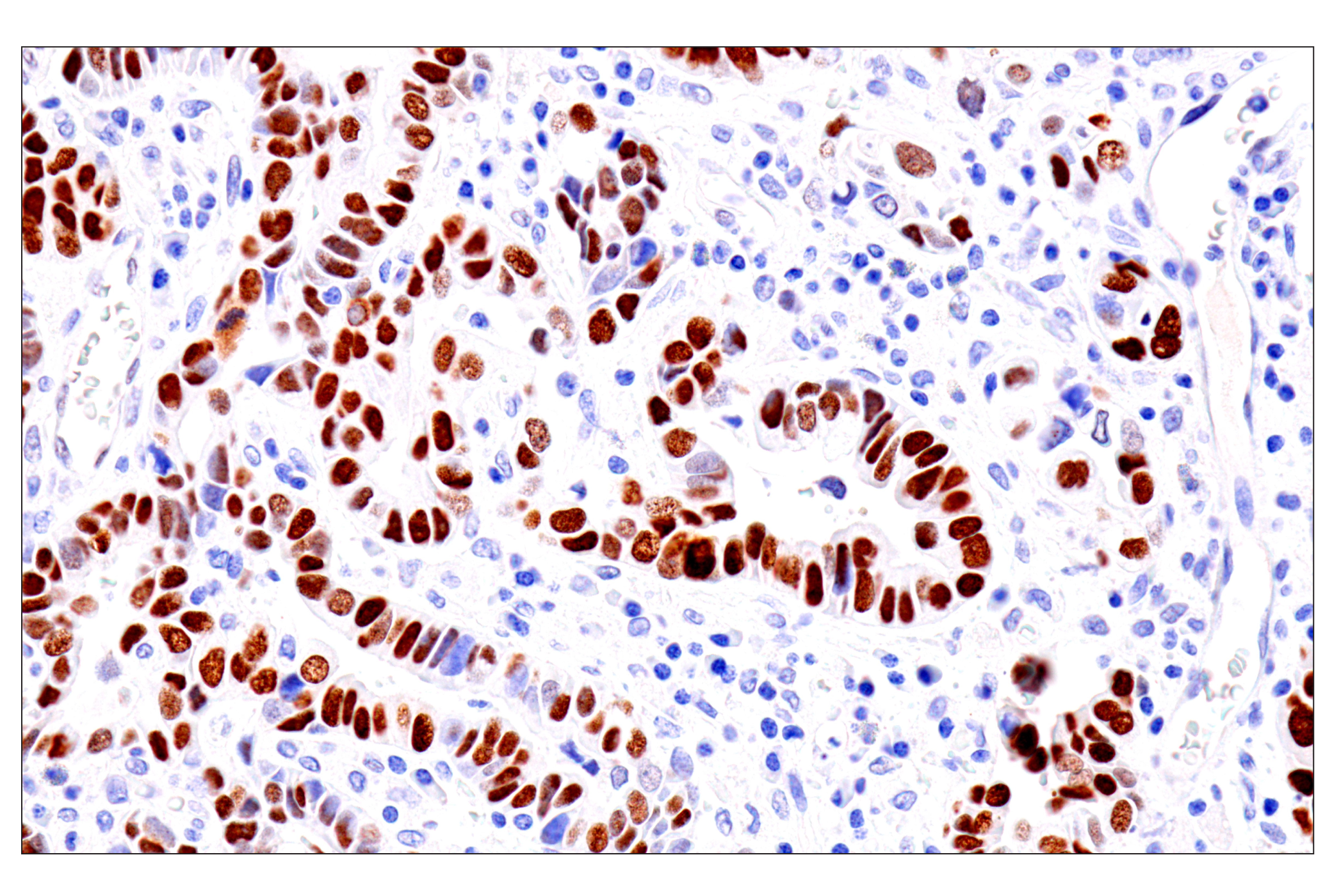
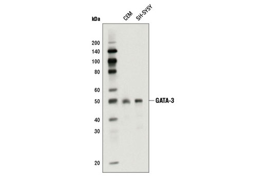
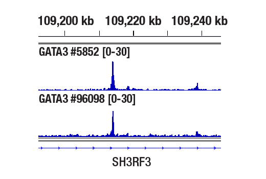
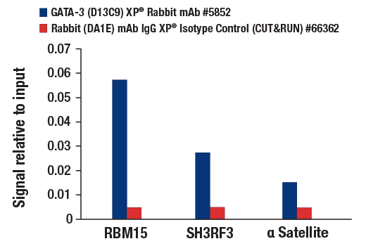
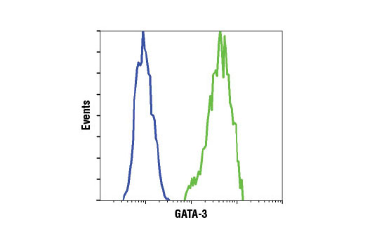
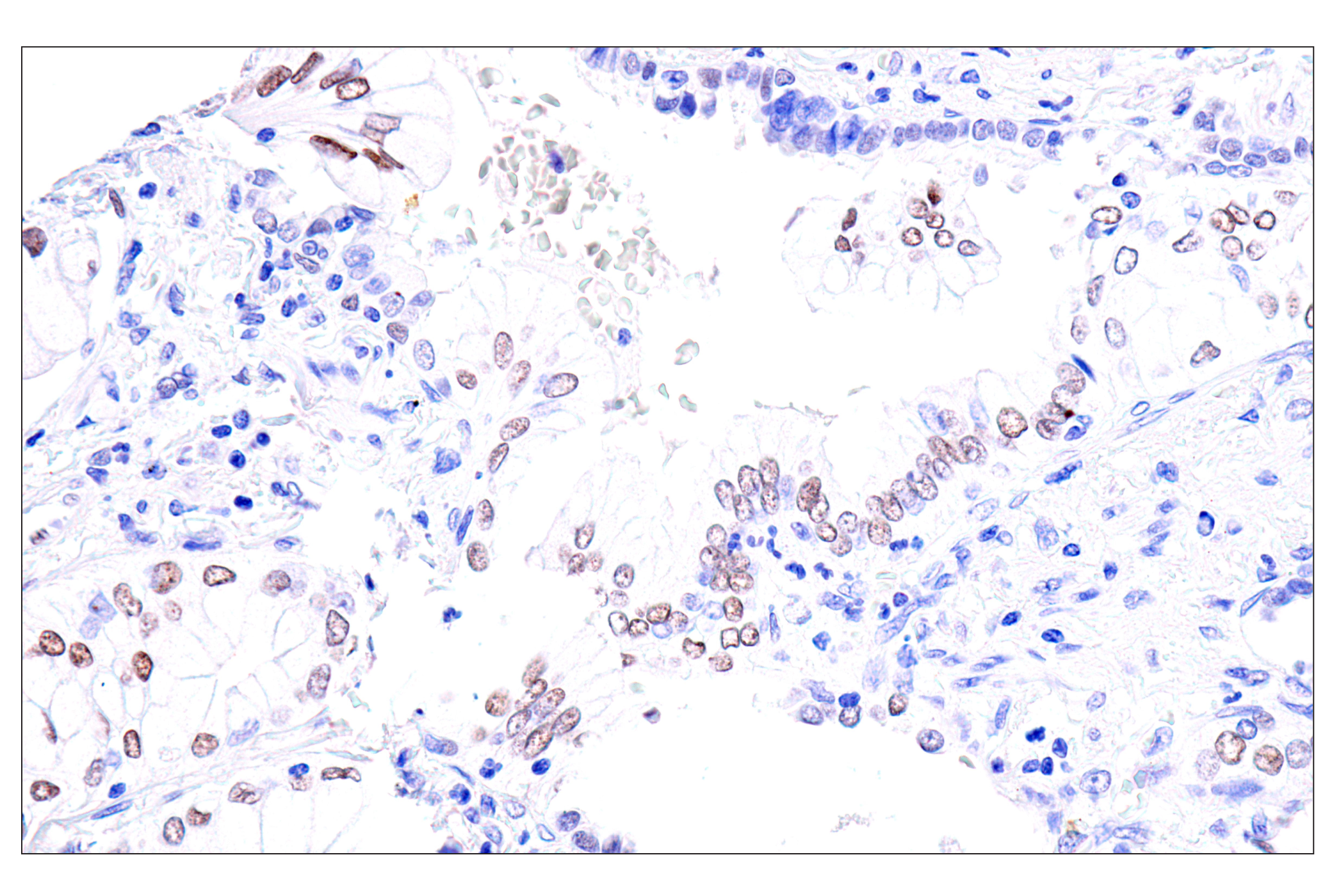
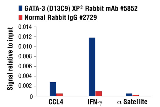
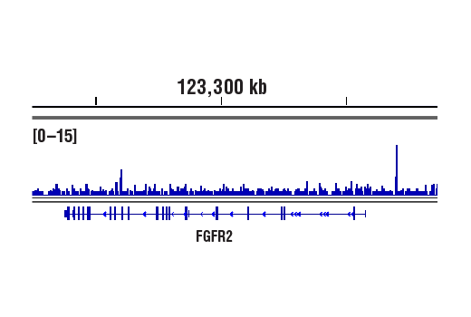
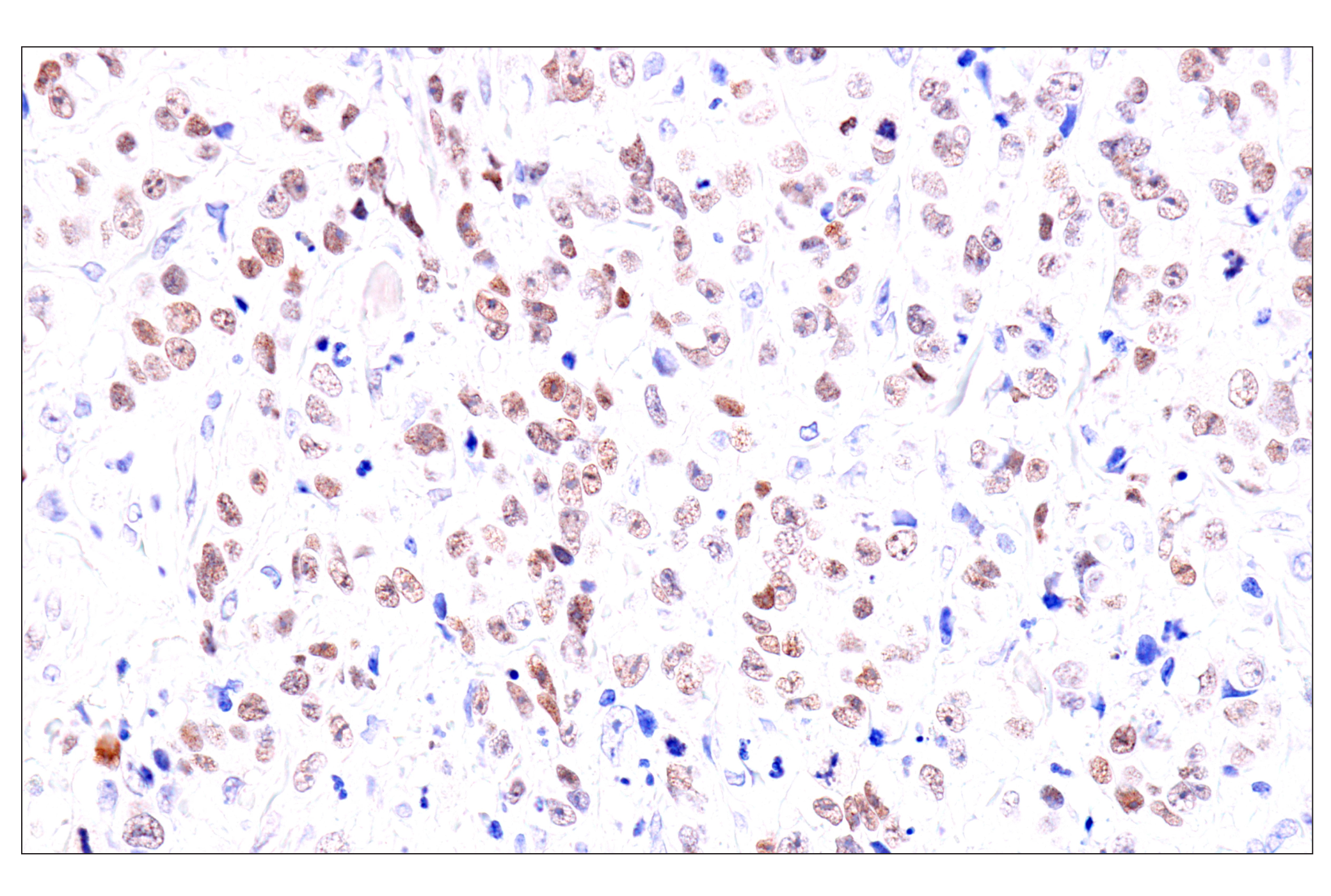
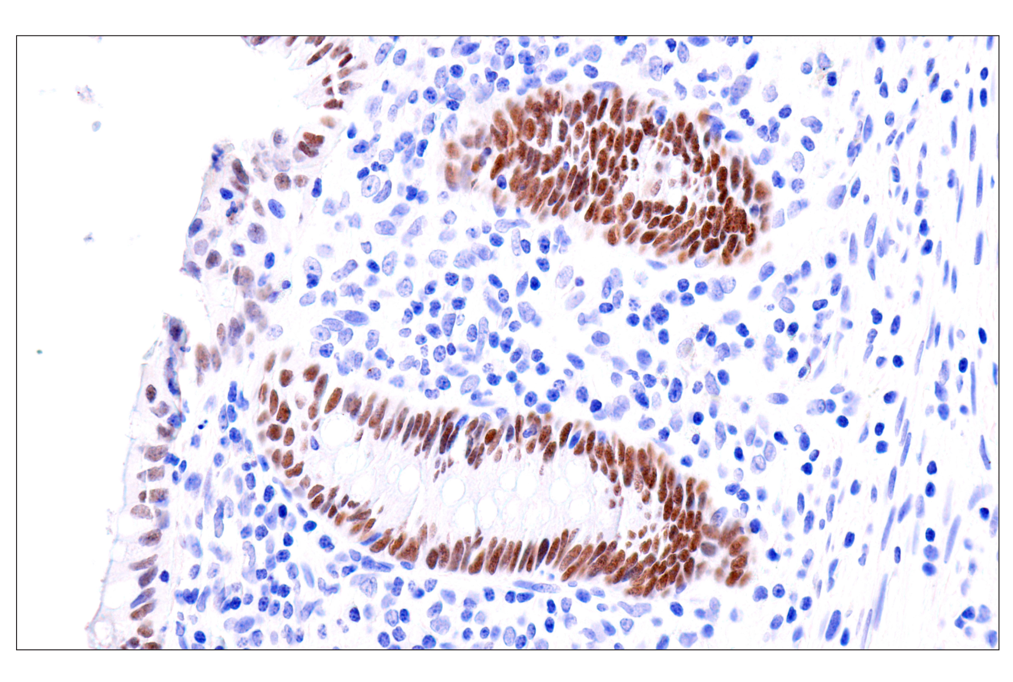
| GATA-3 (D13C9) XP® Rabbit mAb | 5852 | 20 µl | 48 kDa | Rabbit IgG |
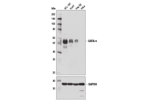
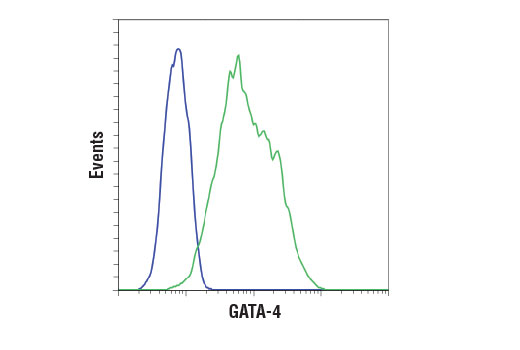
| GATA-4 (D3A3M) Rabbit mAb | 36966 | 20 µl | 55 kDa | Rabbit IgG |
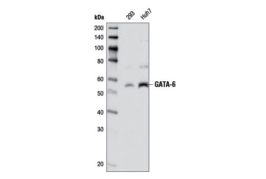
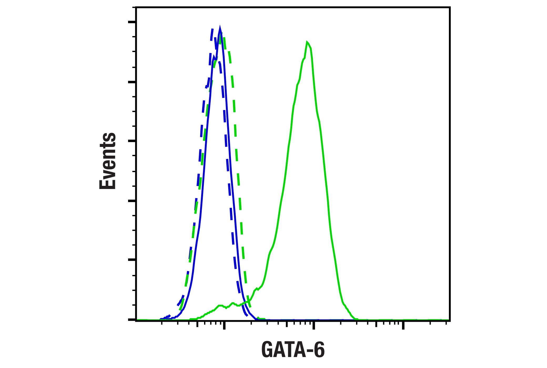
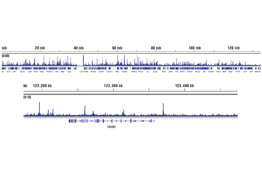
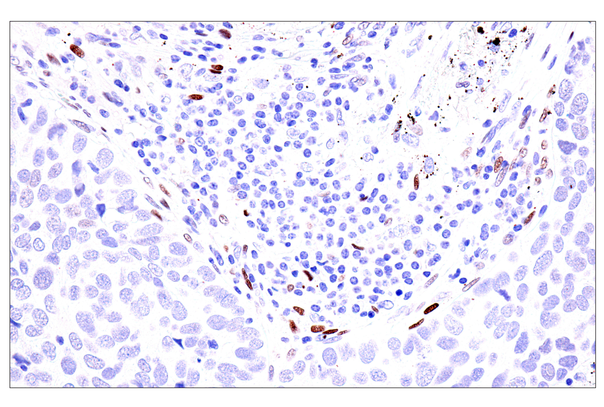
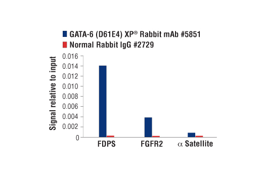
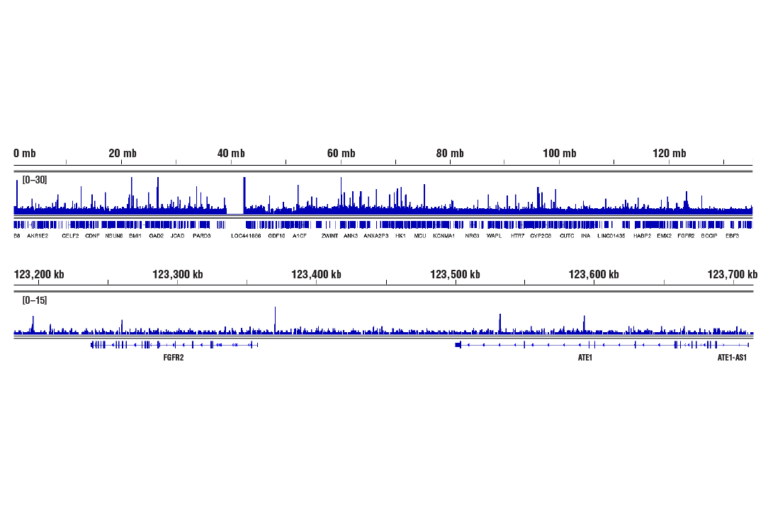
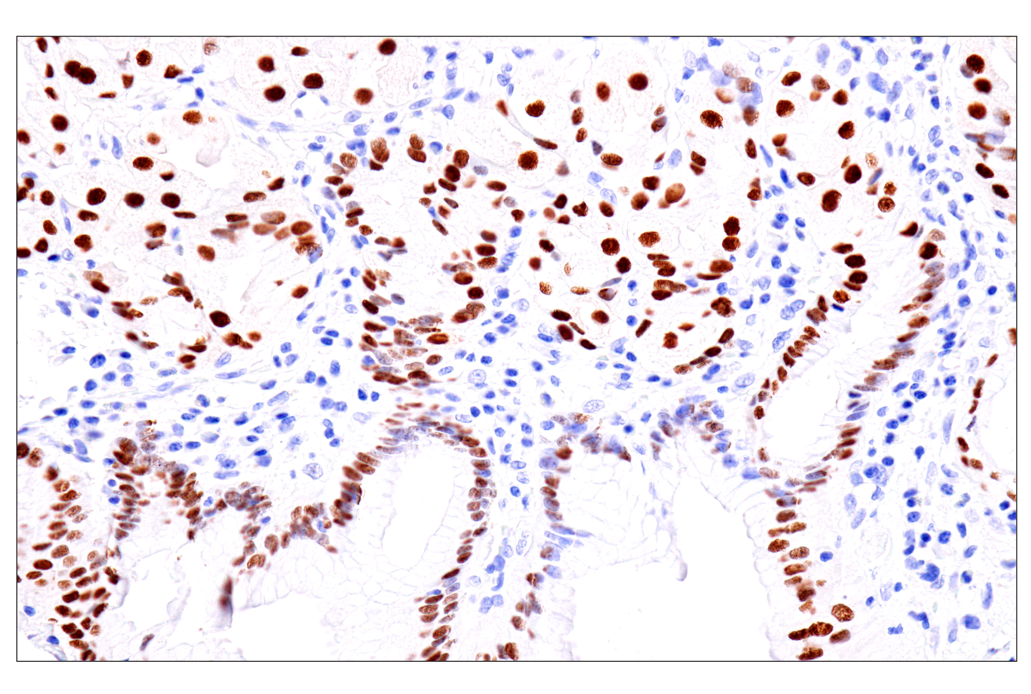
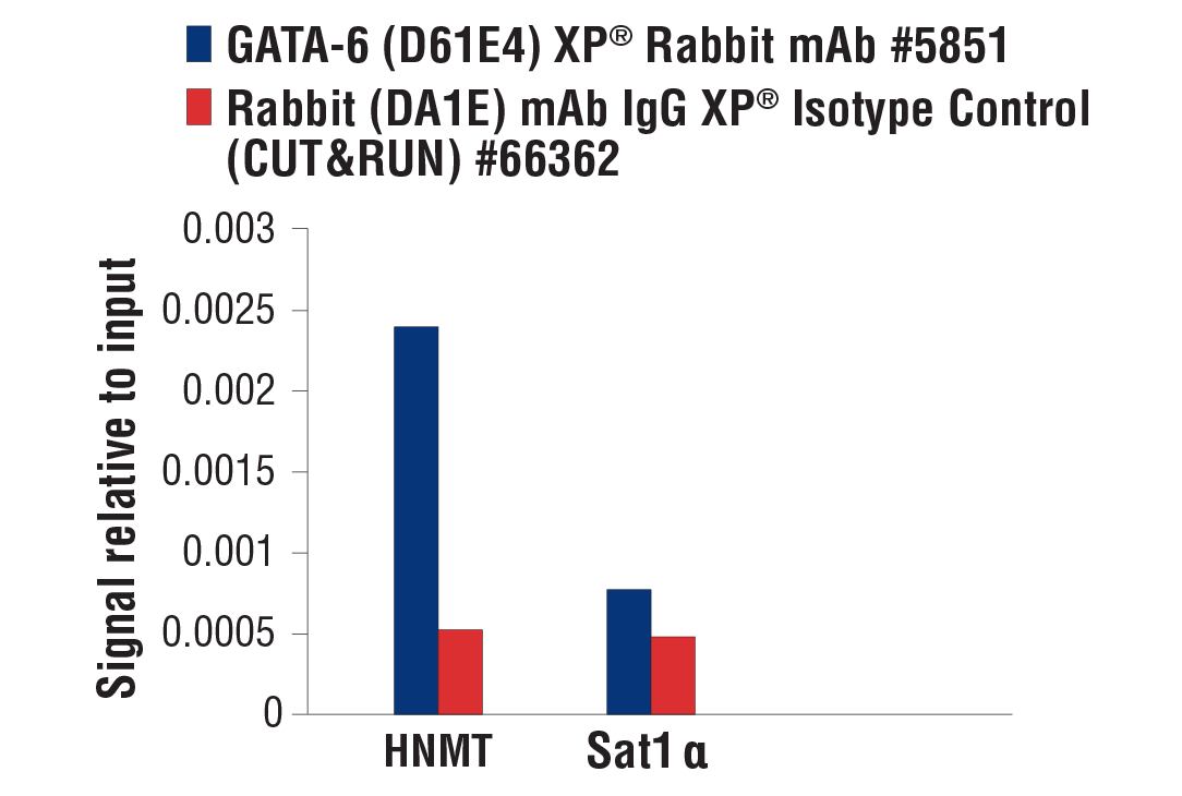
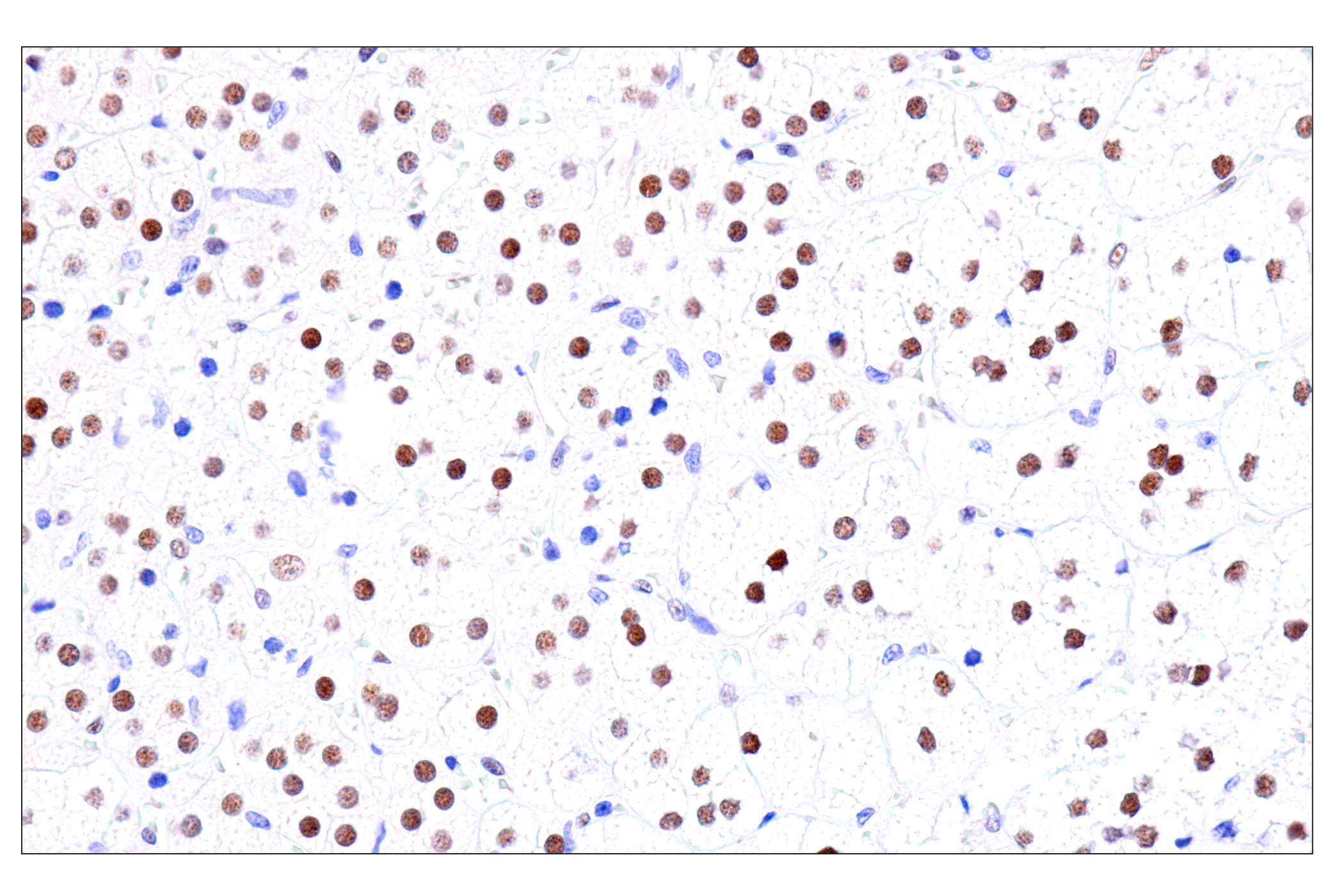
| GATA-6 (D61E4) XP® Rabbit mAb | 5851 | 20 µl | 55 kDa | Rabbit IgG |
| Anti-rabbit IgG, HRP-linked Antibody | 7074 | 100 µl | Goat |
Please visit cellsignal.com for individual component applications, species cross-reactivity, dilutions, protocols, and additional product information.
Description
The GATA Transcription Factor Antibody Sampler Kit provides an economical means of evaluating total levels of GATA family proteins. The kit includes enough antibodies to perform two western blot experiments with each primary antibody.
Storage
Background
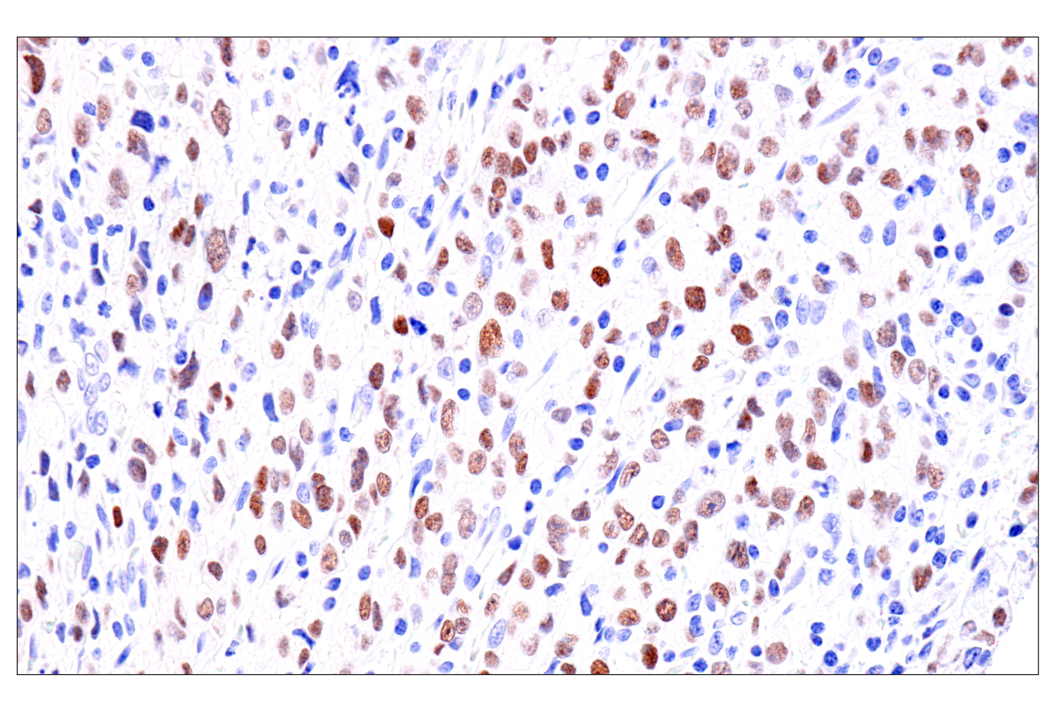
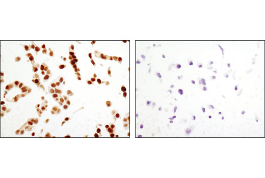
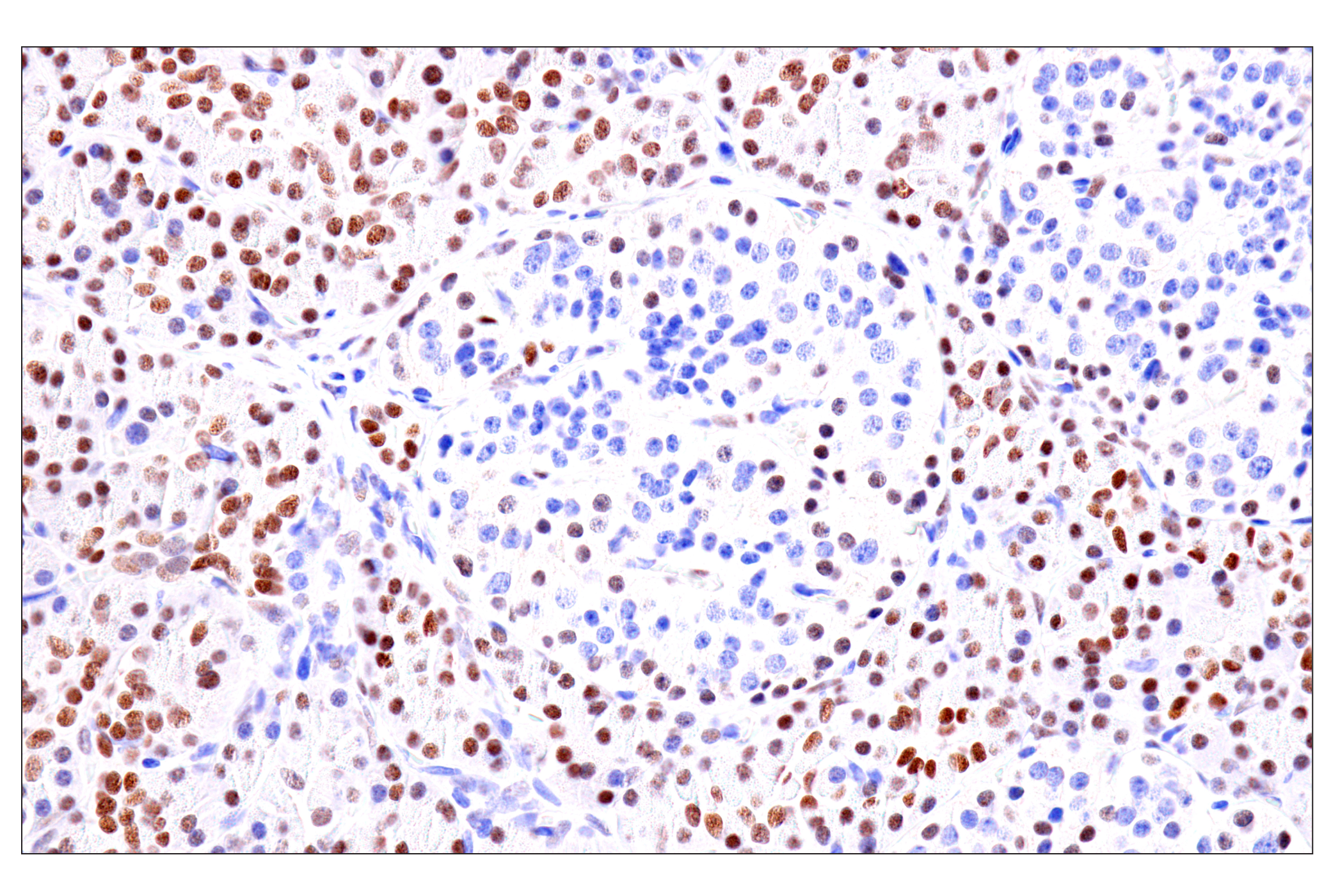
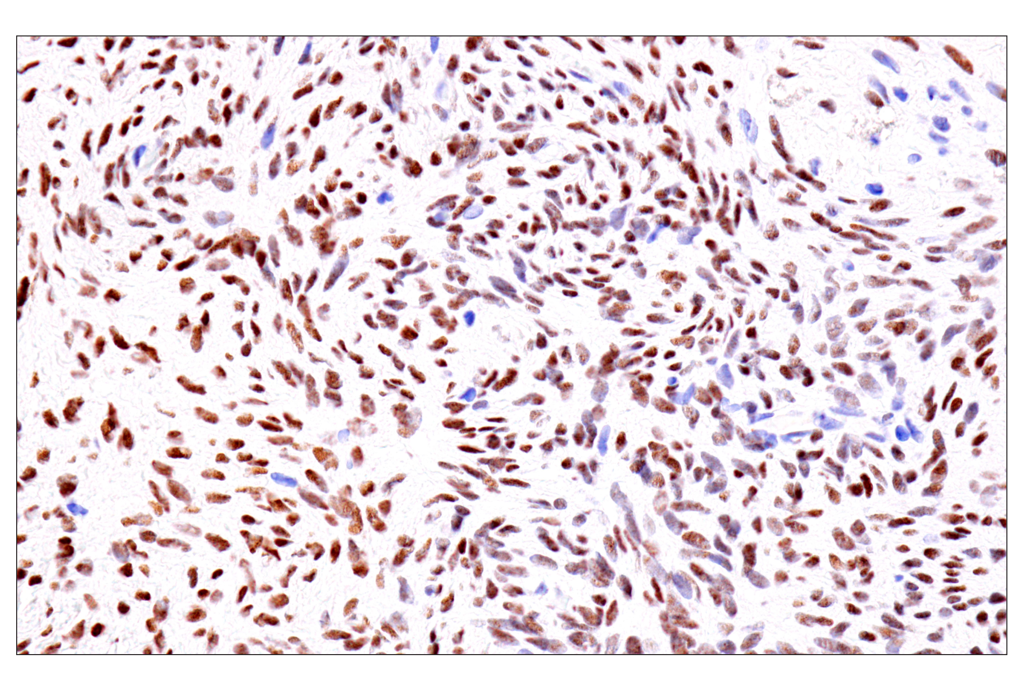
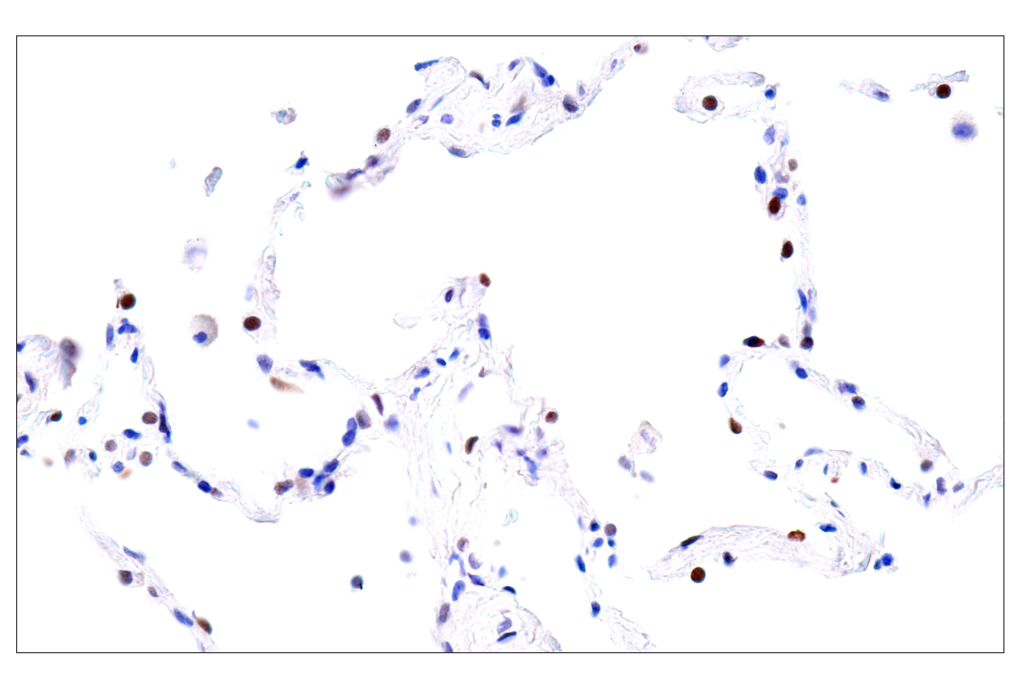
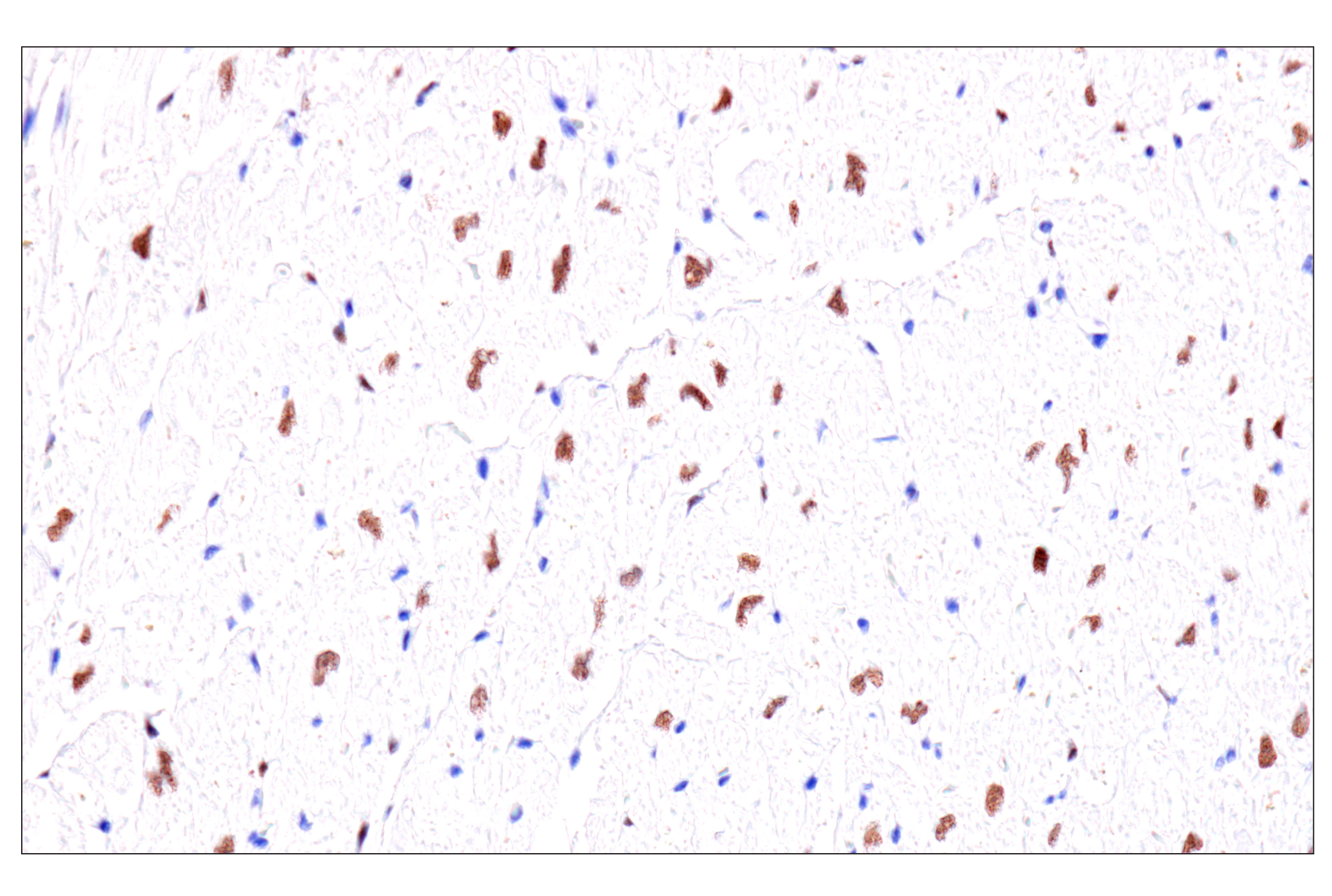
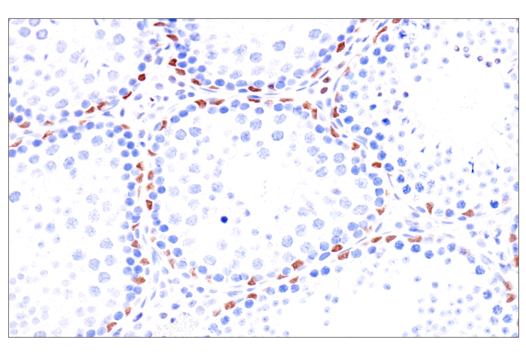
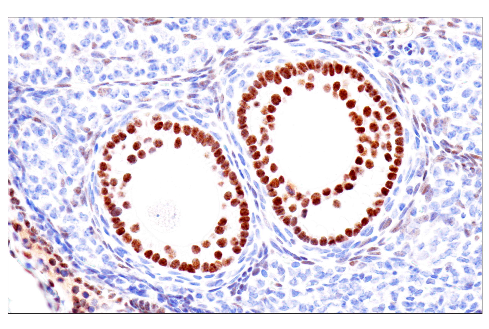
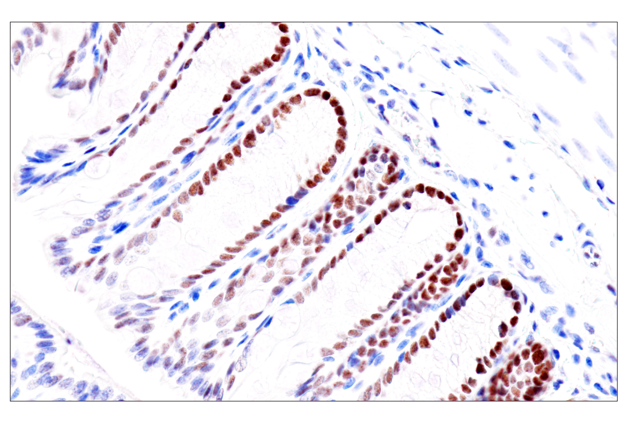
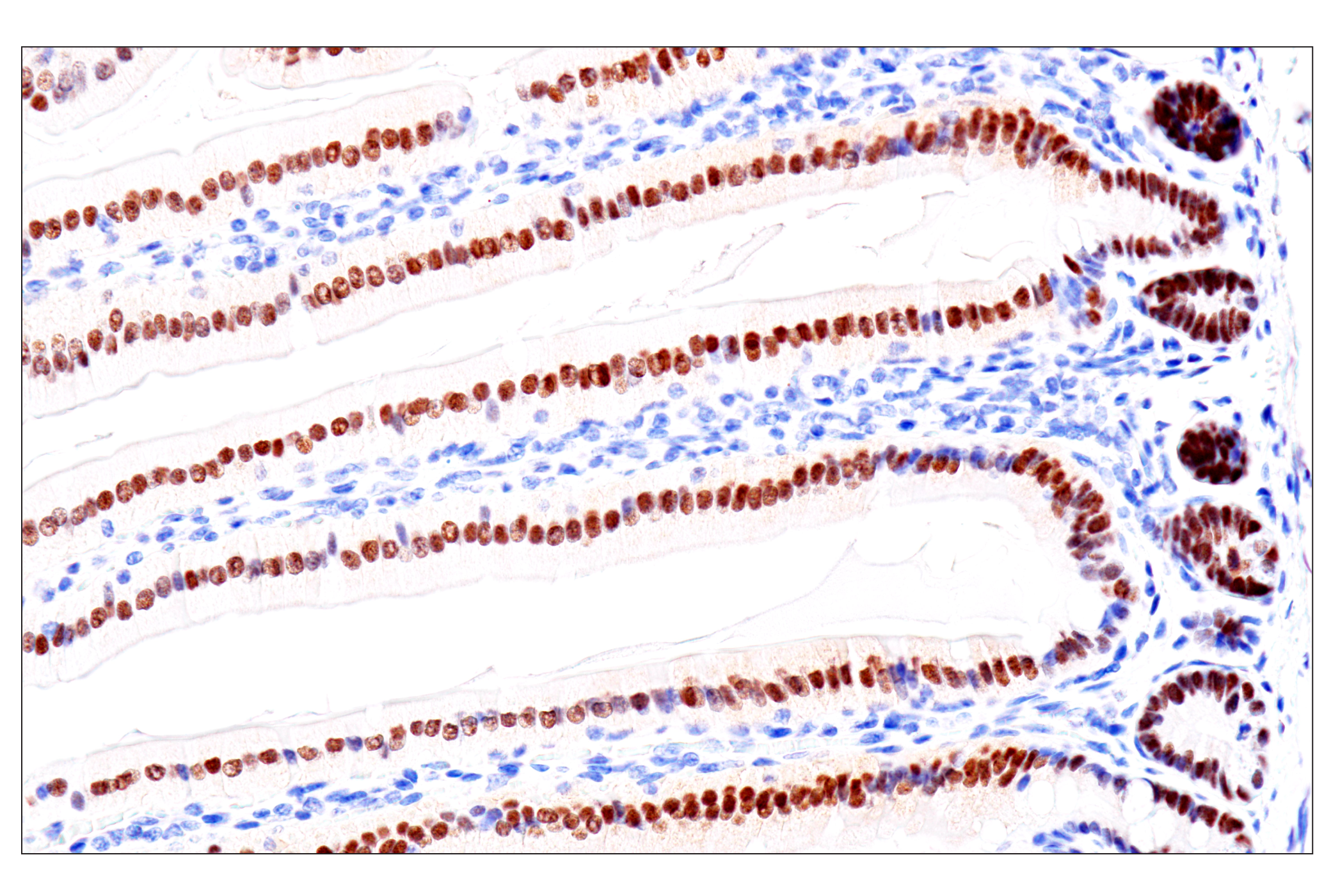
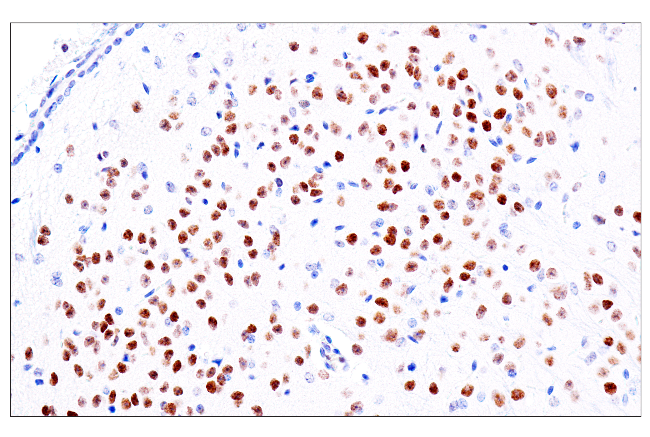
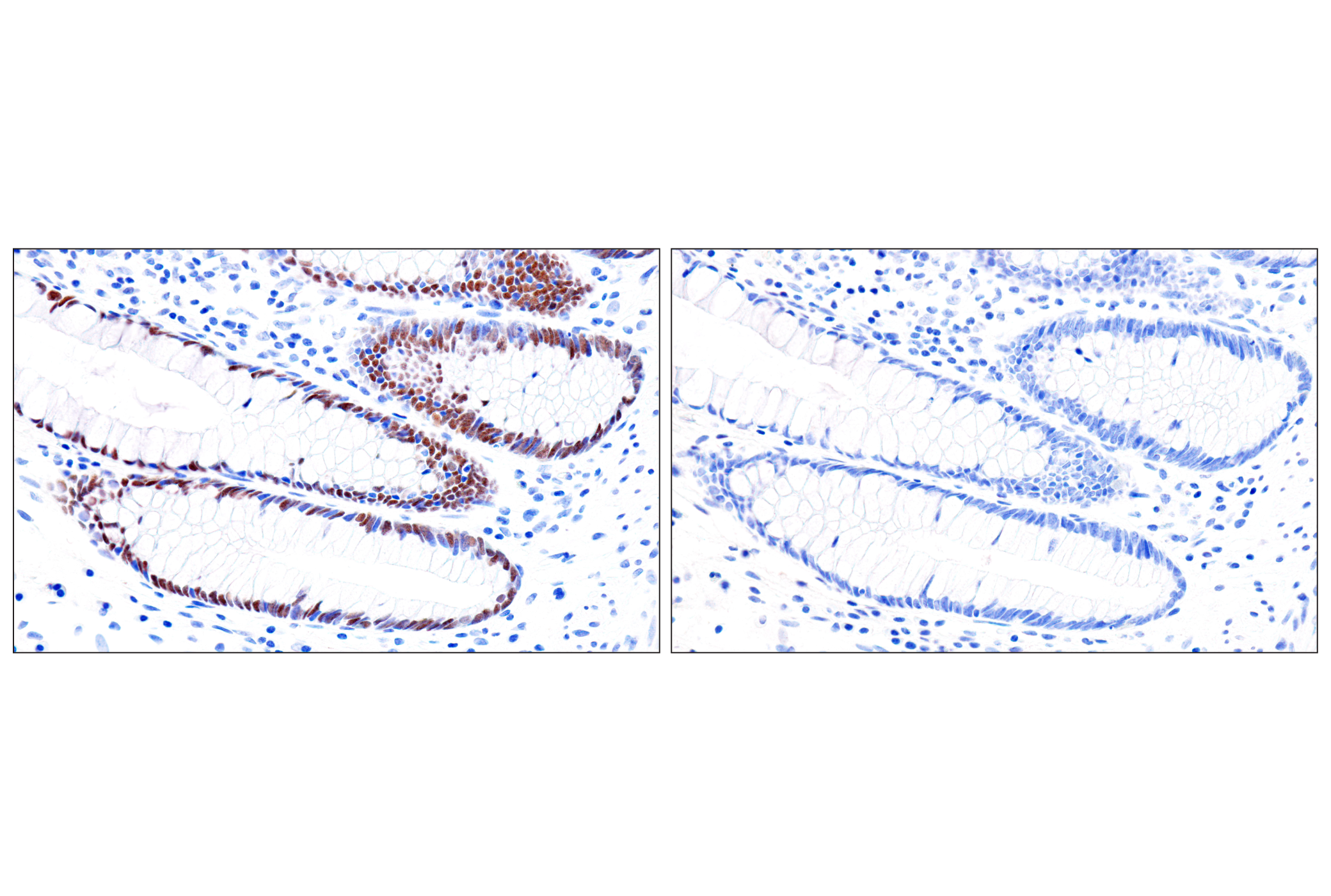
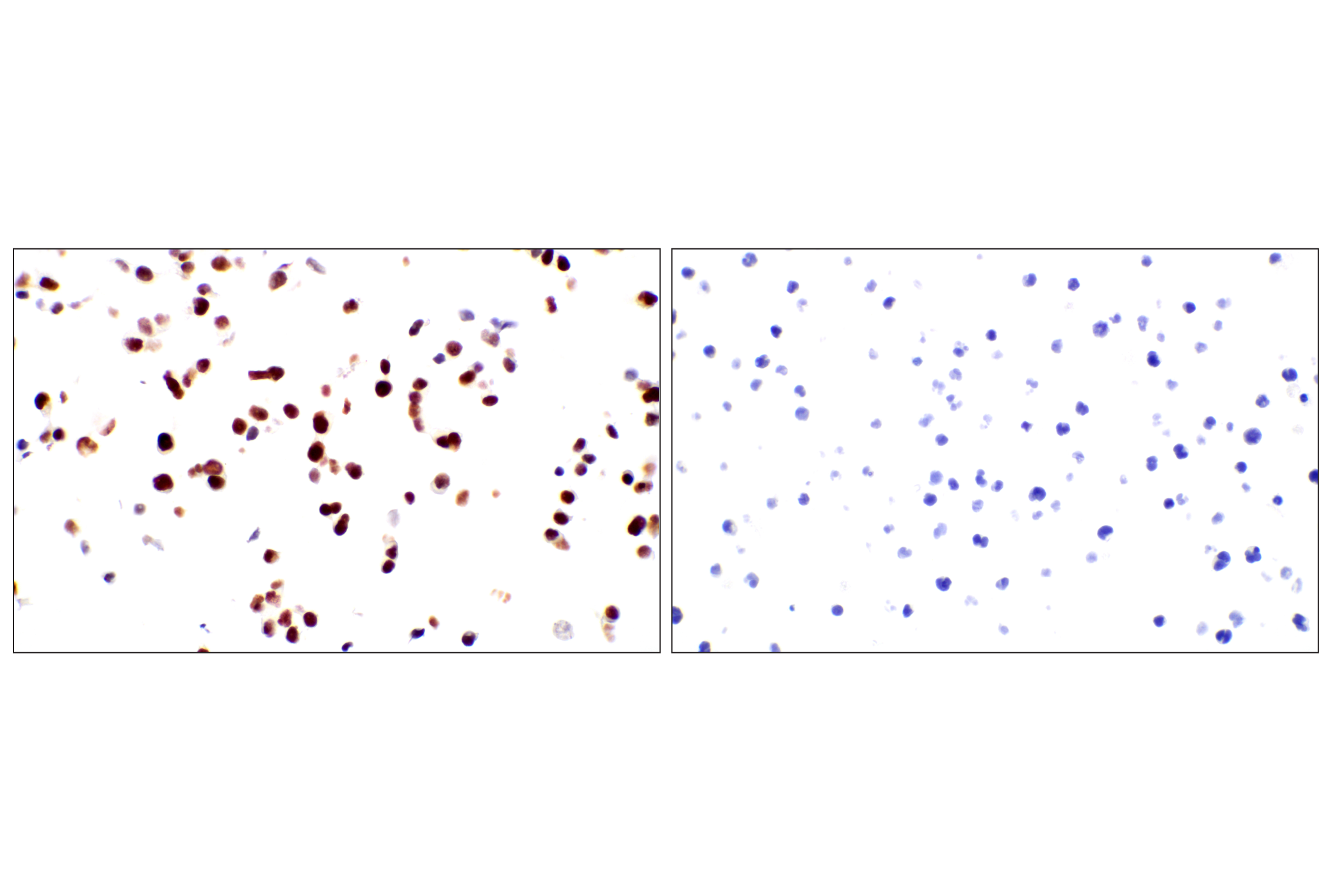
GATA proteins comprise a group of transcription factors that are related by the presence of conserved zinc finger DNA-binding domains, which bind directly to the nucleotide sequence core element GATA (1-3). There are six vertebrate GATA proteins, designated GATA-1 to GATA-6 (3). Although they are commonly divided as hematopoietic (GATA-1-3) or cardiac (GATA-4-6) factors, GATA proteins are expressed in a wide variety of tissue and play critical roles in embryonic development and organ differentiation (4). GATA-1 is the founding member of the GATA family and is required for erythroid and megakaryocytic cell development (5,6). Mutations in the corresponding GATA-1 gene are linked to many human diseases, including acute megakaryoblastic leukemia in Down Syndrome children (DS-AMKL), X-linked thrombocytopenia, and gray platelet syndrome (7-10). GATA-2 is widely expressed and plays an essential role in many developmental processes (11). Studies on GATA-2 knockout mice indicate that this protein is required in hematopoiesis (12). GATA-2 also inhibits the differentiation of white and brown adipocytes and has been shown to suppress the proliferation of neuronal progenitor cells (13-15). GATA-3 is a critical regulator of development and is expressed in both hematopoietic and non-hematopoietic tissues, including the kidney, skin, mammary gland, and central nervous system (16-19). GATA-3 knockout mouse embryos die between E11 and E12 due to growth retardation and deformities in the brain and spinal cord (20). The function of GATA-3 has also been extensively studied in T cell development and has been shown to be a downstream target of Notch in Notch-mediated differentiation of TH2 cells (21,22). GATA-4 is crucial for cardiomyocyte differentiation, and not surprisingly, mutations in the GATA-4 gene are implicated in many cardiac diseases, such as tetralogy of Fallot, familial and sporadic dilated cardiomyopathy, and atrial septal defect (23-27). GATA-4 and GATA-6 together maintain intestinal epithelial structure by regulating enterocyte gene expression (28). They also have overlapping roles in steroidogenesis and genital ridge formation during gonadal development (29). GATA-6 plays a critical role in endoderm development and is essential for the development of the heart, gut, and other organs (30-32). Knockout of GATA-6 is embryonic lethal due to defects in the formation of the heart tube and a failure to develop extraembryonic endoderm (30).
- Ko, L.J. and Engel, J.D. (1993) Mol Cell Biol 13, 4011-22.
- Merika, M. and Orkin, S.H. (1993) Mol Cell Biol 13, 3999-4010.
- Lowry, J.A. and Atchley, W.R. (2000) J Mol Evol 50, 103-15.
- Tremblay, M. et al. (2018) Development 145, dev164384. doi: 10.1242/dev.164384.
- Pevny, L. et al. (1991) Nature 349, 257-60.
- Fujiwara, Y. et al. (1996) Proc Natl Acad Sci USA 93, 12355-8.
- Wechsler, J. et al. (2002) Nat Genet 32, 148-52.
- Cantor, A.B. (2005) Int J Hematol 81, 378-84.
- Mehaffey, M.G. et al. (2001) Blood 98, 2681-8.
- Tubman, V.N. et al. (2007) Blood 109, 3297-9.
- Tong, Q. et al. (2003) Drug News Perspect 16, 585-8.
- Tsai, F.Y. et al. (1994) Nature 371, 221-6.
- Tong, Q. et al. (2005) Mol Cell Biol 25, 706-15.
- Tsai, J. et al. (2005) EMBO Rep 6, 879-84.
- El Wakil, A. et al. (2006) Development 133, 2155-65.
- Debacker, C. et al. (1999) Mech Dev 85, 183-7.
- Grote, D. et al. (2008) PLoS Genet 4, e1000316.
- Kaufman, C.K. et al. (2003) Genes Dev 17, 2108-22.
- Kouros-Mehr, H. et al. (2006) Cell 127, 1041-55.
- Pandolfi, P.P. et al. (1995) Nat Genet 11, 40-4.
- Ho, I.C. et al. (2009) Nat Rev Immunol 9, 125-35.
- Amsen, D. et al. (2007) Immunity 27, 89-99.
- Gan, L. et al. (2014) Gene Expr Patterns 16, 8-22.
- Yang, Y.Q. et al. (2013) Hum Mutat 34, 1662-71.
- Li, R.G. et al. (2013) Biochem Biophys Res Commun 439, 591-6.
- Li, J. et al. (2014) Gene 548, 174-81.
- Mohan, R.A. et al. (2014) Am J Med Genet A 164A, 2732-8.
- Walker, E.M. et al. (2014) Dev Biol 392, 283-94.
- LaVoie, H.A. (2014) Biol Reprod 91, 38.
- Cai, K.Q. et al. (2008) Dev Dyn 237, 2820-9.
- Charron, F. and Nemer, M. (1999) Semin Cell Dev Biol 10, 85-91.
- Haveri, H. et al. (2008) BMC Gastroenterol 8, 9.
Background References
Trademarks and Patents
使用に関する制限
法的な権限を与えられたCSTの担当者が署名した書面によって別途明示的に合意された場合を除き、 CST、その関連会社または代理店が提供する製品には以下の条件が適用されます。お客様が定める条件でここに定められた条件に含まれるものを超えるもの、 または、ここに定められた条件と異なるものは、法的な権限を与えられたCSTの担当者が別途書面にて受諾した場合を除き、拒絶され、 いかなる効力も効果も有しません。
研究専用 (For Research Use Only) またはこれに類似する表示がされた製品は、 いかなる目的についても FDA または外国もしくは国内のその他の規制機関により承認、認可または許可を受けていません。 お客様は製品を診断もしくは治療目的で使用してはならず、また、製品に表示された内容に違反する方法で使用してはなりません。 CST が販売または使用許諾する製品は、エンドユーザーであるお客様に対し、使途を研究および開発のみに限定して提供されるものです。 診断、予防もしくは治療目的で製品を使用することまたは製品を再販売 (単独であるか他の製品等の一部であるかを問いません) もしくはその他の商業的利用の目的で購入することについては、CST から別途許諾を得る必要があります。 お客様は以下の事項を遵守しなければなりません。(a) CST の製品 (単独であるか他の資材と一緒であるかを問いません) を販売、使用許諾、貸与、寄付もしくはその他の態様で第三者に譲渡したり使用させたりしてはなりません。また、商用の製品を製造するために CST の製品を使用してはなりません。(b) 複製、改変、リバースエンジニアリング、逆コンパイル、 分解または他の方法により製品の構造または技術を解明しようとしてはなりません。また、 CST の製品またはサービスと競合する製品またはサービスを開発する目的で CST の製品を使用してはなりません。(c) CST の製品の商標、商号、ロゴ、特許または著作権に関する通知または表示を除去したり改変したりしてはなりません。(d) CST の製品をCST 製品販売条件(CST’s Product Terms of Sale) および該当する書面のみに従って使用しなければなりません。(e) CST の製品に関連してお客様が使用する第三者の製品またはサービスに関する使用許諾条件、 サービス提供条件またはこれに類する合意事項を遵守しなければなりません。