WB, IP, IHC-P, IF-F, IF-IC, FC-FP
H M R Mk
Endogenous
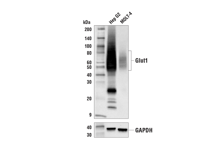
45-60
Rabbit IgG
#P11166
6513
Product Information
Product Usage Information
| Application | Dilution |
|---|---|
| Western Blotting | 1:1000 |
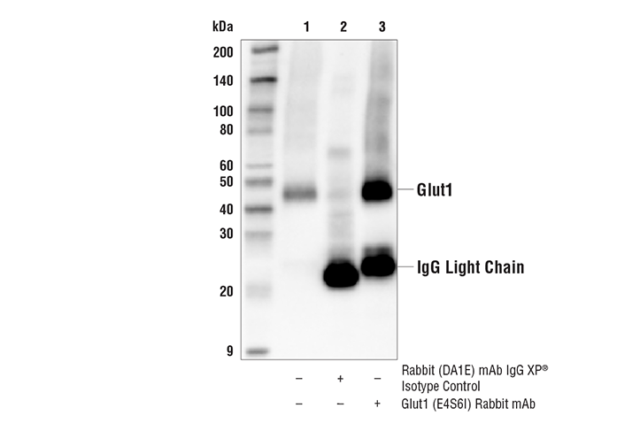
| Immunoprecipitation | 1:50 |
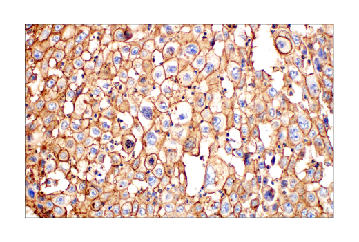
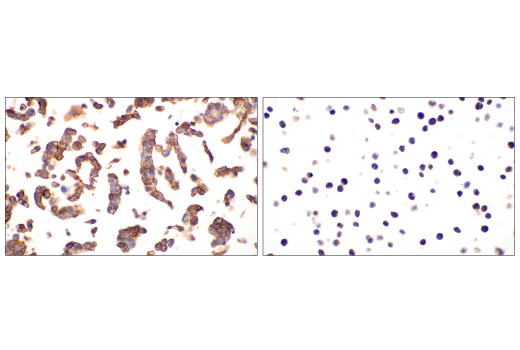
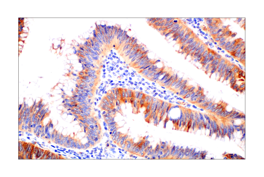
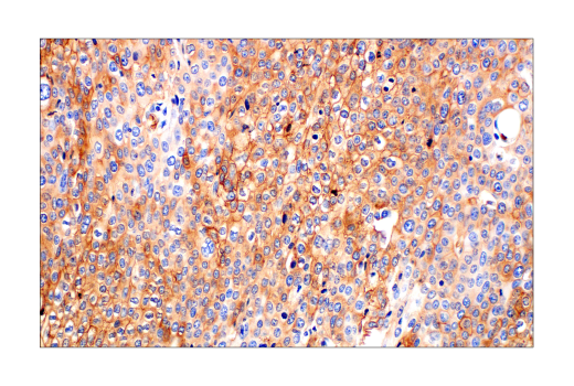
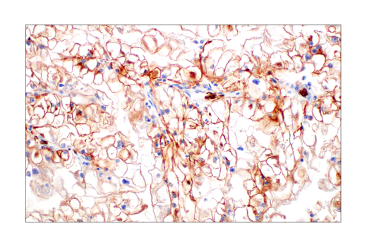
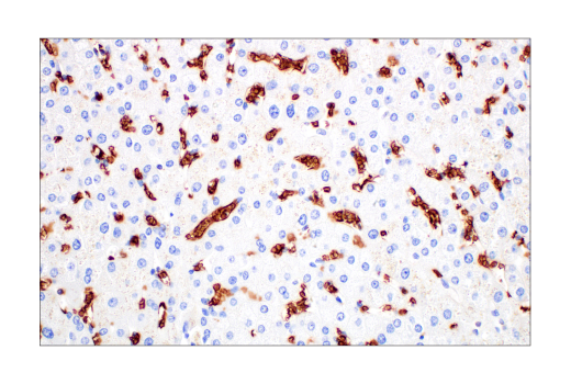
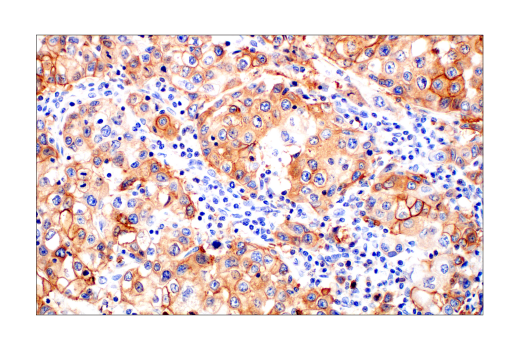
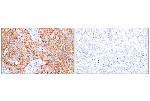
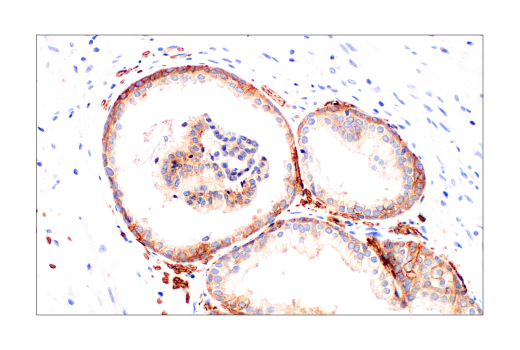
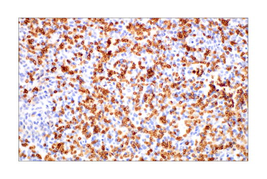
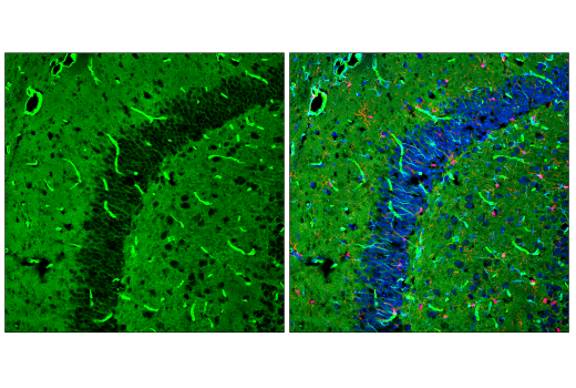
| Immunohistochemistry (Paraffin) | 1:250 - 1:1000 |
| Immunofluorescence (Frozen) | 1:50 - 1:200 |
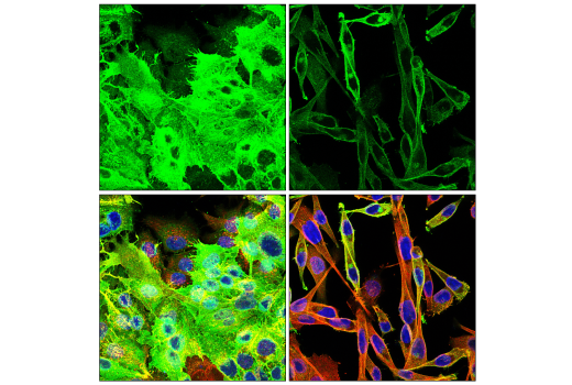
| Immunofluorescence (Immunocytochemistry) | 1:50 - 1:200 |
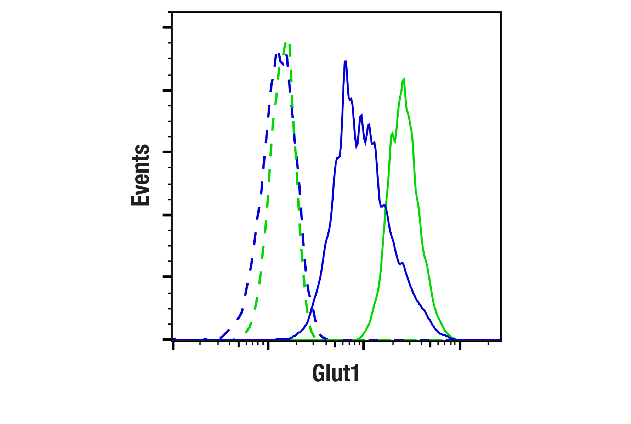
| Flow Cytometry (Fixed/Permeabilized) | 1:50 - 1:200 |
Storage
For a carrier free (BSA and azide free) version of this product see product #84221.
Specificity / Sensitivity
Species Reactivity:
Human, Mouse, Rat, Monkey
Source / Purification
Monoclonal antibody is produced by immunizing animals with a synthetic peptide corresponding to residues near the carboxy terminus of human Glut1 protein.
Background
Glucose transporter 1 (Glut1, SLC2A1) is a widely expressed transport protein that displays a broad range of substrate specificity in transporting a number of different aldose sugars as well as an oxidized form of vitamin C into cells (1,2). Glut1 is responsible for the basal-level uptake of glucose from the blood through facilitated diffusion (2). Research studies show that Glut1 and the transcription factor HIF-1α mediate the regulation of glycolysis by O-GlcNAcylation in cancer cells (3). Additional studies demonstrate that Glut1 is required for CD4 T cell activation and is critical for the expansion and survival of T effector (Teff) cells (4). Mutations in the corresponding SLC2A1 gene cause GLUT1 deficiency syndromes (GLUT1DS1, GLUT1DS2), a pair of neurologic disorders characterized by delayed development, seizures, spasticity, paroxysmal exercise-induced dyskinesia, and acquired microcephaly (5,6). Two other neurologic disorders - dystonia-9 (DYT9) and susceptibility to idiopathic generalized epilepsy 12 (EIG12) - are also caused by mutations in the SLC2A1 gene (7,8).
- Ferrer, C.M. et al. (2014) Mol Cell 54, 820-31.
- Deng, D. et al. (2014) Nature 510, 121-5.
- Agus, D.B. et al. (1997) J Clin Invest 100, 2842-8.
- Macintyre, A.N. et al. (2014) Cell Metab 20, 61-72.
- Wang, D. et al. (2005) Ann Neurol 57, 111-8.
- Schneider, S.A. et al. (2009) Mov Disord 24, 1684-8.
- Weber, Y.G. et al. (2011) Neurology 77, 959-64.
- Suls, A. et al. (2009) Ann Neurol 66, 415-9.
Species Reactivity
Species reactivity is determined by testing in at least one approved application (e.g., western blot).
Western Blot Buffer
IMPORTANT: For western blots, incubate membrane with diluted primary antibody in 5% w/v BSA, 1X TBS, 0.1% Tween® 20 at 4°C with gentle shaking, overnight.
Applications Key
WB: Western Blotting IP: Immunoprecipitation IHC-P: Immunohistochemistry (Paraffin) IF-F: Immunofluorescence (Frozen) IF-IC: Immunofluorescence (Immunocytochemistry) FC-FP: Flow Cytometry (Fixed/Permeabilized)
Cross-Reactivity Key
H: human M: mouse R: rat Hm: hamster Mk: monkey Vir: virus Mi: mink C: chicken Dm: D. melanogaster X: Xenopus Z: zebrafish B: bovine Dg: dog Pg: pig Sc: S. cerevisiae Ce: C. elegans Hr: horse GP: Guinea Pig Rab: rabbit All: all species expected
Trademarks and Patents
使用に関する制限
法的な権限を与えられたCSTの担当者が署名した書面によって別途明示的に合意された場合を除き、 CST、その関連会社または代理店が提供する製品には以下の条件が適用されます。お客様が定める条件でここに定められた条件に含まれるものを超えるもの、 または、ここに定められた条件と異なるものは、法的な権限を与えられたCSTの担当者が別途書面にて受諾した場合を除き、拒絶され、 いかなる効力も効果も有しません。
研究専用 (For Research Use Only) またはこれに類似する表示がされた製品は、 いかなる目的についても FDA または外国もしくは国内のその他の規制機関により承認、認可または許可を受けていません。 お客様は製品を診断もしくは治療目的で使用してはならず、また、製品に表示された内容に違反する方法で使用してはなりません。 CST が販売または使用許諾する製品は、エンドユーザーであるお客様に対し、使途を研究および開発のみに限定して提供されるものです。 診断、予防もしくは治療目的で製品を使用することまたは製品を再販売 (単独であるか他の製品等の一部であるかを問いません) もしくはその他の商業的利用の目的で購入することについては、CST から別途許諾を得る必要があります。 お客様は以下の事項を遵守しなければなりません。(a) CST の製品 (単独であるか他の資材と一緒であるかを問いません) を販売、使用許諾、貸与、寄付もしくはその他の態様で第三者に譲渡したり使用させたりしてはなりません。また、商用の製品を製造するために CST の製品を使用してはなりません。(b) 複製、改変、リバースエンジニアリング、逆コンパイル、 分解または他の方法により製品の構造または技術を解明しようとしてはなりません。また、 CST の製品またはサービスと競合する製品またはサービスを開発する目的で CST の製品を使用してはなりません。(c) CST の製品の商標、商号、ロゴ、特許または著作権に関する通知または表示を除去したり改変したりしてはなりません。(d) CST の製品をCST 製品販売条件(CST’s Product Terms of Sale) および該当する書面のみに従って使用しなければなりません。(e) CST の製品に関連してお客様が使用する第三者の製品またはサービスに関する使用許諾条件、 サービス提供条件またはこれに類する合意事項を遵守しなければなりません。