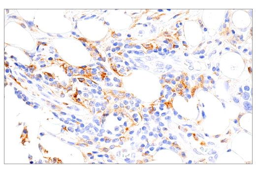
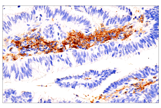
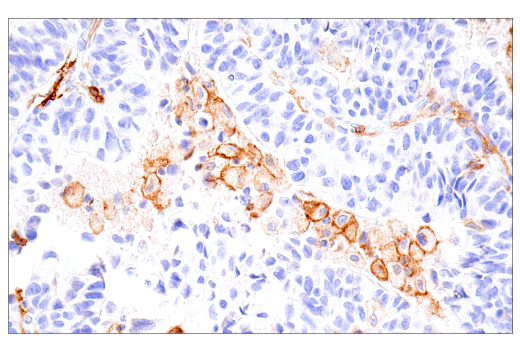
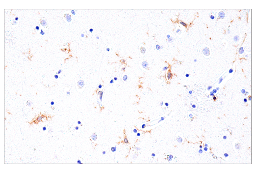
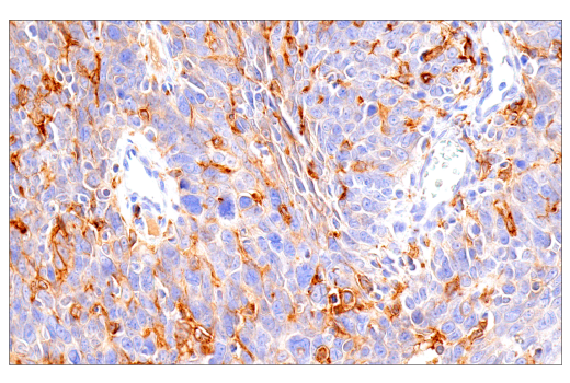
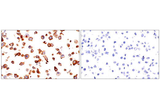
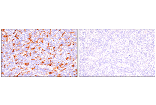
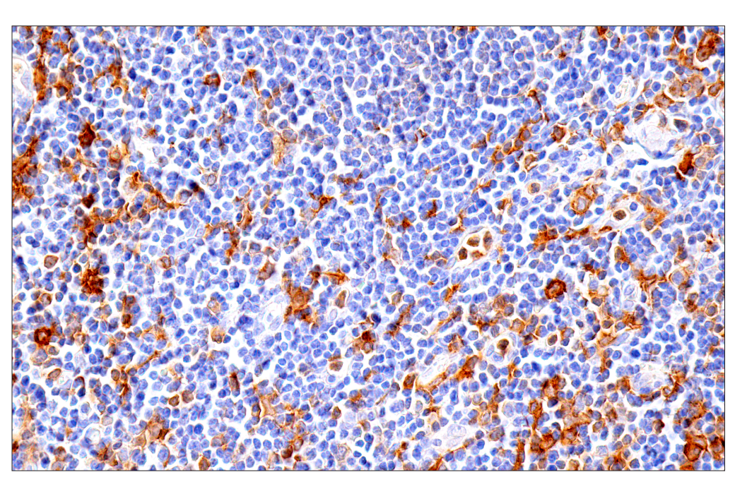
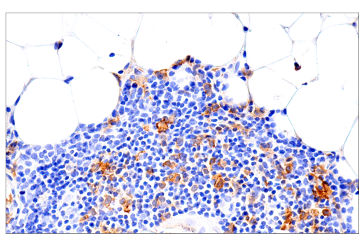
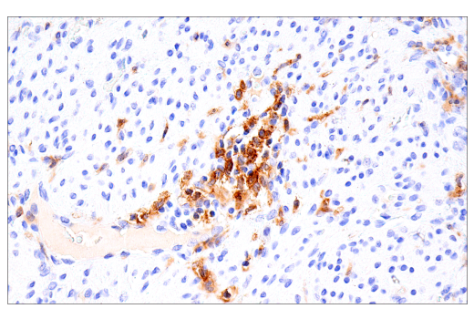
Revision 3
Cell Signaling Technology

Orders: 877-616-CELL (2355) [email protected]

Support: 877-678-TECH (8324)

Web: [email protected] cellsignal.com

3 Trask LaneDanversMassachusetts01923USA
For Research Use Only. Not for Use in Diagnostic Procedures.
Applications:

WB, IHC-Bond, IHC-P

REACTIVITY:

H

SENSITIVITY:

Endogenous

MW (kDa):

70-80

Source/Isotype:

Rabbit IgG

UniProt ID:

#P15509

Entrez-Gene Id:

1438

Product Information

Product Usage Information

Application Dilution
Western Blotting 1:1000
IHC Leica Bond 1:200 - 1:800
Immunohistochemistry (Paraffin) 1:200 - 1:800

Storage

Supplied in 10 mM sodium HEPES (pH 7.5), 150 mM NaCl, 100 µg/ml BSA, 50% glycerol and less than 0.02% sodium azide. Store at –20°C. Do not aliquot the antibody.

For a carrier free (BSA and azide free) version of this product see product #63107.

Specificity / Sensitivity

GM-CSF Receptor α (E8Z3S) Rabbit mAb recognizes endogenous levels of total GM-CSF Receptor α protein. This antibody does not cross-react with GM-CSF Receptor β protein. Based on the antigen sequence, this antibody is predicted to recognize multiple isoforms of GM-CSF Receptor α protein, including secreted isoforms.

Species Reactivity:

Human

Source / Purification

Monoclonal antibody is produced by immunizing animals with recombinant protein specific to the amino terminus of human GM-CSF Receptor α protein.

Background

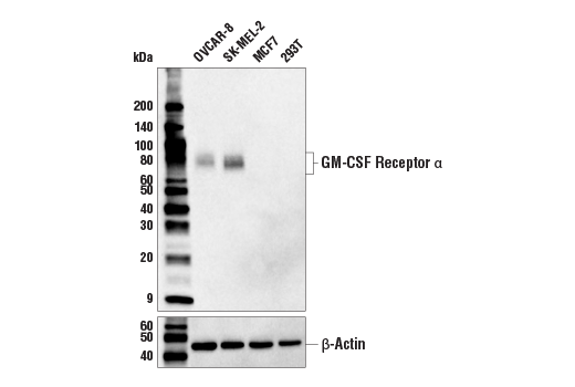
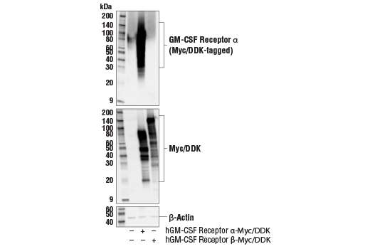
Granulocyte-macrophage colony-stimulating factor receptor subunit alpha (GM-CSF Receptor α), also known as CSF2RA and CD116, is a type 1 transmembrane protein with low affinity for GM-CSF. GM-CSF Receptor α is primarily expressed on myeloid cells, including granulocytes, monocytes, macrophages, and dendritic cells (1). GM-CSF Receptor α forms a heterodimeric receptor complex with GM-CSF Receptor β, creating the high affinity GM-CSF receptor (2). Upon dimerization and ligand binding, the β subunit becomes phosphorylated and transduces signals via Jak/STAT and MAPK pathways for cell proliferation, differentiation, and survival (3,4). Oligomerization of the high affinity GM-CSF Receptor α/β complex is required for optimal intracellular signal transduction (5). Soluble GM-CSF Receptor α, either secreted or cleaved from the cell surface, can competitively bind GM-CSF, preventing membrane receptor signaling (1,6,7). Multiple isoforms of GM-CSF Receptor α have been described.

  1. Barreda, D.R. et al. (2004) Dev Comp Immunol 28, 509-54.
  2. Hansen, G. et al. (2008) Cell 134, 496-507.
  3. Doyle, S.E. and Gasson, J.C. (1998) Blood 92, 867-76.
  4. Lopez, A.F. et al. (2010) IUBMB Life 62, 509-18.
  5. McClure, B.J. et al. (2003) Blood 101, 1308-15.
  6. Raines, M.A. et al. (1991) Proc Natl Acad Sci U S A 88, 8203-7.
  7. Geijsen, N. et al. (2001) Cytokine Growth Factor Rev 12, 19-25.

Species Reactivity

Species reactivity is determined by testing in at least one approved application (e.g., western blot).

Western Blot Buffer

IMPORTANT: For western blots, incubate membrane with diluted primary antibody in 5% w/v nonfat dry milk, 1X TBS, 0.1% Tween® 20 at 4°C with gentle shaking, overnight.

Applications Key

WB: Western Blotting IHC-Bond: IHC Leica Bond IHC-P: Immunohistochemistry (Paraffin)

Cross-Reactivity Key

H: human M: mouse R: rat Hm: hamster Mk: monkey Vir: virus Mi: mink C: chicken Dm: D. melanogaster X: Xenopus Z: zebrafish B: bovine Dg: dog Pg: pig Sc: S. cerevisiae Ce: C. elegans Hr: horse GP: Guinea Pig Rab: rabbit All: all species expected

Trademarks and Patents

Cell Signaling Technology is a trademark of Cell Signaling Technology, Inc.
SignalStain is a registered trademark of Cell Signaling Technology, Inc.
XP is a registered trademark of Cell Signaling Technology, Inc.
All other trademarks are the property of their respective owners. Visit cellsignal.com/trademarks for more information.

使用に関する制限

法的な権限を与えられたCSTの担当者が署名した書面によって別途明示的に合意された場合を除き、 CST、その関連会社または代理店が提供する製品には以下の条件が適用されます。お客様が定める条件でここに定められた条件に含まれるものを超えるもの、 または、ここに定められた条件と異なるものは、法的な権限を与えられたCSTの担当者が別途書面にて受諾した場合を除き、拒絶され、 いかなる効力も効果も有しません。

研究専用 (For Research Use Only) またはこれに類似する表示がされた製品は、 いかなる目的についても FDA または外国もしくは国内のその他の規制機関により承認、認可または許可を受けていません。 お客様は製品を診断もしくは治療目的で使用してはならず、また、製品に表示された内容に違反する方法で使用してはなりません。 CST が販売または使用許諾する製品は、エンドユーザーであるお客様に対し、使途を研究および開発のみに限定して提供されるものです。 診断、予防もしくは治療目的で製品を使用することまたは製品を再販売 (単独であるか他の製品等の一部であるかを問いません) もしくはその他の商業的利用の目的で購入することについては、CST から別途許諾を得る必要があります。 お客様は以下の事項を遵守しなければなりません。(a) CST の製品 (単独であるか他の資材と一緒であるかを問いません) を販売、使用許諾、貸与、寄付もしくはその他の態様で第三者に譲渡したり使用させたりしてはなりません。また、商用の製品を製造するために CST の製品を使用してはなりません。(b) 複製、改変、リバースエンジニアリング、逆コンパイル、 分解または他の方法により製品の構造または技術を解明しようとしてはなりません。また、 CST の製品またはサービスと競合する製品またはサービスを開発する目的で CST の製品を使用してはなりません。(c) CST の製品の商標、商号、ロゴ、特許または著作権に関する通知または表示を除去したり改変したりしてはなりません。(d) CST の製品をCST 製品販売条件(CST’s Product Terms of Sale) および該当する書面のみに従って使用しなければなりません。(e) CST の製品に関連してお客様が使用する第三者の製品またはサービスに関する使用許諾条件、 サービス提供条件またはこれに類する合意事項を遵守しなければなりません。

Revision 3
#69817

GM-CSF Receptor α (E8Z3S) Rabbit mAb

Western Blotting Image 1: GM-CSF Receptor α (E8Z3S) Rabbit mAb Expand Image
Western blot analysis of extracts from various human cell lines using GM-CSF Receptor α (E8Z3S) Rabbit mAb (upper) and β-Actin (D6A8) Rabbit mAb #8457 (lower).
Western Blotting Image 2: GM-CSF Receptor α (E8Z3S) Rabbit mAb Expand Image
Western blot analysis of extracts from 293T cells, mock transfected (-) or transfected with constructs expressing full-length human GM-CSF Receptor α protein (hGM-CSF Receptor α-Myc/DDK; +) and human GM-CSF Receptor β protein (hGM-CSF Receptor β-Myc/DDK; +), using GM-CSF Receptor α (E8Z3S) Rabbit mAb (upper), Myc-Tag (71D10) Rabbit mAb #2278 (middle), and β-Actin (D6A8) Rabbit mAb #8457 (lower).
Immunohistochemistry Image 1: GM-CSF Receptor α (E8Z3S) Rabbit mAb Expand Image
Immunohistochemical analysis of paraffin-embedded human ductal breast carcinoma using GM-CSF Receptor α (E8Z3S) Rabbit mAb performed on the Leica® BOND Rx.
Immunohistochemistry Image 2: GM-CSF Receptor α (E8Z3S) Rabbit mAb Expand Image
Immunohistochemical analysis of paraffin-embedded human colon carcinoma using GM-CSF Receptor α (E8Z3S) Rabbit mAb performed on the Leica® BOND Rx.
Immunohistochemistry Image 3: GM-CSF Receptor α (E8Z3S) Rabbit mAb Expand Image
Immunohistochemical analysis of paraffin-embedded human esophageal adenocarcinoma using GM-CSF Receptor α (E8Z3S) Rabbit mAb performed on the Leica® BOND Rx.
Immunohistochemistry Image 4: GM-CSF Receptor α (E8Z3S) Rabbit mAb Expand Image
Immunohistochemical analysis of paraffin-embedded human non-small cell lung carcinoma using GM-CSF Receptor α (E8Z3S) Rabbit mAb performed on the Leica® BOND Rx.
Immunohistochemistry Image 5: GM-CSF Receptor α (E8Z3S) Rabbit mAb Expand Image
Immunohistochemical analysis of paraffin-embedded normal human placenta using GM-CSF Receptor α (E8Z3S) Rabbit mAb performed on the Leica® BOND Rx.
Immunohistochemistry Image 6: GM-CSF Receptor α (E8Z3S) Rabbit mAb Expand Image
Immunohistochemical analysis of paraffin-embedded normal human spleen using GM-CSF Receptor α (E8Z3S) Rabbit mAb performed on the Leica® BOND Rx.
Immunohistochemistry Image 1: GM-CSF Receptor α (E8Z3S) Rabbit mAb Expand Image
Immunohistochemical analysis of paraffin-embedded human urothelial carcinoma using GM-CSF Receptor α (E8Z3S) Rabbit mAb.
Immunohistochemistry Image 2: GM-CSF Receptor α (E8Z3S) Rabbit mAb Expand Image
Immunohistochemical analysis of paraffin-embedded normal human brain using GM-CSF Receptor α (E8Z3S) Rabbit mAb.
Immunohistochemistry Image 3: GM-CSF Receptor α (E8Z3S) Rabbit mAb Expand Image
Immunohistochemical analysis of paraffin-embedded human squamous cell carcinoma of the tonsil using GM-CSF Receptor α (E8Z3S) Rabbit mAb.
Immunohistochemistry Image 4: GM-CSF Receptor α (E8Z3S) Rabbit mAb Expand Image
Immunohistochemical analysis of paraffin-embedded SK-MEL-2 cell pellet (left, positive) or 293T cell pellet (right, negative) using GM-CSF Receptor α (E8Z3S) Rabbit mAb.
Immunohistochemistry Image 5: GM-CSF Receptor α (E8Z3S) Rabbit mAb Expand Image
Immunohistochemical analysis of paraffin-embedded human B-cell non-Hodgkin lymphoma using GM-CSF Receptor α (E8Z3S) Rabbit mAb (left) compared to concentration-matched Rabbit (DA1E) mAb IgG XP® Isotype Control #3900 (right).
Immunohistochemistry Image 6: GM-CSF Receptor α (E8Z3S) Rabbit mAb Expand Image
Immunohistochemical analysis of paraffin-embedded normal human lymph node using GM-CSF Receptor α (E8Z3S) Rabbit mAb.
Immunohistochemistry Image 7: GM-CSF Receptor α (E8Z3S) Rabbit mAb Expand Image
Immunohistochemical analysis of paraffin-embedded normal human thymus using GM-CSF Receptor α (E8Z3S) Rabbit mAb.
Immunohistochemistry Image 8: GM-CSF Receptor α (E8Z3S) Rabbit mAb Expand Image
Immunohistochemical analysis of paraffin-embedded normal human uterus using GM-CSF Receptor α (E8Z3S) Rabbit mAb.