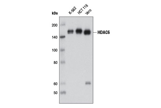
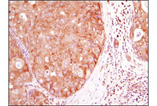
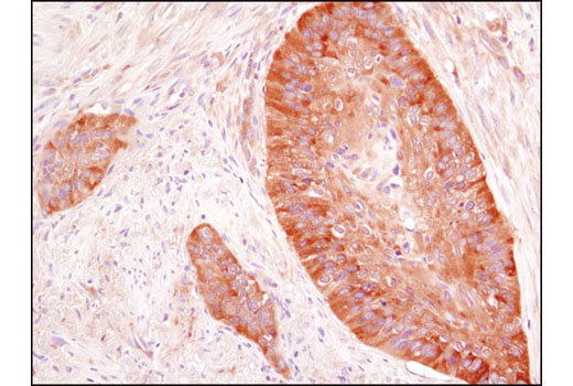
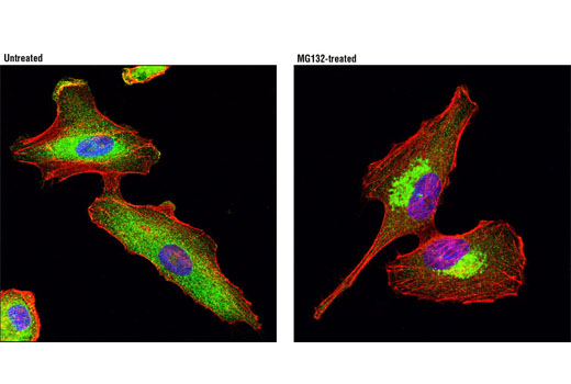
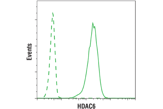
WB, IP, IHC-P, IF-IC, FC-FP
H Mk
Endogenous
160
Rabbit IgG
#Q9UBN7
10013
Product Information
Product Usage Information
| Application | Dilution |
|---|---|
| Western Blotting | 1:1000 |
| Immunoprecipitation | 1:100 |
| Immunohistochemistry (Paraffin) | 1:200 - 1:800 |
| Immunofluorescence (Immunocytochemistry) | 1:100 - 1:400 |
| Flow Cytometry (Fixed/Permeabilized) | 1:100 - 1:400 |
Storage
Specificity / Sensitivity
Species Reactivity:
Human, Monkey
Source / Purification
Monoclonal antibody is produced by immunizing animals with a recombinant protein specific to the carboxy terminus of human HDAC6 protein.
Background
HDAC6 is a class II histone deacetylase enzyme localized to the cytoplasm and associated with the microtubule network (1). It is involved in the regulation of many cellular processes, including cell migration, immune synapse formation, viral infection, and degradation of misfolded proteins (1). HDAC6 contains two tandem catalytic domains that facilitate the deacetylation of multiple protein substrates, including histones and non-histone proteins such as tubulin, cortactin, and HSP90. Despite the ability to deacetylate histone proteins in vitro, there is no evidence for HDAC6-mediated deacetylation of histones in vivo (2,3). The acetylation/deacetylation of tubulin on Lys40 regulates binding and motility of the kinesin-1 motor protein and subsequent transport of cargo proteins such as JNK-interacting protein 1 (JIP1) (4). The acetylation/deacetylation of cortactin regulates cell motility by modulating the binding of cortactin to F-actin (5). Acetylation/deacetylation of HSP90 modulates chaperone complex activity by regulating the binding of an essential cochaperone protein, p23 (6,7). In addition to its role as a protein deacetylase, HDAC6 functions as a component of the aggresome, a proteinaceous inclusion body that forms in response to an accumulation of misfolded or partially denatured proteins (8). Formation of the aggresome is a protective response that sequesters cytotoxic protein aggregates for eventual autophagic clearance from the cell. HDAC6 contains a zinc finger ubiquitin-binding domain that binds both mono- and poly-ubiquitinated proteins (8). HDAC6 binds to both poly-ubiquitinated misfolded proteins and dynein motors, facilitating the transport of misfolded proteins to the aggresome (9,10). HDAC6 is also required for subsequent recruitment of the autophagic machinery and clearance of aggresomes from the cell (11). Thus, HDAC6 plays a key role in the protection against the deleterious effects of pathological protein aggregation that occurs in various diseases, such as neurodegenerative Huntington’s disease (11).
- Boyault, C. et al. (2007) Oncogene 26, 5468-76.
- Haggarty, S.J. et al. (2003) Proc Natl Acad Sci U S A 100, 4389-94.
- Zhang, Y. et al. (2003) EMBO J 22, 1168-79.
- Reed, N.A. et al. (2006) Curr Biol 16, 2166-72.
- Zhang, X. et al. (2007) Mol Cell 27, 197-213.
- Kovacs, J.J. et al. (2005) Mol Cell 18, 601-7.
- Murphy, P.J. et al. (2005) J Biol Chem 280, 33792-9.
- Seigneurin-Berny, D. et al. (2001) Mol Cell Biol 21, 8035-44.
- Kawaguchi, Y. et al. (2003) Cell 115, 727-38.
- Boyault, C. et al. (2006) EMBO J 25, 3357-66.
- Iwata, A. et al. (2005) J Biol Chem 280, 40282-92.
Species Reactivity
Species reactivity is determined by testing in at least one approved application (e.g., western blot).
Western Blot Buffer
IMPORTANT: For western blots, incubate membrane with diluted primary antibody in 5% w/v BSA, 1X TBS, 0.1% Tween® 20 at 4°C with gentle shaking, overnight.
Applications Key
WB: Western Blotting IP: Immunoprecipitation IHC-P: Immunohistochemistry (Paraffin) IF-IC: Immunofluorescence (Immunocytochemistry) FC-FP: Flow Cytometry (Fixed/Permeabilized)
Cross-Reactivity Key
H: human M: mouse R: rat Hm: hamster Mk: monkey Vir: virus Mi: mink C: chicken Dm: D. melanogaster X: Xenopus Z: zebrafish B: bovine Dg: dog Pg: pig Sc: S. cerevisiae Ce: C. elegans Hr: horse GP: Guinea Pig Rab: rabbit All: all species expected
Trademarks and Patents
使用に関する制限
法的な権限を与えられたCSTの担当者が署名した書面によって別途明示的に合意された場合を除き、 CST、その関連会社または代理店が提供する製品には以下の条件が適用されます。お客様が定める条件でここに定められた条件に含まれるものを超えるもの、 または、ここに定められた条件と異なるものは、法的な権限を与えられたCSTの担当者が別途書面にて受諾した場合を除き、拒絶され、 いかなる効力も効果も有しません。
研究専用 (For Research Use Only) またはこれに類似する表示がされた製品は、 いかなる目的についても FDA または外国もしくは国内のその他の規制機関により承認、認可または許可を受けていません。 お客様は製品を診断もしくは治療目的で使用してはならず、また、製品に表示された内容に違反する方法で使用してはなりません。 CST が販売または使用許諾する製品は、エンドユーザーであるお客様に対し、使途を研究および開発のみに限定して提供されるものです。 診断、予防もしくは治療目的で製品を使用することまたは製品を再販売 (単独であるか他の製品等の一部であるかを問いません) もしくはその他の商業的利用の目的で購入することについては、CST から別途許諾を得る必要があります。 お客様は以下の事項を遵守しなければなりません。(a) CST の製品 (単独であるか他の資材と一緒であるかを問いません) を販売、使用許諾、貸与、寄付もしくはその他の態様で第三者に譲渡したり使用させたりしてはなりません。また、商用の製品を製造するために CST の製品を使用してはなりません。(b) 複製、改変、リバースエンジニアリング、逆コンパイル、 分解または他の方法により製品の構造または技術を解明しようとしてはなりません。また、 CST の製品またはサービスと競合する製品またはサービスを開発する目的で CST の製品を使用してはなりません。(c) CST の製品の商標、商号、ロゴ、特許または著作権に関する通知または表示を除去したり改変したりしてはなりません。(d) CST の製品をCST 製品販売条件(CST’s Product Terms of Sale) および該当する書面のみに従って使用しなければなりません。(e) CST の製品に関連してお客様が使用する第三者の製品またはサービスに関する使用許諾条件、 サービス提供条件またはこれに類する合意事項を遵守しなければなりません。