Revision 1
Cell Signaling Technology

Orders: 877-616-CELL (2355) [email protected]

Support: 877-678-TECH (8324)

Web: [email protected] cellsignal.com

3 Trask LaneDanversMassachusetts01923USA
For Research Use Only. Not for Use in Diagnostic Procedures.
Applications:

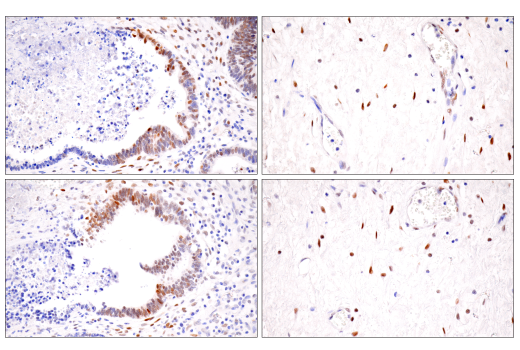
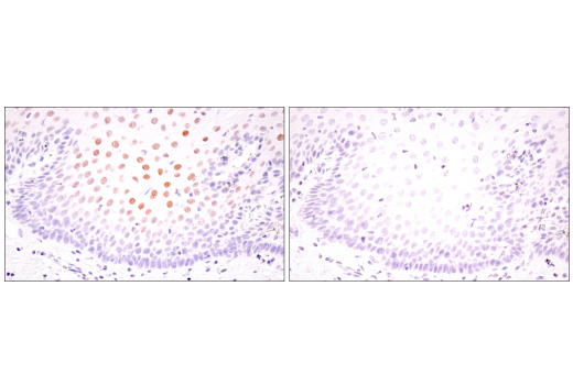
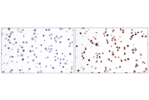
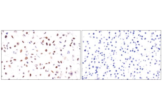
WB, IHC-Bond, IHC-P, IF-IC

REACTIVITY:

H M

SENSITIVITY:

Endogenous

MW (kDa):

120

Source/Isotype:

Rabbit IgG

UniProt ID:

#Q16665

Entrez-Gene Id:

3091

Product Information

Product Usage Information

This product is the carrier free version of product #48085. All data were generated using the same antibody clone in the standard formulation which contains BSA and glycerol.

This formulation is ideal for use with technologies requiring specialized or custom antibody labeling, including fluorophores, metals, lanthanides, and oligonucleotides. It is not recommended for ChIP, ChIP-seq, CUT&RUN or CUT&Tag assays. If you require a carrier free formulation for chromatin profiling, please contact us. Optimal dilutions/concentrations should be determined by the end user.

Formulation

Supplied in 1X PBS, BSA and Azide Free.

For standard formulation of this product see product #48085.

Storage

Store at -20°C. This product will freeze at -20°C so it is recommended to aliquot into single-use vials to avoid multiple freeze/thaw cycles. A slight precipitate may be present and can be dissolved by gently vortexing. This will not interfere with antibody performance.

Specificity / Sensitivity

HIF-1α (E1V6A) Rabbit mAb (BSA and Azide Free) recognizes endogenous levels of total HIF-1α protein. This antibody does not cross-react with HIF-2α protein.

Species Reactivity:

Human, Mouse

Source / Purification

Monoclonal antibody is produced by immunizing animals with a synthetic peptide corresponding to residues surrounding Ala475 of human HIF-1α protein.

Background

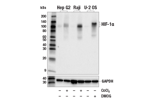
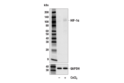
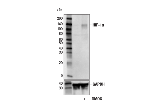
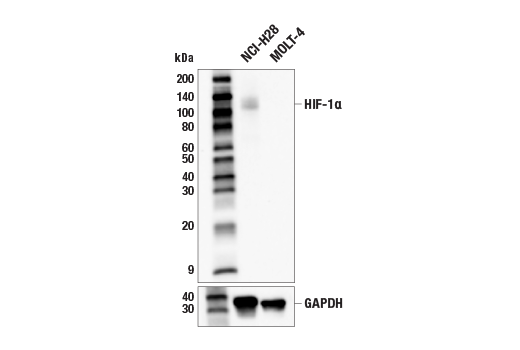
Hypoxia-inducible factor 1 (HIF1) is a heterodimeric transcription factor that plays a critical role in the cellular response to hypoxia (1). The HIF1 complex consists of two subunits, HIF-1α and HIF-1β, which are basic helix-loop-helix proteins of the PAS (Per, ARNT, Sim) family (2). HIF1 regulates the transcription of a broad range of genes that facilitate responses to the hypoxic environment, including genes regulating angiogenesis, erythropoiesis, cell cycle, metabolism, and apoptosis. The widely expressed HIF-1α is typically degraded rapidly in normoxic cells by the ubiquitin/proteasomal pathway. Under normoxic conditions, HIF-1α is proline hydroxylated leading to a conformational change that promotes binding to the von Hippel-Lindau protein (VHL) E3 ligase complex; ubiquitination and proteasomal degradation follows (3,4). Both hypoxic conditions and chemical hydroxylase inhibitors (such as desferrioxamine and cobalt) inhibit HIF-1α degradation and lead to its stabilization. In addition, HIF-1α can be induced in an oxygen-independent manner by various cytokines through the PI3K-AKT-mTOR pathway (5-7).

HIF-1β is also known as AhR nuclear translocator (ARNT) due to its ability to partner with the aryl hydrocarbon receptor (AhR) to form a heterodimeric transcription factor complex (8). Together with AhR, HIF-1β plays an important role in xenobiotics metabolism (8). In addition, a chromosomal translocation leading to a TEL-ARNT fusion protein is associated with acute myeloblastic leukemia (9). Studies also found that ARNT/HIF-1β expression levels decrease significantly in pancreatic islets from patients with type 2 diabetes, suggesting that HIF-1β plays an important role in pancreatic β-cell function (10).

  1. Sharp, F.R. and Bernaudin, M. (2004) Nat Rev Neurosci 5, 437-48.
  2. Wang, G.L. et al. (1995) Proc Natl Acad Sci U S A 92, 5510-4.
  3. Jaakkola, P. et al. (2001) Science 292, 468-72.
  4. Maxwell, P.H. et al. (1999) Nature 399, 271-5.
  5. Fukuda, R. et al. (2002) J Biol Chem 277, 38205-11.
  6. Jiang, B.H. et al. (2001) Cell Growth Differ 12, 363-9.
  7. Laughner, E. et al. (2001) Mol Cell Biol 21, 3995-4004.
  8. Walisser, J.A. et al. (2004) Proc Natl Acad Sci U S A 101, 16677-82.
  9. Salomon-Nguyen, F. et al. (2000) Proc Natl Acad Sci U S A 97, 6757-62.
  10. Gunton, J.E. et al. (2005) Cell 122, 337-49.

Species Reactivity

Species reactivity is determined by testing in at least one approved application (e.g., western blot).

Applications Key

WB: Western Blotting IHC-Bond: IHC Leica Bond IHC-P: Immunohistochemistry (Paraffin) IF-IC: Immunofluorescence (Immunocytochemistry)

Cross-Reactivity Key

H: human M: mouse R: rat Hm: hamster Mk: monkey Vir: virus Mi: mink C: chicken Dm: D. melanogaster X: Xenopus Z: zebrafish B: bovine Dg: dog Pg: pig Sc: S. cerevisiae Ce: C. elegans Hr: horse GP: Guinea Pig Rab: rabbit All: all species expected

Trademarks and Patents

Cell Signaling Technology is a trademark of Cell Signaling Technology, Inc.
SignalStain is a registered trademark of Cell Signaling Technology, Inc.
XP is a registered trademark of Cell Signaling Technology, Inc.
All other trademarks are the property of their respective owners. Visit cellsignal.com/trademarks for more information.

使用に関する制限

法的な権限を与えられたCSTの担当者が署名した書面によって別途明示的に合意された場合を除き、 CST、その関連会社または代理店が提供する製品には以下の条件が適用されます。お客様が定める条件でここに定められた条件に含まれるものを超えるもの、 または、ここに定められた条件と異なるものは、法的な権限を与えられたCSTの担当者が別途書面にて受諾した場合を除き、拒絶され、 いかなる効力も効果も有しません。

研究専用 (For Research Use Only) またはこれに類似する表示がされた製品は、 いかなる目的についても FDA または外国もしくは国内のその他の規制機関により承認、認可または許可を受けていません。 お客様は製品を診断もしくは治療目的で使用してはならず、また、製品に表示された内容に違反する方法で使用してはなりません。 CST が販売または使用許諾する製品は、エンドユーザーであるお客様に対し、使途を研究および開発のみに限定して提供されるものです。 診断、予防もしくは治療目的で製品を使用することまたは製品を再販売 (単独であるか他の製品等の一部であるかを問いません) もしくはその他の商業的利用の目的で購入することについては、CST から別途許諾を得る必要があります。 お客様は以下の事項を遵守しなければなりません。(a) CST の製品 (単独であるか他の資材と一緒であるかを問いません) を販売、使用許諾、貸与、寄付もしくはその他の態様で第三者に譲渡したり使用させたりしてはなりません。また、商用の製品を製造するために CST の製品を使用してはなりません。(b) 複製、改変、リバースエンジニアリング、逆コンパイル、 分解または他の方法により製品の構造または技術を解明しようとしてはなりません。また、 CST の製品またはサービスと競合する製品またはサービスを開発する目的で CST の製品を使用してはなりません。(c) CST の製品の商標、商号、ロゴ、特許または著作権に関する通知または表示を除去したり改変したりしてはなりません。(d) CST の製品をCST 製品販売条件(CST’s Product Terms of Sale) および該当する書面のみに従って使用しなければなりません。(e) CST の製品に関連してお客様が使用する第三者の製品またはサービスに関する使用許諾条件、 サービス提供条件またはこれに類する合意事項を遵守しなければなりません。

Revision 1
#87697

HIF-1α (E1V6A) Rabbit mAb (BSA and Azide Free)

Western Blotting Image 1: HIF-1α (E1V6A) Rabbit mAb (BSA and Azide Free) Expand Image
Western blot analysis of extracts from Hep G2 cells untreated (-) or treated with cobalt chloride (CoCl2) (0.1 mM, 4 hr; +), Raji cells untreated (-) or treated with CoCl2 (0.1 mM, 4 hr; +), and U-2 OS cells untreated (-) or treated with DMOG (1 mM, 6 hr; +), using HIF-1α (E1V6A) Rabbit mAb (upper) or GAPDH (D16H11) XP® Rabbit mAb #5174 (lower). Data were generated using the standard formulation of this product.
Western Blotting Image 2: HIF-1α (E1V6A) Rabbit mAb (BSA and Azide Free) Expand Image
Western blot analysis of extracts from C2C12 cells, untreated (-) or treated with cobalt chloride (CoCl2) (0.1 mM, 4 hr; +), using HIF-1α (E1V6A) Rabbit mAb (upper) or GAPDH (D16H11) XP® Rabbit mAb #5174 (lower). Data were generated using the standard formulation of this product.
Western Blotting Image 3: HIF-1α (E1V6A) Rabbit mAb (BSA and Azide Free) Expand Image
Western blot analysis of extracts from NCI-H3122 cells, untreated (-) or treated with DMOG (1 mM, 6 hr; +), using HIF-1α (E1V6A) Rabbit mAb (upper) or GAPDH (D16H11) XP® Rabbit mAb #5174 (lower). Data were generated using the standard formulation of this product.
Western Blotting Image 4: HIF-1α (E1V6A) Rabbit mAb (BSA and Azide Free) Expand Image
Western blot analysis of extracts from NCI-H28 and MOLT-4 cells using HIF-1α (E1V6A) Rabbit mAb (upper) or GAPDH (D16H11) XP® Rabbit mAb #5174 (lower). Data were generated using the standard formulation of this product.
Immunohistochemistry Image 1: HIF-1α (E1V6A) Rabbit mAb (BSA and Azide Free) Expand Image
Immunohistochemical analysis of paraffin-embedded human renal cell carcinoma using HIF-1α (E1V6A) Rabbit mAb performed on the Leica® BOND Rx. Data were generated using the standard formulation of this product.
Immunohistochemistry Image 2: HIF-1α (E1V6A) Rabbit mAb (BSA and Azide Free) Expand Image
Immunohistochemical analysis of paraffin-embedded human gastrointestinal stromal tumor using HIF-1α (E1V6A) Rabbit mAb performed on the Leica® BOND Rx. Data were generated using the standard formulation of this product.
Immunohistochemistry Image 1: HIF-1α (E1V6A) Rabbit mAb (BSA and Azide Free) Expand Image
Immunohistochemical analysis of paraffin-embedded human esophageal adenocarcinoma using HIF-1α (E1V6A) Rabbit mAb. Data were generated using the standard formulation of this product.
Immunohistochemistry Image 2: HIF-1α (E1V6A) Rabbit mAb (BSA and Azide Free) Expand Image
Immunohistochemical analysis of paraffin-embedded human endometrioid adenocarcinoma using HIF-1α (E1V6A) Rabbit mAb. Data were generated using the standard formulation of this product.
Immunohistochemistry Image 3: HIF-1α (E1V6A) Rabbit mAb (BSA and Azide Free) Expand Image
Immunohistochemical analysis of paraffin-embedded human ductal breast carcinoma using HIF-1α (E1V6A) Rabbit mAb. Data were generated using the standard formulation of this product.
Immunohistochemistry Image 4: HIF-1α (E1V6A) Rabbit mAb (BSA and Azide Free) Expand Image
Immunohistochemical analysis of paraffin-embedded human colon carcinoma using HIF-1α (E1V6A) Rabbit mAb. Data were generated using the standard formulation of this product.
Immunohistochemistry Image 5: HIF-1α (E1V6A) Rabbit mAb (BSA and Azide Free) Expand Image
Immunohistochemical analysis of paraffin-embedded LL/2 syngeneic tumor using HIF-1α (E1V6A) Rabbit mAb. Data were generated using the standard formulation of this product.
Immunohistochemistry Image 6: HIF-1α (E1V6A) Rabbit mAb (BSA and Azide Free) Expand Image
Immunohistochemical analysis of paraffin-embedded 4T1 syngeneic mammary tumor using HIF-1α (E1V6A) Rabbit mAb. Data were generated using the standard formulation of this product.
Immunohistochemistry Image 7: HIF-1α (E1V6A) Rabbit mAb (BSA and Azide Free) Expand Image
Immunohistochemical analysis of paraffin-embedded human endometrioid adenocarcinoma (left) and gastric carcinoma (right) using HIF-1α (E1V6A) Rabbit mAb (top) or a HIF-1α Rabbit mAb (bottom). These two antibodies detect unique, non-overlapping epitopes on human HIF-1α. The similar staining patterns obtained with both antibodies help to confirm the specificity of the staining. Data were generated using the standard formulation of this product.
Immunohistochemistry Image 8: HIF-1α (E1V6A) Rabbit mAb (BSA and Azide Free) Expand Image
Immunohistochemical analysis of paraffin-embedded normal human esophagus using HIF-1α (E1V6A) Rabbit mAb (left) compared to concentration-matched Rabbit (DA1E) mAb IgG XP® Isotype Control #3900 (right). Data were generated using the standard formulation of this product.
Immunohistochemistry Image 9: HIF-1α (E1V6A) Rabbit mAb (BSA and Azide Free) Expand Image
Immunohistochemical analysis of paraffin-embedded NCI-H3122 cell pellets, untreated (left) or treated with DMOG (1 mM, 6 hr; right), using HIF-1α (E1V6A) Rabbit mAb. Data were generated using the standard formulation of this product.
Immunohistochemistry Image 10: HIF-1α (E1V6A) Rabbit mAb (BSA and Azide Free) Expand Image
Immunohistochemical analysis of paraffin-embedded NCI-H28 cell pellet (left, positive) or MOLT-4 cell pellet (right, negative) using HIF-1α (E1V6A) Rabbit mAb. Data were generated using the standard formulation of this product.
Immunohistochemistry Image 11: HIF-1α (E1V6A) Rabbit mAb (BSA and Azide Free) Expand Image
Immunohistochemical analysis of paraffin-embedded Raji cell pellets, untreated (left) or treated with cobalt chloride (CoCl2) (100 μM, 4 hr; right), using HIF-1α (E1V6A) Rabbit mAb. Data were generated using the standard formulation of this product.
Immunofluorescence Image 1: HIF-1α (E1V6A) Rabbit mAb (BSA and Azide Free) Expand Image
Confocal immunofluorescent analysis of Hep G2 cells, untreated (left) or treated with cobalt chloride (500 μM, 24 h; right), using HIF-1α (E1V6A) Rabbit mAb (green) and DyLight 650 Phalloidin #12956 (red). Data were generated using the standard formulation of this product.