Revision 1
Cell Signaling Technology

Orders: 877-616-CELL (2355) [email protected]

Support: 877-678-TECH (8324)

Web: [email protected] cellsignal.com

3 Trask LaneDanversMassachusetts01923USA
For Research Use Only. Not for Use in Diagnostic Procedures.
:

:

:

:

UniProt ID:

#P16104

Entrez-Gene Id:

3014

Product Includes Quantity Reactivity MW(kDa) Isotype
Phospho-Histone H2A.X (Ser139) (20E3) Rabbit mAb 9718 100 µl H M R Mk 15 Rabbit IgG
Histone H2A.X (D17A3) XP® Rabbit mAb 7631 100 µl H M R Mk 15 Rabbit IgG

Please visit cellsignal.com for individual component applications, species cross-reactivity, dilutions, protocols, and additional product information.

Description

PhosphoPlus® Duets from Cell Signaling Technology (CST) provide a means to assess protein activation status. Each Duet contains an activation-state and total protein antibody to your target of interest. These antibodies have been selected from CST's product offering based upon superior performance in specified applications.

Storage

Supplied in 10 mM sodium HEPES (pH 7.5), 150 mM NaCl, 100 µg/ml BSA, 50% glycerol and less than 0.02% sodium azide. Store at –20°C. Do not aliquot the antibodies.

Background

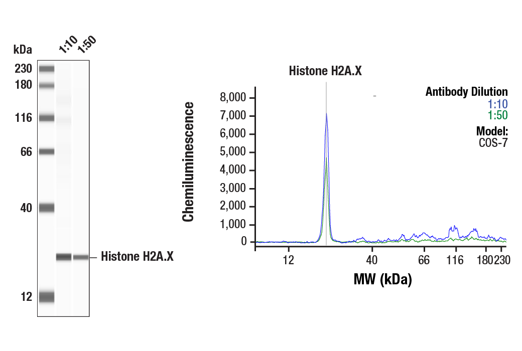
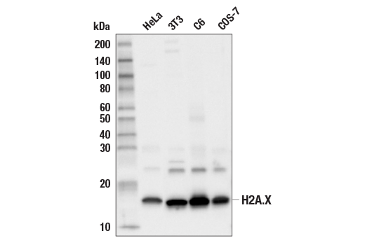
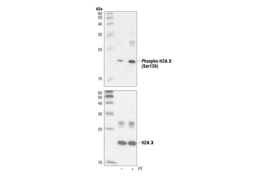
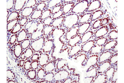
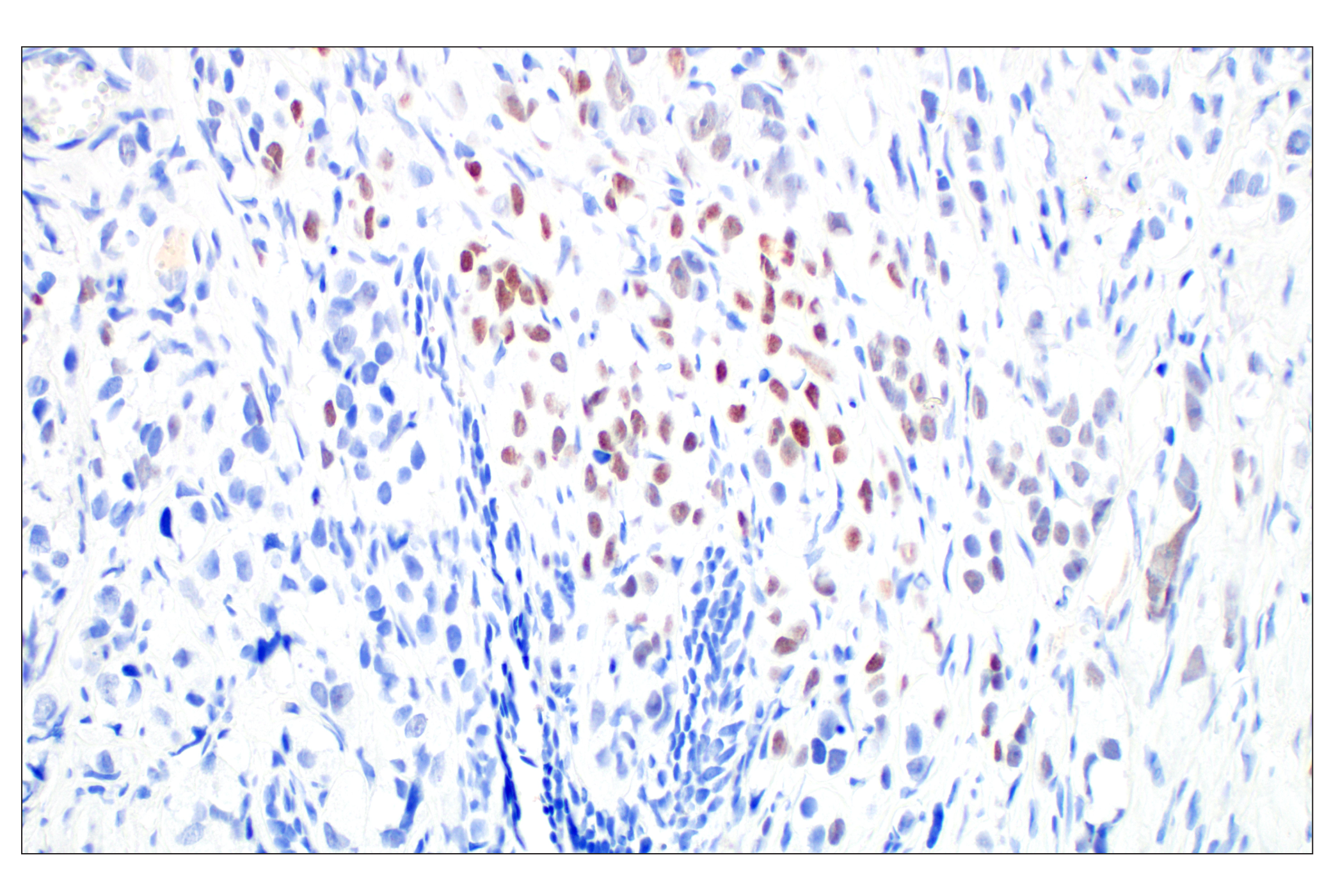
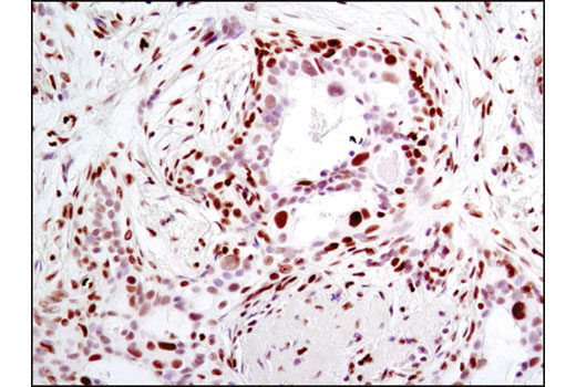
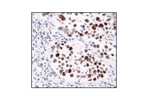
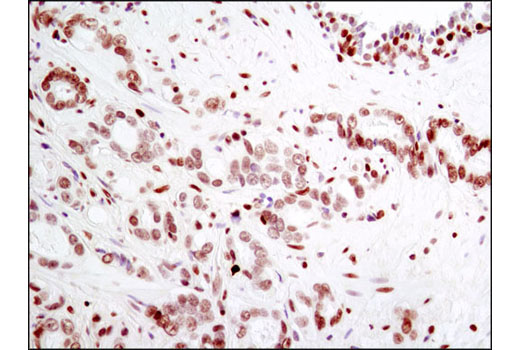
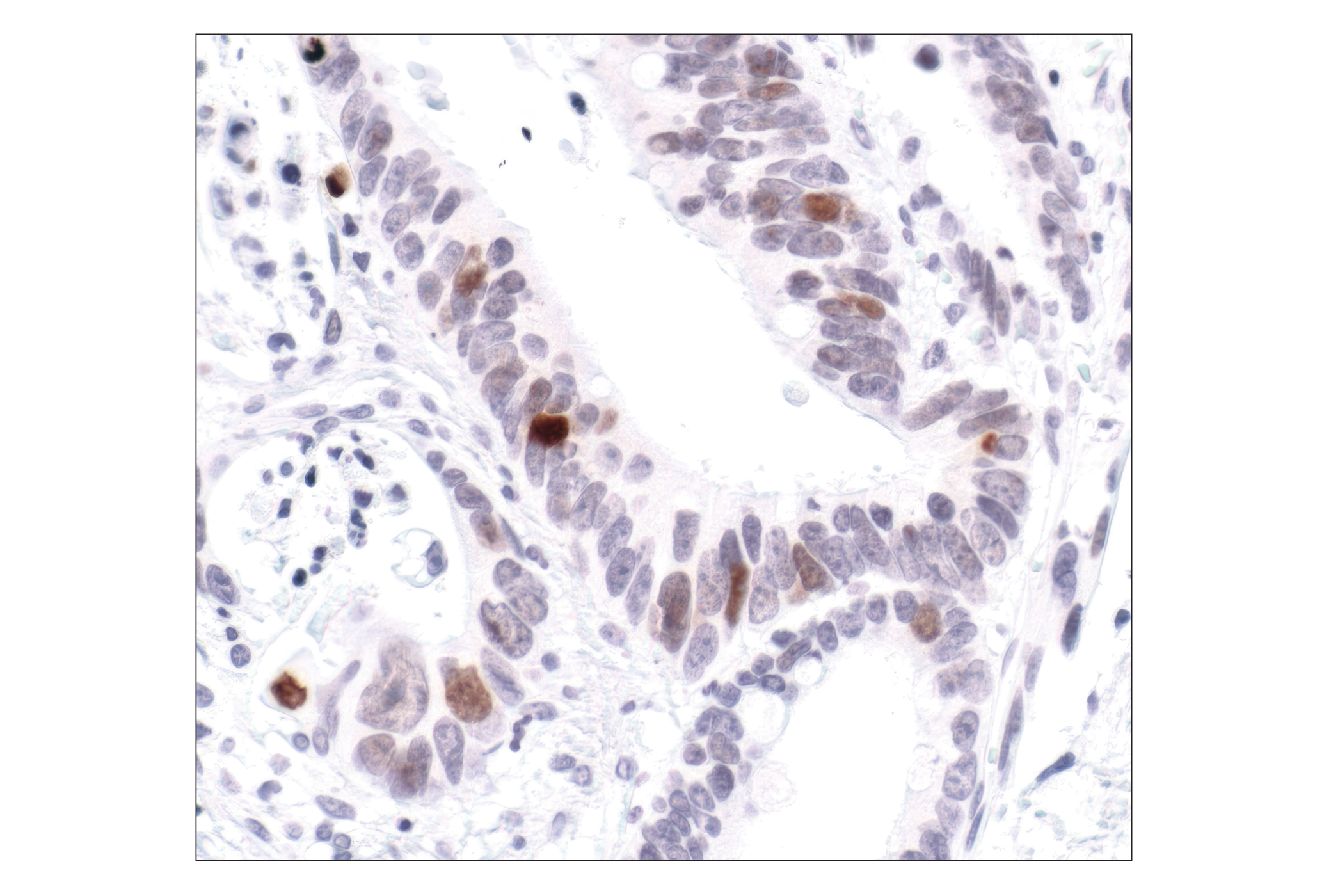
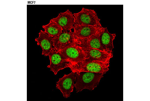
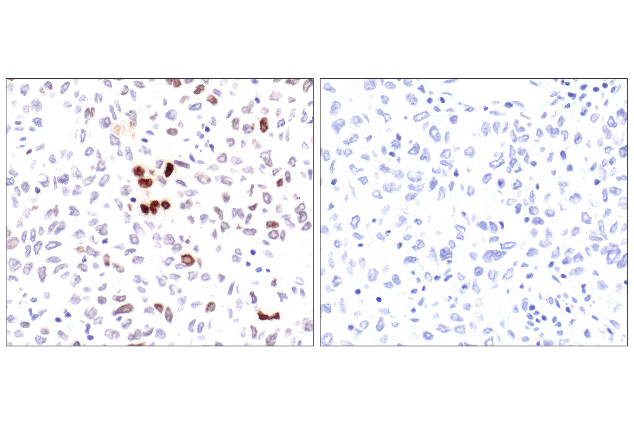
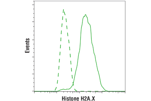
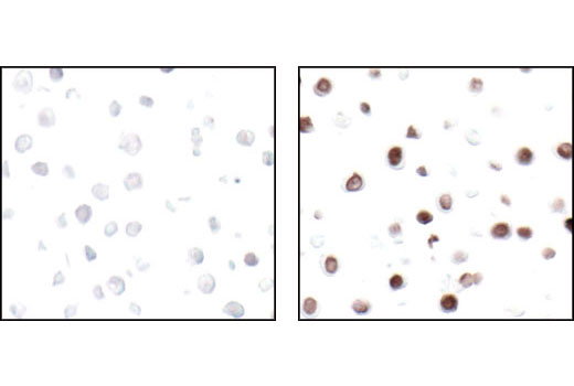
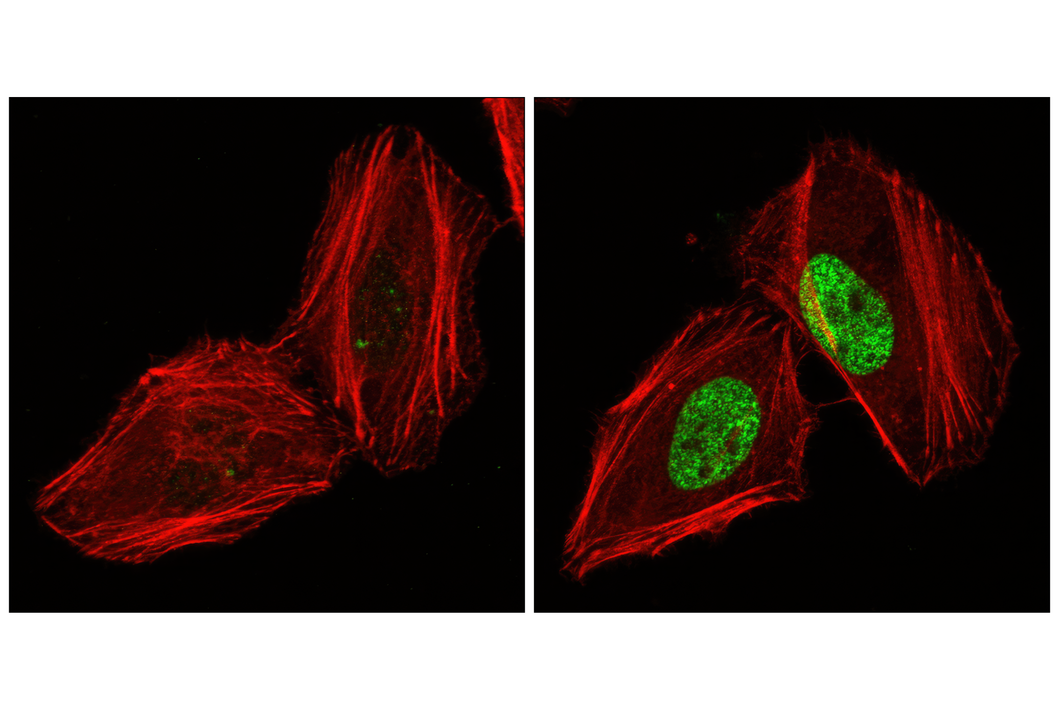
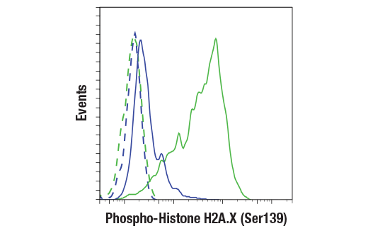
Histone H2A.X is a variant histone that represents approximately 10% of the total H2A histone proteins in normal human fibroblasts (1). H2A.X is required for checkpoint-mediated cell cycle arrest and DNA repair following double-stranded DNA breaks (1). DNA damage, caused by ionizing radiation, UV-light, or radiomimetic agents, results in rapid phosphorylation of H2A.X at Ser139 by PI3K-like kinases, including ATM, ATR, and DNA-PK (2,3). Within minutes following DNA damage, H2A.X is phosphorylated at Ser139 at sites of DNA damage (4). This very early event in the DNA-damage response is required for recruitment of a multitude of DNA-damage response proteins, including MDC1, NBS1, RAD50, MRE11, 53BP1, and BRCA1 (1). In addition to its role in DNA-damage repair, H2A.X is required for DNA fragmentation during apoptosis and is phosphorylated by various kinases in response to apoptotic signals. H2A.X is phosphorylated at Ser139 by DNA-PK in response to cell death receptor activation, c-Jun N-terminal Kinase (JNK1) in response to UV-A irradiation, and p38 MAPK in response to serum starvation (5-8). H2A.X is constitutively phosphorylated on Tyr142 in undamaged cells by WSTF (Williams-Beuren syndrome transcription factor) (9,10). Upon DNA damage, and concurrent with phosphorylation of Ser139, Tyr142 is dephosphorylated at sites of DNA damage by recruited EYA1 and EYA3 phosphatases (9). While phosphorylation at Ser139 facilitates the recruitment of DNA repair proteins and apoptotic proteins to sites of DNA damage, phosphorylation at Tyr142 appears to determine which set of proteins are recruited. Phosphorylation of H2A.X at Tyr142 inhibits the recruitment of DNA repair proteins and promotes binding of pro-apoptotic factors such as JNK1 (9). Mouse embryonic fibroblasts expressing only mutant H2A.X Y142F, which favors recruitment of DNA repair proteins over apoptotic proteins, show a reduced apoptotic response to ionizing radiation (9). Thus, it appears that the balance of H2A.X Tyr142 phosphorylation and dephosphorylation provides a switch mechanism to determine cell fate after DNA damage.

  1. Yuan, J. et al. (2010) FEBS Lett 584, 3717-24.
  2. Rogakou, E.P. et al. (1998) J Biol Chem 273, 5858-68.
  3. Burma, S. et al. (2001) J Biol Chem 276, 42462-7.
  4. Rogakou, E.P. et al. (1999) J Cell Biol 146, 905-16.
  5. Mukherjee, B. et al. (2006) DNA Repair (Amst) 5, 575-90.
  6. Solier, S. et al. (2009) Mol Cell Biol 29, 68-82.
  7. Lu, C. et al. (2006) Mol Cell 23, 121-32.
  8. Lu, C. et al. (2008) FEBS Lett 582, 2703-8.
  9. Cook, P.J. et al. (2009) Nature 458, 591-6.
  10. Xiao, A. et al. (2009) Nature 457, 57-62.

Background References

    Trademarks and Patents

    Cell Signaling Technology is a trademark of Cell Signaling Technology, Inc.
    PhosphoPlus is a registered trademark of Cell Signaling Technology, Inc.
    XP is a registered trademark of Cell Signaling Technology, Inc.
    All other trademarks are the property of their respective owners. Visit cellsignal.com/trademarks for more information.

    使用に関する制限

    法的な権限を与えられたCSTの担当者が署名した書面によって別途明示的に合意された場合を除き、 CST、その関連会社または代理店が提供する製品には以下の条件が適用されます。お客様が定める条件でここに定められた条件に含まれるものを超えるもの、 または、ここに定められた条件と異なるものは、法的な権限を与えられたCSTの担当者が別途書面にて受諾した場合を除き、拒絶され、 いかなる効力も効果も有しません。

    研究専用 (For Research Use Only) またはこれに類似する表示がされた製品は、 いかなる目的についても FDA または外国もしくは国内のその他の規制機関により承認、認可または許可を受けていません。 お客様は製品を診断もしくは治療目的で使用してはならず、また、製品に表示された内容に違反する方法で使用してはなりません。 CST が販売または使用許諾する製品は、エンドユーザーであるお客様に対し、使途を研究および開発のみに限定して提供されるものです。 診断、予防もしくは治療目的で製品を使用することまたは製品を再販売 (単独であるか他の製品等の一部であるかを問いません) もしくはその他の商業的利用の目的で購入することについては、CST から別途許諾を得る必要があります。 お客様は以下の事項を遵守しなければなりません。(a) CST の製品 (単独であるか他の資材と一緒であるかを問いません) を販売、使用許諾、貸与、寄付もしくはその他の態様で第三者に譲渡したり使用させたりしてはなりません。また、商用の製品を製造するために CST の製品を使用してはなりません。(b) 複製、改変、リバースエンジニアリング、逆コンパイル、 分解または他の方法により製品の構造または技術を解明しようとしてはなりません。また、 CST の製品またはサービスと競合する製品またはサービスを開発する目的で CST の製品を使用してはなりません。(c) CST の製品の商標、商号、ロゴ、特許または著作権に関する通知または表示を除去したり改変したりしてはなりません。(d) CST の製品をCST 製品販売条件(CST’s Product Terms of Sale) および該当する書面のみに従って使用しなければなりません。(e) CST の製品に関連してお客様が使用する第三者の製品またはサービスに関する使用許諾条件、 サービス提供条件またはこれに類する合意事項を遵守しなければなりません。

    Revision 1
    #45456

    PhosphoPlus® Histone H2A.X (Ser139) Antibody Duet

    PhosphoPlus® Histone H2A.X (Ser139) Antibody Duet: Image 1 Expand Image
    Simple Western analysis of lysates (0.1 mg/mL) from COS-7 cells using Histone H2A.X (D17A3) XP® Rabbit mAb #7361. The virtual lane view (left) shows the target band (as indicated) and a band corresponding to Histone H2A.X (as indicated) at 1:10 and 1:50 dilutions of primary antibody. The corresponding electropherogram view (right) plots chemiluminescence by molecular weight along the capillary at 1:10 (blue line) and 1:50 (green line) dilutions of primary antibody. This experiment was performed under reducing conditions on the Jess Simple Western instrument from ProteinSimple, a BioTechne brand, using the 12-230 kDa separation module.
    PhosphoPlus® Histone H2A.X (Ser139) Antibody Duet: Image 2 Expand Image
    Western blot analysis of extracts from various cell lines using Histone H2A.X (D17A3) XP® Rabbit mAb.
    PhosphoPlus® Histone H2A.X (Ser139) Antibody Duet: Image 3 Expand Image
    Western blot analysis of extracts from untreated or UV-treated 293 cells, using Phospho-Histone H2A.X (Ser139) (20E3) Rabbit mAb (upper) or Histone H2A.X Antibody #2595 (lower).
    PhosphoPlus® Histone H2A.X (Ser139) Antibody Duet: Image 4 Expand Image
    Immunohistochemical analysis of paraffin-embedded human ovarian clear cell carcinoma using Phospho-Histone H2A.X (Ser139) (20E3) Rabbit mAb performed on the Leica BOND Rx.
    PhosphoPlus® Histone H2A.X (Ser139) Antibody Duet: Image 5 Expand Image
    Immunohistochemical analysis of paraffin-embedded mouse colon using H2A.X (D17A3) Rabbit mAb.
    PhosphoPlus® Histone H2A.X (Ser139) Antibody Duet: Image 6 Expand Image
    Immunohistochemical analysis of paraffin-embedded human prostate adenocarcinoma using Phospho-Histone H2A.X (Ser139) (20E3) Rabbit mAb performed on the Leica BOND Rx.
    PhosphoPlus® Histone H2A.X (Ser139) Antibody Duet: Image 7 Expand Image
    Immunohistochemical analysis of paraffin-embedded human breast carcinoma using Histone H2A.X (D17A3) XP® Rabbit mAb.
    PhosphoPlus® Histone H2A.X (Ser139) Antibody Duet: Image 8 Expand Image
    Immunohistochemical analysis of paraffin-embedded human lung carcinoma, using Phospho-Histone H2A.X (Ser139) (20E3) Rabbit mAb, showing nuclear localization.
    PhosphoPlus® Histone H2A.X (Ser139) Antibody Duet: Image 9 Expand Image
    Immunohistochemical analysis of paraffin-embedded human prostate carcinoma using Histone H2A.X (D17A3) XP® Rabbit mAb.
    PhosphoPlus® Histone H2A.X (Ser139) Antibody Duet: Image 10 Expand Image
    Immunohistochemical analysis of paraffin-embedded human colon carcinoma, using Phospho-Histone H2A.X (Ser139) (20E3) Rabbit mAb.
    PhosphoPlus® Histone H2A.X (Ser139) Antibody Duet: Image 11 Expand Image
    Confocal immunofluorescent analysis of MCF7 cells using Histone H2A.X (D17A3) XP® Rabbit mAb (green). Actin filaments were labeled with DY-554 phalloidin (red).
    PhosphoPlus® Histone H2A.X (Ser139) Antibody Duet: Image 12 Expand Image
    Immunohistochemical analysis of paraffin-embedded human lung carcinoma untreated (left) or lambda-phosphatase-treated (right), using Phospho-Histone H2A.X (Ser139) (20E3) Rabbit mAb.
    PhosphoPlus® Histone H2A.X (Ser139) Antibody Duet: Image 13 Expand Image
    Flow cytometric analysis of MCF7 cells (green) using Histone H2A.X (D17A3) XP® Rabbit mAb (solid lines) or a concentration matched Rabbit (DA1E) mAb IgG XP® Isotype Control #3900 (dashed lines). Anti-rabbit IgG (H+L), F(ab')2 Fragment (Alexa Fluor® 488 Conjugate) #4412 was used as a secondary antibody.
    PhosphoPlus® Histone H2A.X (Ser139) Antibody Duet: Image 14 Expand Image
    Immunohistochemical analysis of paraffin-embedded HT-29 cells untreated (left) or UV-treated (right), using Phospho-Histone H2A.X (Ser139) (20E3) Rabbit mAb.
    PhosphoPlus® Histone H2A.X (Ser139) Antibody Duet: Image 15 Expand Image
    Confocal immunofluorescent analysis of HeLa cells, untreated (left) or UV-treated (right), using Phospho-Histone H2A.X (Ser139) (20E3) Rabbit mAb (green). Actin filaments have been labeled with DY-554 phalloidin (red).
    PhosphoPlus® Histone H2A.X (Ser139) Antibody Duet: Image 16 Expand Image
    Flow cytometric analysis of HeLa cells, untreated (blue) or treated with UV (100 mJ, 2hr recovery; green) using Phospho-H2A.X (Ser139) (20E3) Rabbit mAb (solid lines) or concentration-matched Rabbit (DA1E) mAb IgG XP® isotype control #3900 (dashed lines). Anti-rabbit IgG (H+L), F(ab')2 Fragment (Alexa Fluor® 488 Conjugate) #4412 was used as a secondary antibody.