| Product Includes | Product # | Quantity | Mol. Wt | Isotype/Source |
|---|---|---|---|---|
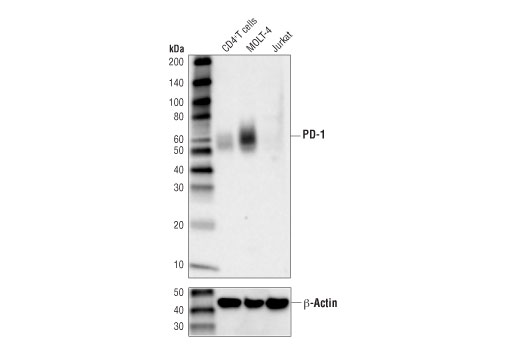
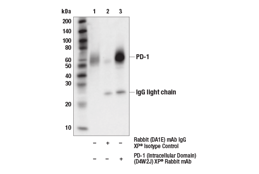
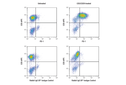
| PD-1 (Intracellular Domain) (D4W2J) XP® Rabbit mAb | 86163 | 20 µl | 52-65 kDa | Rabbit IgG |
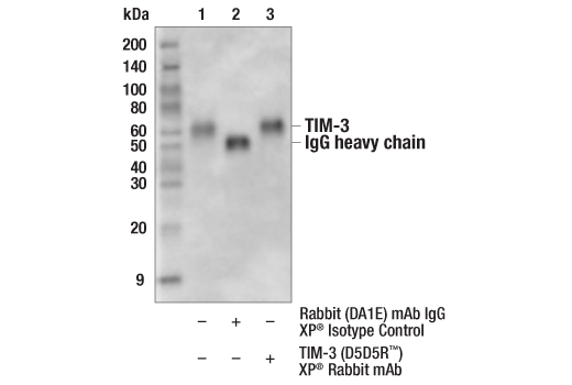
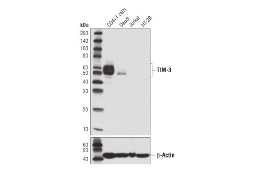
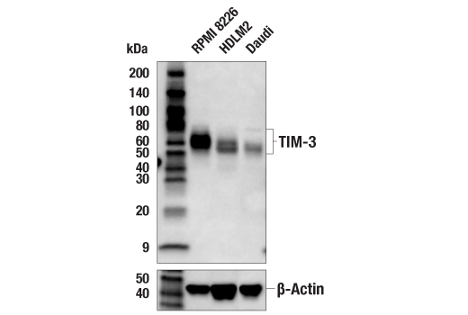
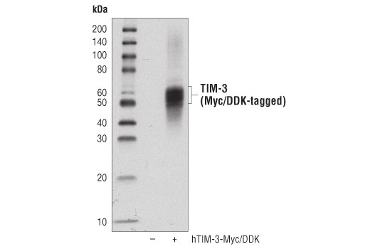
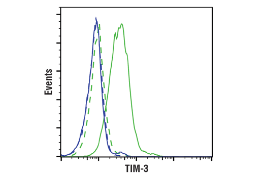
| TIM-3 (D5D5R™) XP® Rabbit mAb | 45208 | 20 µl | 45-70 kDa | Rabbit IgG |
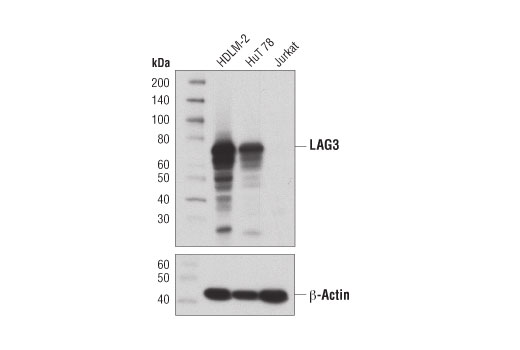
| LAG3 (D2G4O) XP® Rabbit mAb | 15372 | 20 µl | 60-80 kDa | Rabbit IgG |
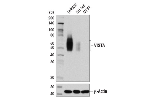
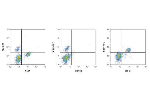
| VISTA (D1L2G™) XP® Rabbit mAb | 64953 | 20 µl | 45-65 kDa | Rabbit IgG |
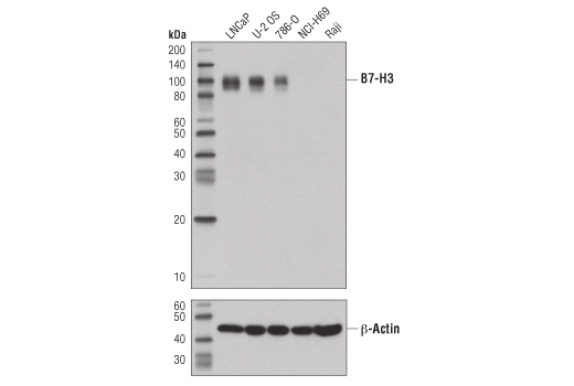
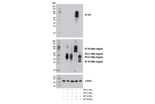
| B7-H3 (D9M2L) XP® Rabbit mAb | 14058 | 20 µl | 90 kDa | Rabbit IgG |
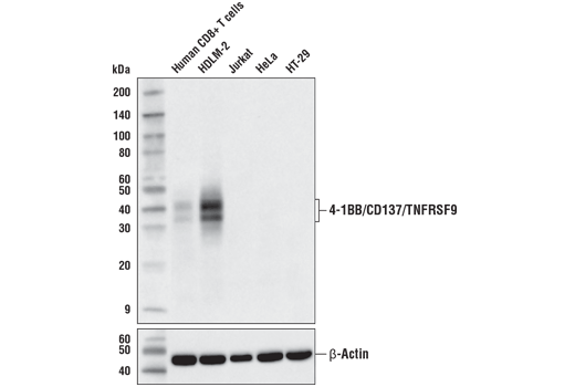
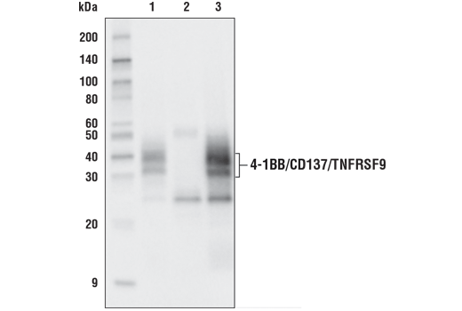
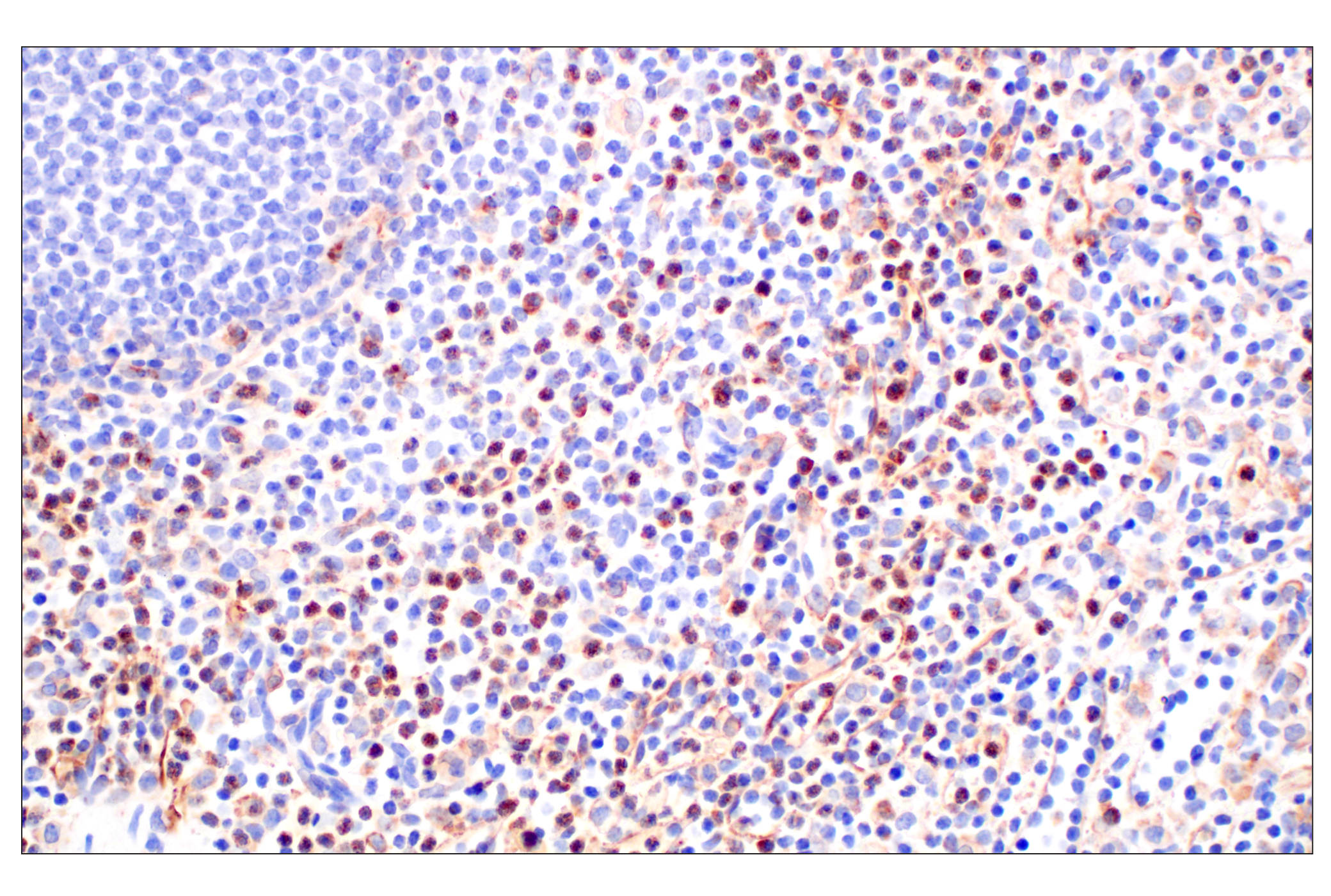
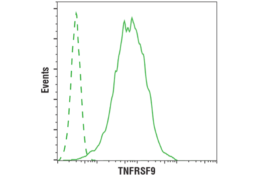
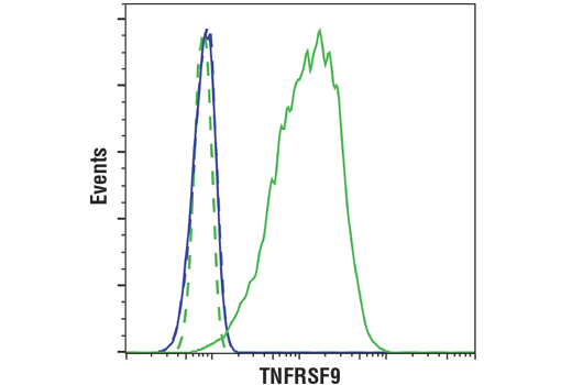
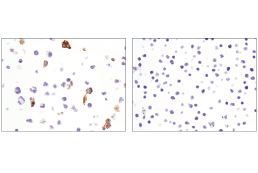
| 4-1BB/CD137/TNFRSF9 (D2Z4Y) Rabbit mAb | 34594 | 20 µl | 32, 40 kDa | Rabbit IgG |
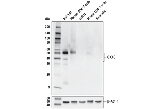
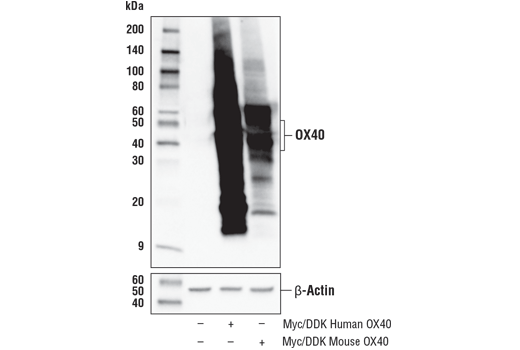
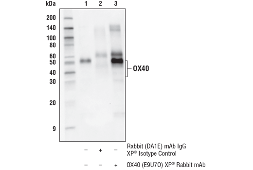
| OX40 (E9U7O) XP® Rabbit mAb | 61637 | 20 µl | 35-50 kDa | Rabbit IgG |
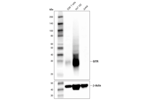
| GITR (D9I9D) Rabbit mAb | 68014 | 20 µl | 25 kDa | Rabbit IgG |
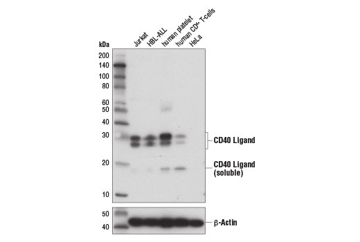
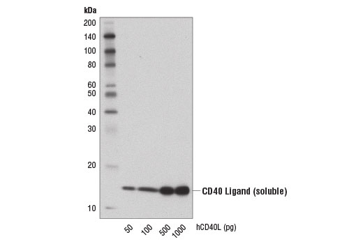
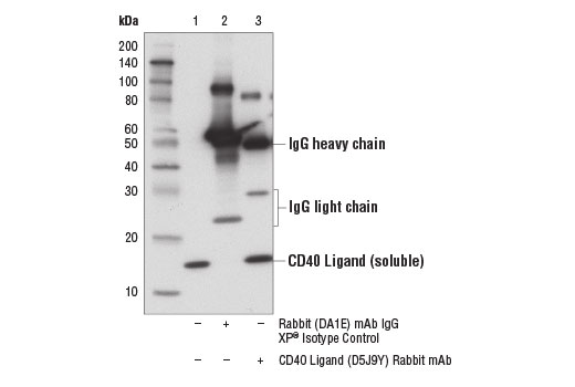
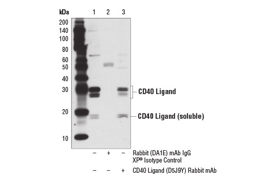
| CD40 Ligand (D5J9Y) Rabbit mAb | 15094 | 20 µl | 25-30 (membrane bound); 17 (soluble) kDa | Rabbit IgG |
Please visit cellsignal.com for individual component applications, species cross-reactivity, dilutions, protocols, and additional product information.
Description
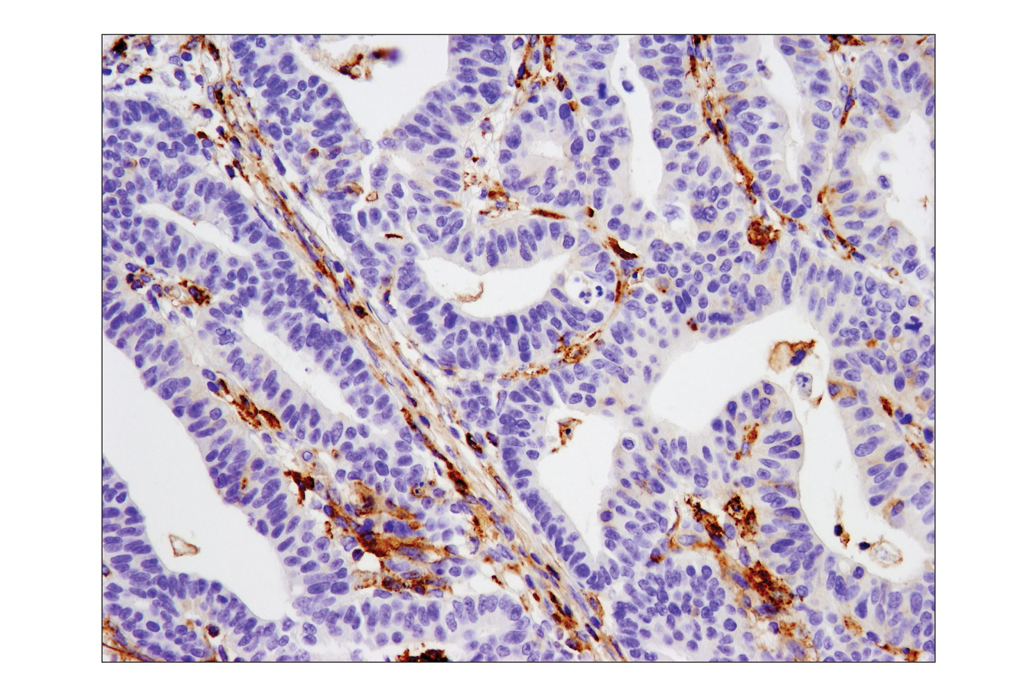
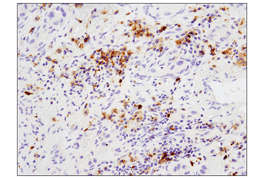
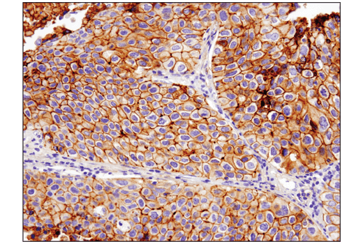
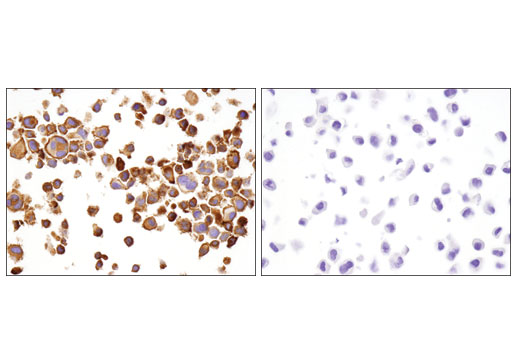
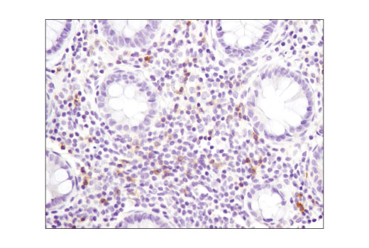
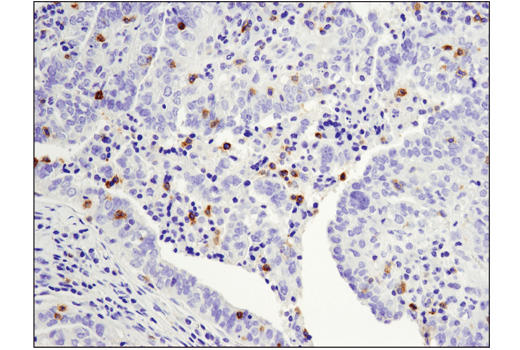
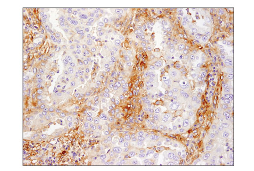
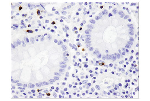
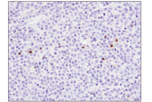
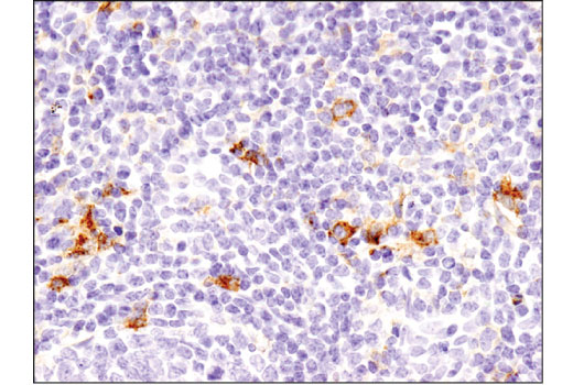
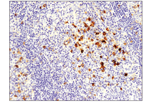
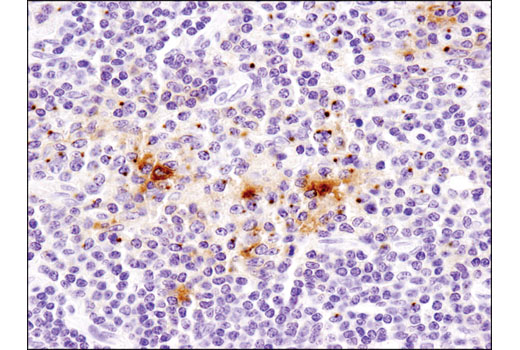
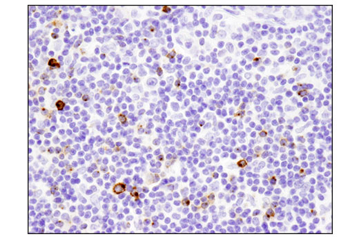
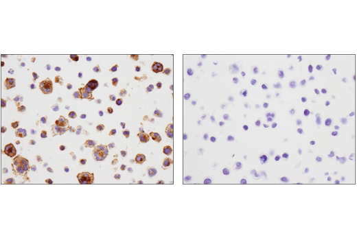
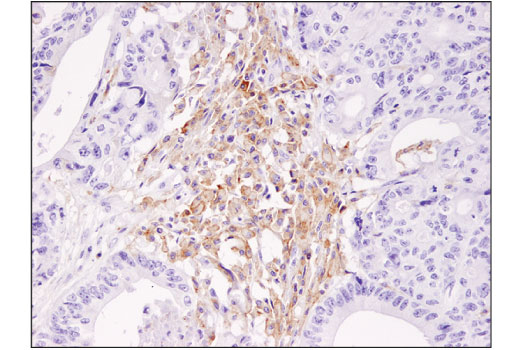
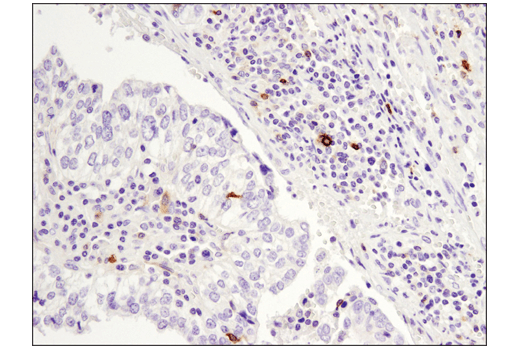
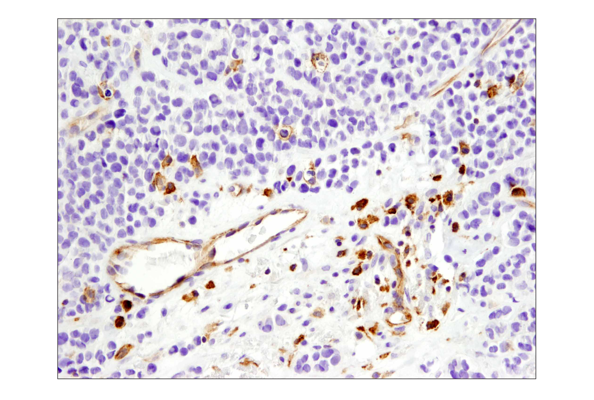
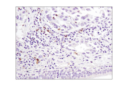
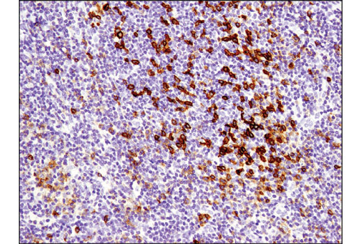
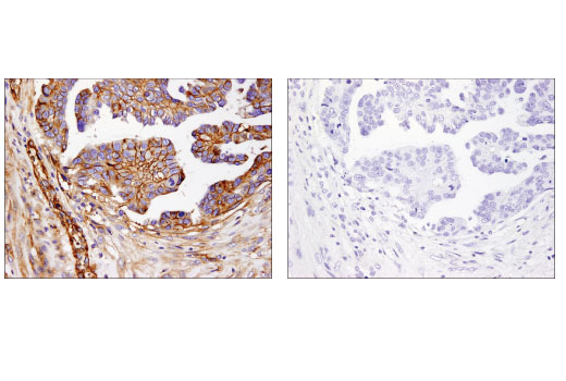
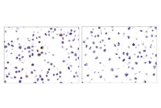
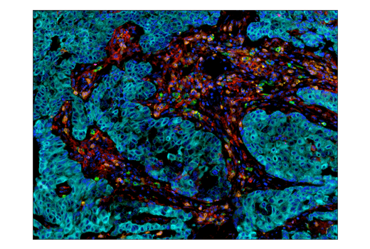
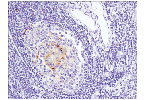
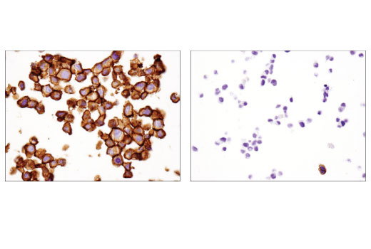
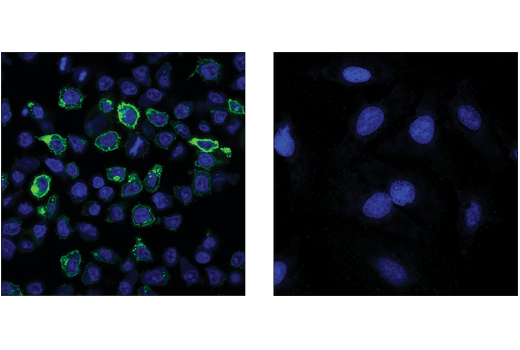
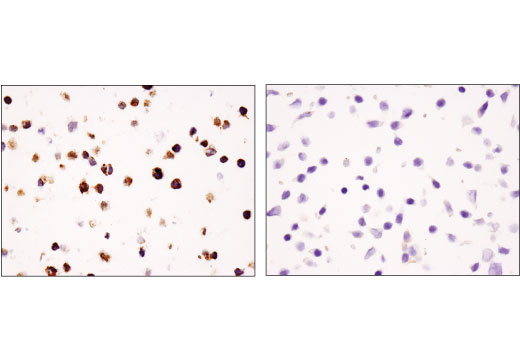
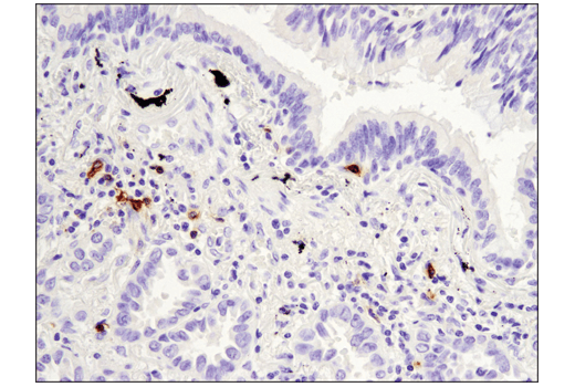
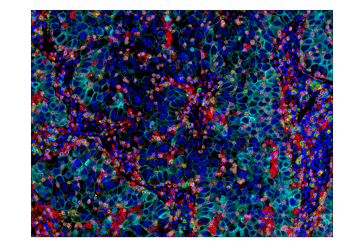
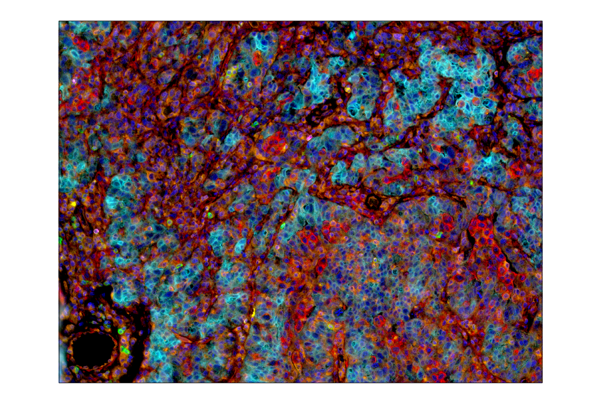
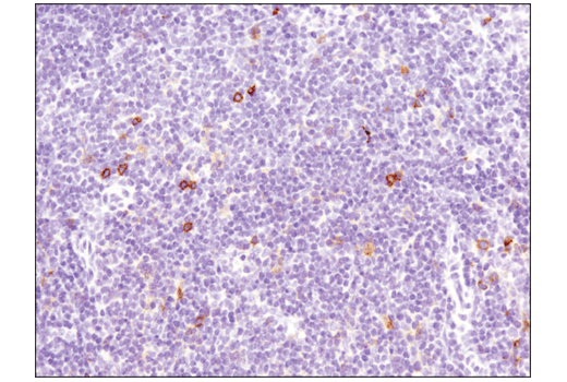
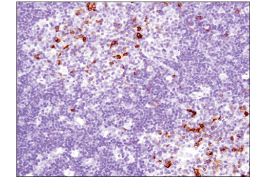
The Human T Cell Co-inhibitory and Co-stimulatory Receptor IHC Antibody Sampler Kit provides an economical means of detecting expression of receptors that modulate T cell activity in formalin-fixed, paraffin-embedded tissue samples.
Storage
Background
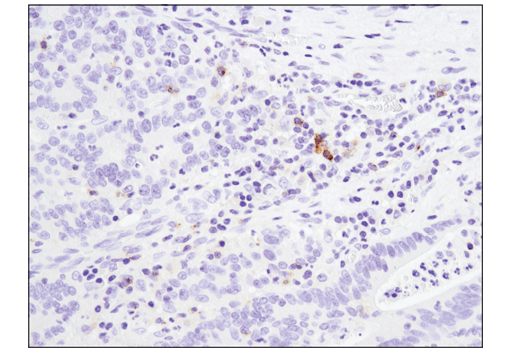
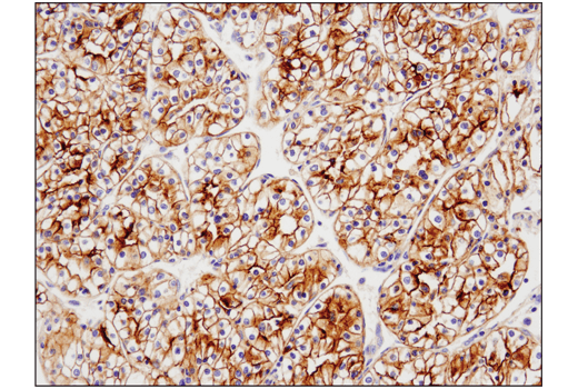
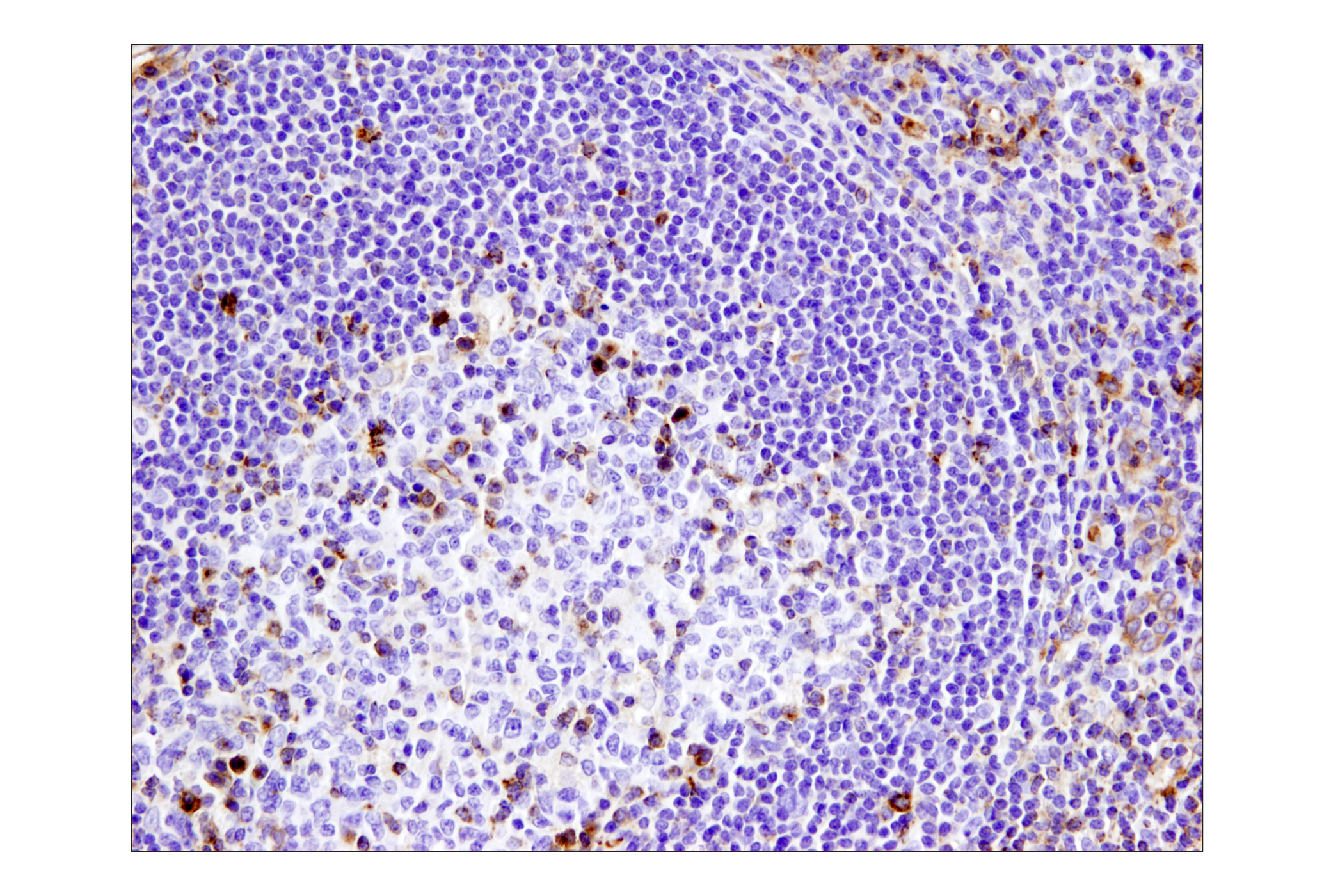
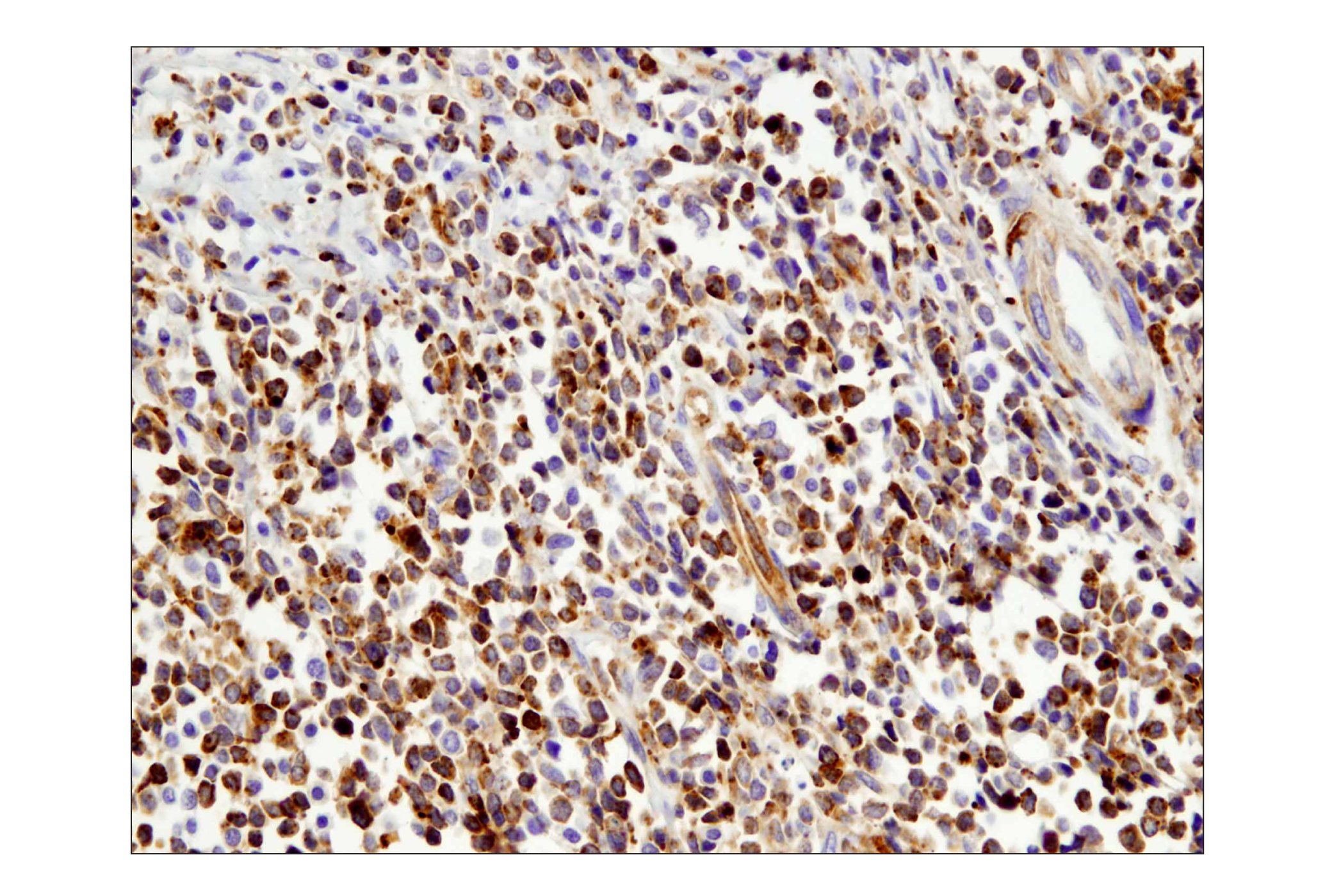
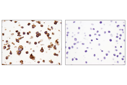
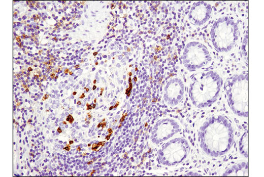
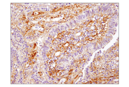
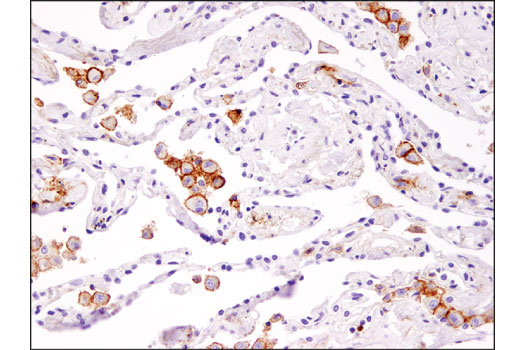
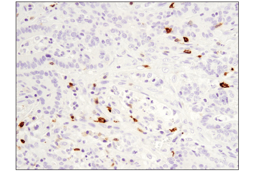
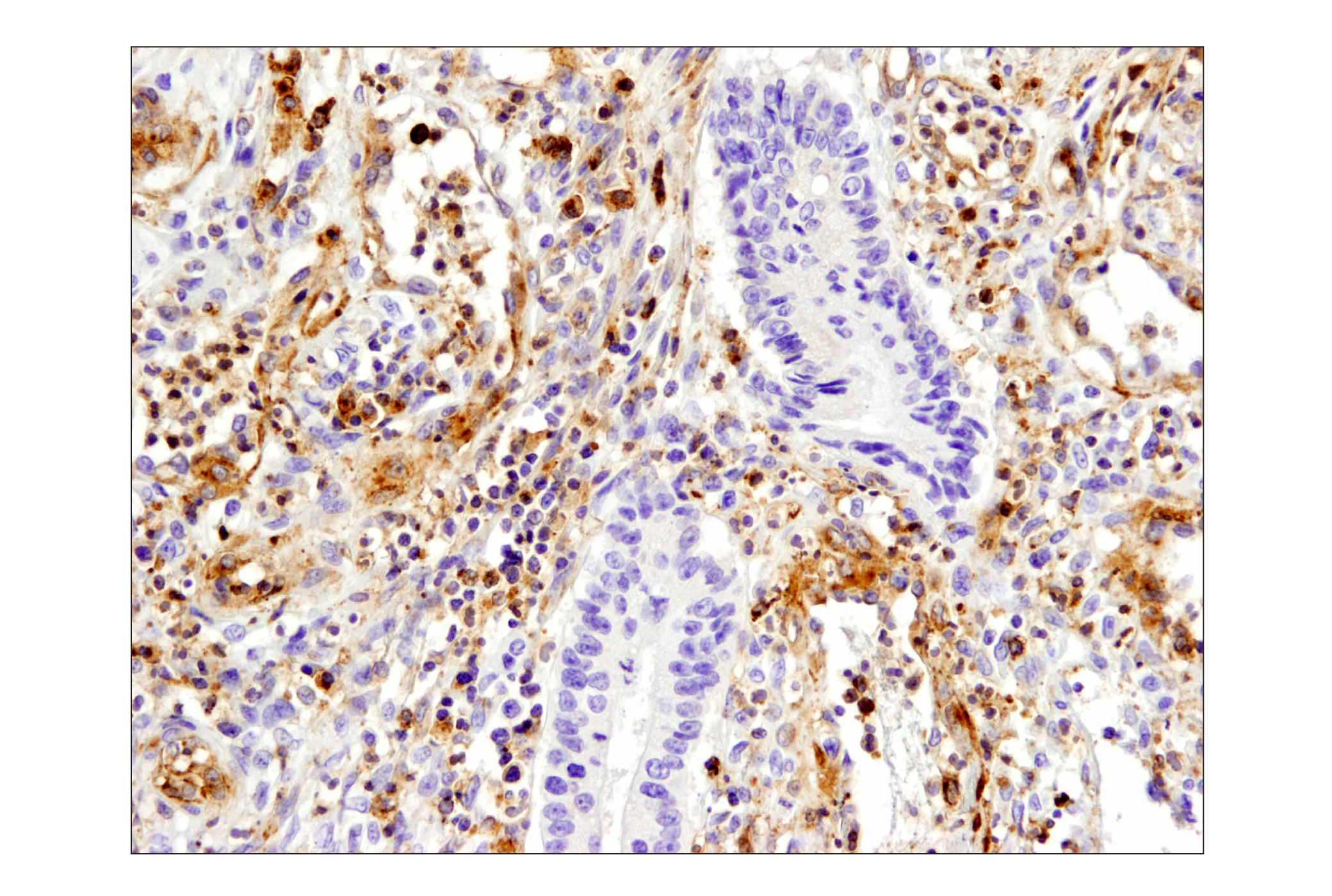
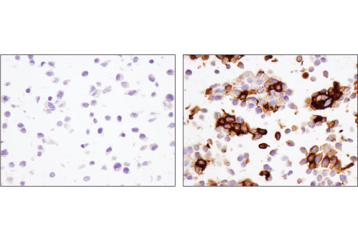
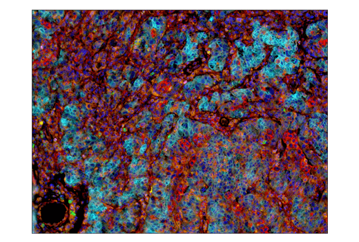
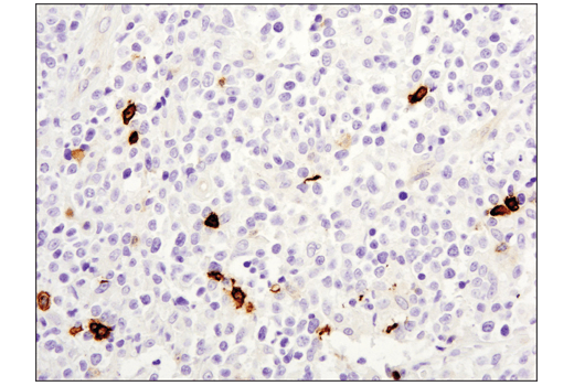
PD-1 (PDCD1, CD279), TIM-3 (HAVCR2), LAG3 (CD223), VISTA (PD-H1), and B7-H3 (CD276) are immune cell co-inhibitory receptors (also known as immune checkpoints) that negatively regulate T cell function, and dampen the immune response to pathogens and cancer. In addition to activated T cells, PD-1 is expressed by activated B-cells and monocytes. TIM-3 is expressed by exhausted T cells in the settings of chronic infection and cancer. Tumor-infiltrating macrophages and dendritic cells also express TIM-3. LAG3 is primarily expressed by activated CD4+ T cells, CD8+ T cells, FoxP3+ T regulatory cells (Tregs) and natural killer (NK) cells. Although primarily expressed by myeloid cells, VISTA is also expressed by CD4+, CD8+, and Treg cells. Research examining the biological function of B7-H3 suggested that B7-H3 can be both a positive and negative regulator of T cell response. B7-H3 is expressed by antigen presenting cells, activated T cells, and a few normal tissues, including placenta and prostate. Expression of B7-H3 is seen in several cancer types, including prostate, breast, colon, lung, and gastric cancers, and in endothelial cells from tumor associated vasculature. Therapeutic blockade of these immune checkpoint receptors is a promising strategy for neoplastic intervention by enabling anti-tumor immune responses (1-3).
4-1BB (TNFRSF9, CD137), GITR (TNFRSF18), OX40 (TNFRSF4, CD134), and CD40 ligand (CD40L, CD154, TRAP, gp39) are immune cell co-stimulatory receptors that promote effector T cell survival and activation, and enable optimal immune responses to pathogens. 4-1BB is expressed in activated CD4+ and CD8+ T cells, natural killer cells and dendritic cells. GITR is expressed constitutively at high levels on Tregs, at low levels on naive and memory T cells, and is induced upon T cell activation. Studies show GITR can also be induced on NK cells, macrophages, and DCs. GITR ligation has been shown to induce CD8+ T cell activation, cytoxicity, and memory T cell survival, and conversely inhibit Treg suppressive function while promoting effector T cell resistance to Treg suppression. OX40 is primarily expressed on activated CD4+ and CD8+ T cells, while CD40L is primarily expressed on the surface of T cells, but has also been reported in blood platelets, mast cells, basophils, NK cells, and B cells. Research studies show that agonists of these co-stimulatory receptors augment anti-tumor immunity in several cancer types. Due to the combined effects on both Treg suppression and effector cell activation, GITR represents a unique opportunity for immunotherapeutic intervention in cancer. These pathways are an important area of interest in the study of cancer, vascular diseases, and inflammatory disorders (4-7).
- Schildberg, F.A. et al. (2016) Immunity 44, 955-72.
- Anderson, A.C. et al. (2016) Immunity 44, 989-1004.
- Callahan, M.K. et al. (2016) Immunity 44, 1069-78.
- Ward-Kavanagh, L.K. et al. (2016) Immunity 44, 1005-19.
- Ara, A. et al. (2018) Immunotargets Ther 7, 55-61.
- Knee, D.A. et al. (2016) Eur J Cancer 67, 1-10.
- Chester, C. et al. (2018) Blood 131, 49-57.
Background References
Trademarks and Patents
使用に関する制限
法的な権限を与えられたCSTの担当者が署名した書面によって別途明示的に合意された場合を除き、 CST、その関連会社または代理店が提供する製品には以下の条件が適用されます。お客様が定める条件でここに定められた条件に含まれるものを超えるもの、 または、ここに定められた条件と異なるものは、法的な権限を与えられたCSTの担当者が別途書面にて受諾した場合を除き、拒絶され、 いかなる効力も効果も有しません。
研究専用 (For Research Use Only) またはこれに類似する表示がされた製品は、 いかなる目的についても FDA または外国もしくは国内のその他の規制機関により承認、認可または許可を受けていません。 お客様は製品を診断もしくは治療目的で使用してはならず、また、製品に表示された内容に違反する方法で使用してはなりません。 CST が販売または使用許諾する製品は、エンドユーザーであるお客様に対し、使途を研究および開発のみに限定して提供されるものです。 診断、予防もしくは治療目的で製品を使用することまたは製品を再販売 (単独であるか他の製品等の一部であるかを問いません) もしくはその他の商業的利用の目的で購入することについては、CST から別途許諾を得る必要があります。 お客様は以下の事項を遵守しなければなりません。(a) CST の製品 (単独であるか他の資材と一緒であるかを問いません) を販売、使用許諾、貸与、寄付もしくはその他の態様で第三者に譲渡したり使用させたりしてはなりません。また、商用の製品を製造するために CST の製品を使用してはなりません。(b) 複製、改変、リバースエンジニアリング、逆コンパイル、 分解または他の方法により製品の構造または技術を解明しようとしてはなりません。また、 CST の製品またはサービスと競合する製品またはサービスを開発する目的で CST の製品を使用してはなりません。(c) CST の製品の商標、商号、ロゴ、特許または著作権に関する通知または表示を除去したり改変したりしてはなりません。(d) CST の製品をCST 製品販売条件(CST’s Product Terms of Sale) および該当する書面のみに従って使用しなければなりません。(e) CST の製品に関連してお客様が使用する第三者の製品またはサービスに関する使用許諾条件、 サービス提供条件またはこれに類する合意事項を遵守しなければなりません。