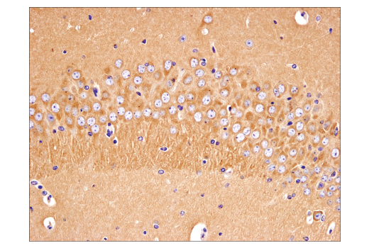
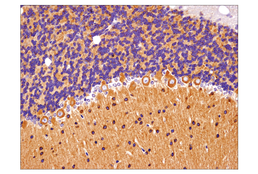
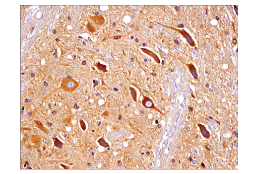
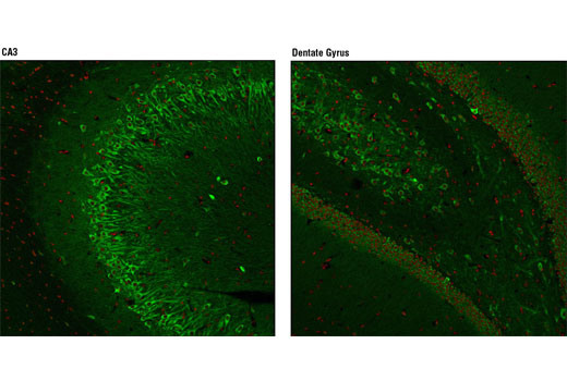
WB, IHC-P, IF-F
H M R
Endogenous
350
Rabbit IgG
#P42858
3064
Product Information
Product Usage Information
This product is the carrier free version of product #5656. All data were generated using the same antibody clone in the standard formulation which contains BSA and glycerol.
This formulation is ideal for use with technologies requiring specialized or custom antibody labeling, including fluorophores, metals, lanthanides, and oligonucleotides. Optimal dilutions/concentrations should be determined by the end user.
Formulation
Storage
Specificity / Sensitivity
Species Reactivity:
Human, Mouse, Rat
Source / Purification
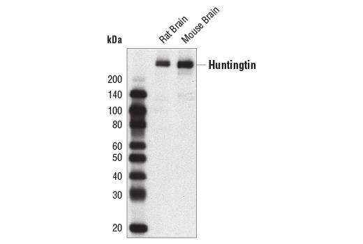
Monoclonal antibody is produced by immunizing animals with a synthetic peptide corresponding to residues surrounding Pro1220 of human huntingtin protein.
Background
Huntington's Disease (HD) is a fatal neurodegenerative disorder characterized by psychiatric, cognitive, and motor dysfunction. Neuropathology of HD involves specific neuronal subpopulations: GABA-ergic neurons of the striatum and neurons within the cerebral cortex selectively degenerate (1,2). The genetic analysis of HD has been the flagship study of inherited neurological diseases from initial chromosomal localization to identification of the gene.
Huntingtin is a large (340-350 kD) cytosolic protein that may be involved in a number of cellular functions such as transcription, gastrulation, neurogenesis, neurotransmission, axonal transport, neural positioning, and apoptosis (2,3). The HD gene from unaffected individuals contains between 6 and 34 CAG trinucleotide repeats, with expansion beyond this range causing the onset of disease symptoms. A strong inverse correlation exists between the age of onset in patients and the number of huntingtin gene CAG repeats encoding a stretch of polyglutamine peptides (1,2). The huntingtin protein undergoes numerous post-translational modifications including phosphorylation, ubiquitination, sumoylation, palmitoylation, and cleavage (2). Phosphorylation of Ser421 by Akt can partially counteract the toxicity that results from the expanded polyglutamine tract. Varying Akt expression in the brain correlates with regional differences in huntingtin protein phosphorylation; this pattern inversely correlates with the regions that are most affected by degeneration in diseased brain (2). A key step in the disease is the proteolytic cleavage of huntingtin protein into amino-terminal fragments that contain expanded glutamine repeats and translocate into the nucleus. Caspase mediated cleavage of huntingtin at Asp513 is associated with increased polyglutamine aggregate formation and toxicity. Phosphorylation of Ser434 by CDK5 protects against cleavage (2,3).
Species Reactivity
Species reactivity is determined by testing in at least one approved application (e.g., western blot).
Applications Key
WB: Western Blotting IHC-P: Immunohistochemistry (Paraffin) IF-F: Immunofluorescence (Frozen)
Cross-Reactivity Key
H: human M: mouse R: rat Hm: hamster Mk: monkey Vir: virus Mi: mink C: chicken Dm: D. melanogaster X: Xenopus Z: zebrafish B: bovine Dg: dog Pg: pig Sc: S. cerevisiae Ce: C. elegans Hr: horse GP: Guinea Pig Rab: rabbit All: all species expected
Trademarks and Patents
使用に関する制限
法的な権限を与えられたCSTの担当者が署名した書面によって別途明示的に合意された場合を除き、 CST、その関連会社または代理店が提供する製品には以下の条件が適用されます。お客様が定める条件でここに定められた条件に含まれるものを超えるもの、 または、ここに定められた条件と異なるものは、法的な権限を与えられたCSTの担当者が別途書面にて受諾した場合を除き、拒絶され、 いかなる効力も効果も有しません。
研究専用 (For Research Use Only) またはこれに類似する表示がされた製品は、 いかなる目的についても FDA または外国もしくは国内のその他の規制機関により承認、認可または許可を受けていません。 お客様は製品を診断もしくは治療目的で使用してはならず、また、製品に表示された内容に違反する方法で使用してはなりません。 CST が販売または使用許諾する製品は、エンドユーザーであるお客様に対し、使途を研究および開発のみに限定して提供されるものです。 診断、予防もしくは治療目的で製品を使用することまたは製品を再販売 (単独であるか他の製品等の一部であるかを問いません) もしくはその他の商業的利用の目的で購入することについては、CST から別途許諾を得る必要があります。 お客様は以下の事項を遵守しなければなりません。(a) CST の製品 (単独であるか他の資材と一緒であるかを問いません) を販売、使用許諾、貸与、寄付もしくはその他の態様で第三者に譲渡したり使用させたりしてはなりません。また、商用の製品を製造するために CST の製品を使用してはなりません。(b) 複製、改変、リバースエンジニアリング、逆コンパイル、 分解または他の方法により製品の構造または技術を解明しようとしてはなりません。また、 CST の製品またはサービスと競合する製品またはサービスを開発する目的で CST の製品を使用してはなりません。(c) CST の製品の商標、商号、ロゴ、特許または著作権に関する通知または表示を除去したり改変したりしてはなりません。(d) CST の製品をCST 製品販売条件(CST’s Product Terms of Sale) および該当する書面のみに従って使用しなければなりません。(e) CST の製品に関連してお客様が使用する第三者の製品またはサービスに関する使用許諾条件、 サービス提供条件またはこれに類する合意事項を遵守しなければなりません。