WB, W-S, IP, IHC-Bond, IHC-P, IF-F, IF-IC, FC-FP
H M R Hm Mk
Endogenous
17
Rabbit IgG
#P55008
199
Product Information
Product Usage Information
| Application | Dilution |
|---|---|
| Western Blotting | 1:1000 |
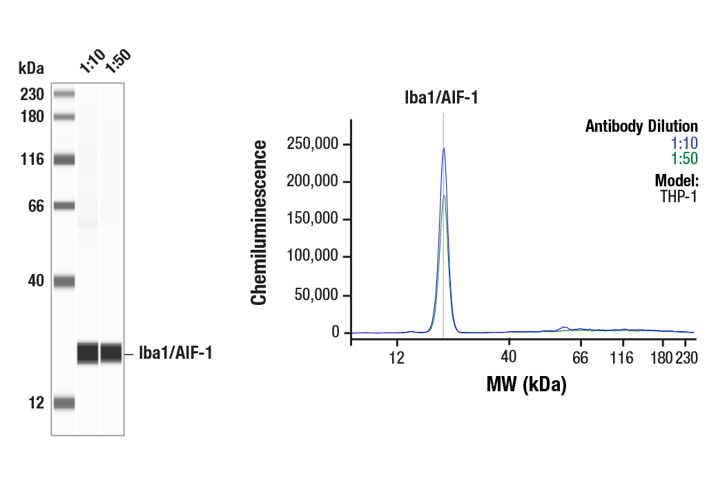
| Simple Western™ | 1:10 - 1:50 |
| Immunoprecipitation | 1:50 |
| IHC Leica Bond | 1:800 - 1:3200 |
| Immunohistochemistry (Paraffin) | 1:800 - 1:3200 |
| Immunofluorescence (Frozen) | 1:50 - 1:200 |
| Immunofluorescence (Immunocytochemistry) | 1:50 - 1:200 |
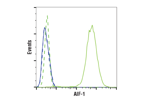
| Flow Cytometry (Fixed/Permeabilized) | 1:50 - 1:200 |
Storage
Supplied in 10 mM sodium HEPES (pH 7.5), 150 mM NaCl, 100 µg/ml BSA, 50% glycerol and less than 0.02% sodium azide. Store at –20°C. Do not aliquot the antibody.
For a carrier-free (BSA and azide free) version of this product see product #79394.
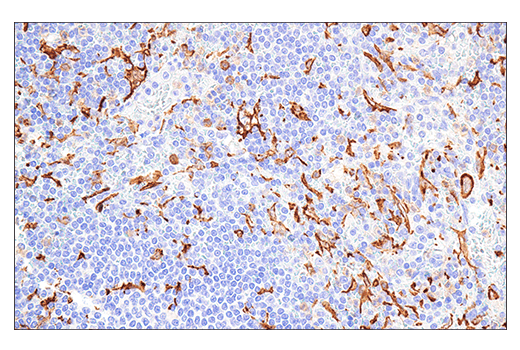
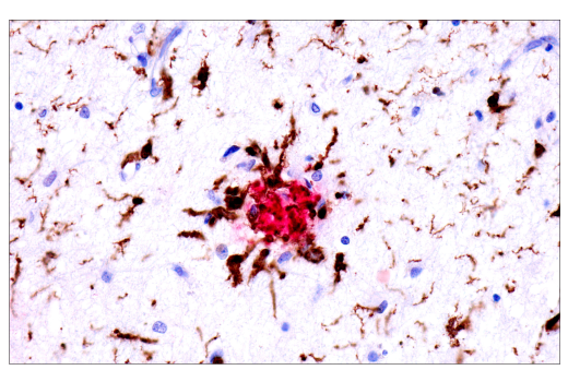
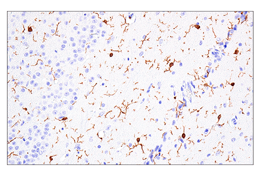
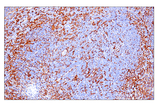
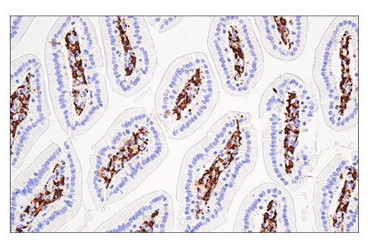
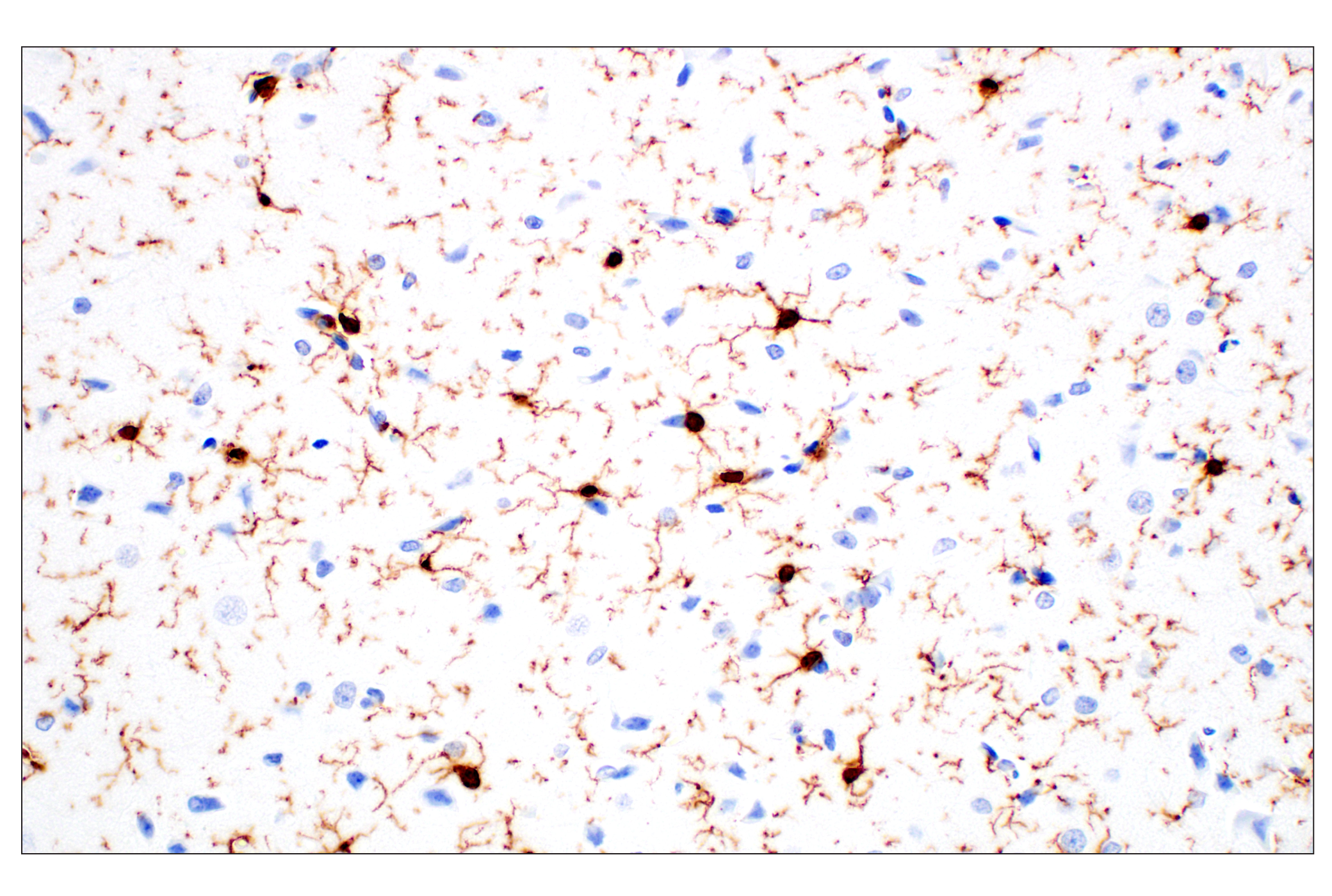
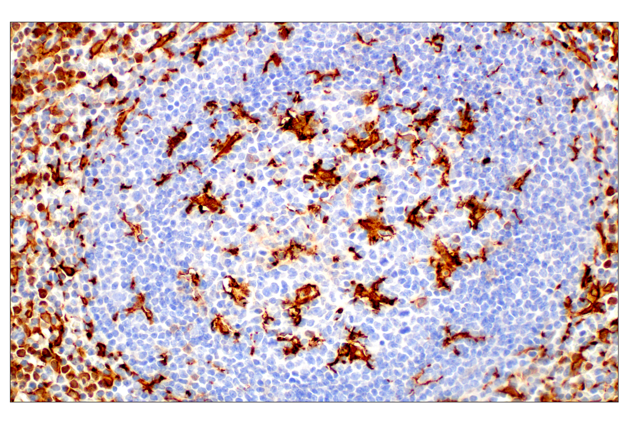
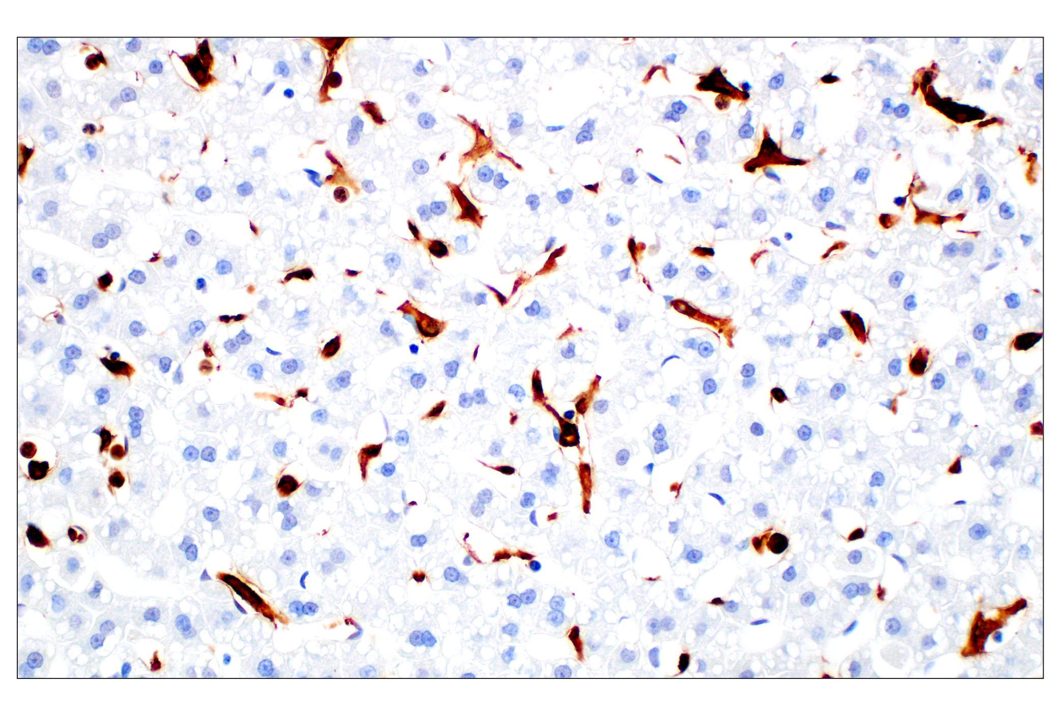
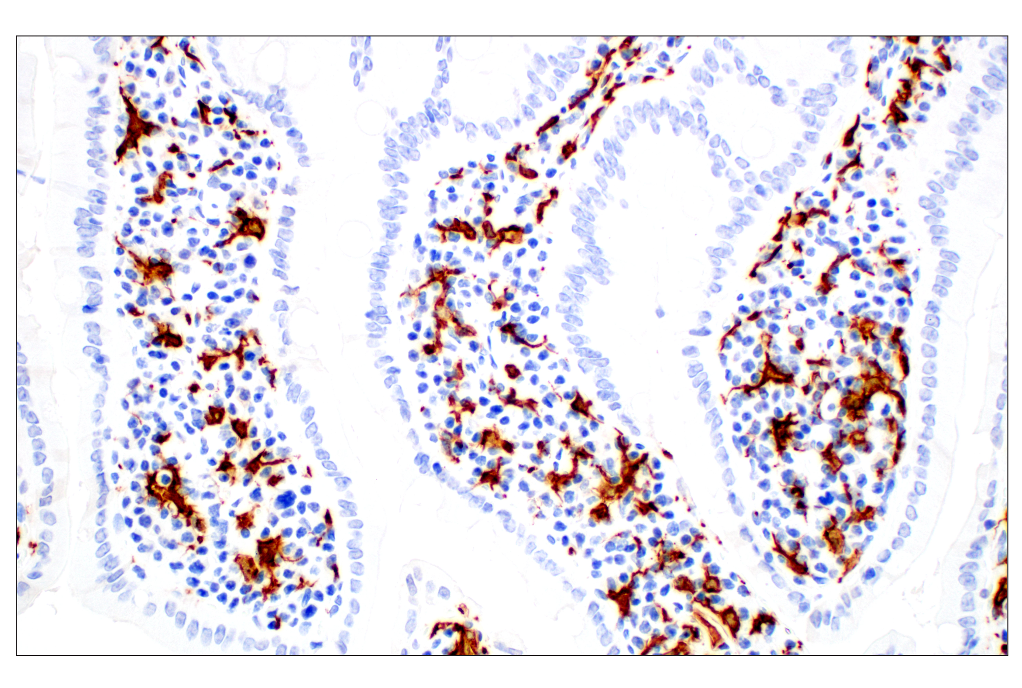
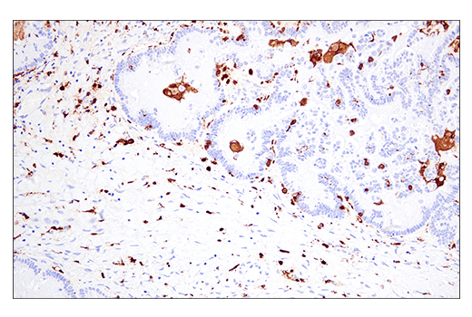
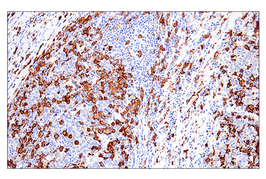
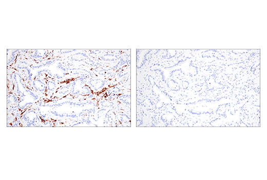
Specificity / Sensitivity
Species Reactivity:
Human, Mouse, Rat, Hamster, Monkey
Source / Purification
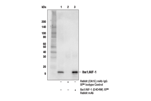
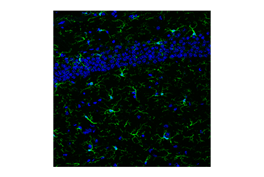
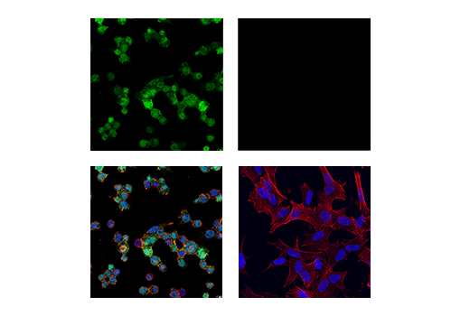
Monoclonal antibody is produced by immunizing animals with a synthetic peptide corresponding to residues surrounding Ala139 of human Iba1/AIF-1 protein.
Background
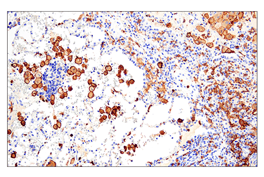
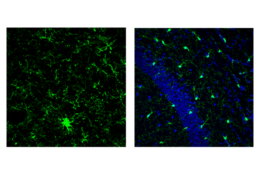
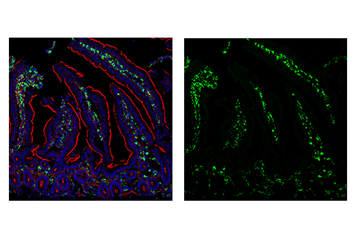
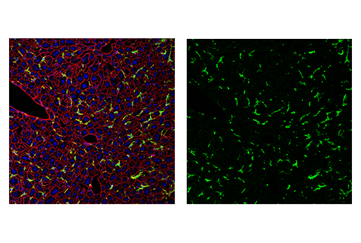
Ionized calcium-binding adaptor molecule 1 (Iba1), also known as allograft inflammatory factor 1 (AIF-1), is an evolutionarily conserved cytoplasmic calcium binding protein containing a central pair of EF-hand calcium binding motifs (1,2). Iba1/AIF-1 was originally cloned from activated macrophages in human atherosclerotic allogenic heart grafts undergoing chronic transplant rejection as well as from rat monocytes (3,4). Its function is not very well understood, but Iba1/AIF-1 expression is upregulated in response to interferon-gamma and, therefore, could modulate macrophage-dependent immune response (3). As an F-actin-binding protein, Iba1/AIF-1 may function to remodel the actin cytoskeleton and contribute to morphological changes that correlate with various microglial/macrophage states (5). Iba1/AIF-1 is also uniquely expressed in cells of monocytic lineage and is, therefore, widely used as a marker for microglia/macrophages in the brain and other tissue (6).
- Schulze, J.O. et al. (2008) FEBS J 275, 4627-40.
- Deininger, M.H. et al. (2002) FEBS Lett 514, 115-21.
- Utans, U. et al. (1995) J Clin Invest 95, 2954-62.
- Imai, Y. et al. (1996) Biochem Biophys Res Commun 224, 855-62.
- Kanazawa, H. et al. (2002) J Biol Chem 277, 20026-32.
- Ito, D. et al. (1998) Brain Res Mol Brain Res 57, 1-9.
Species Reactivity
Species reactivity is determined by testing in at least one approved application (e.g., western blot).
Western Blot Buffer
IMPORTANT: For western blots, incubate membrane with diluted primary antibody in 5% w/v BSA, 1X TBS, 0.1% Tween® 20 at 4°C with gentle shaking, overnight.
Applications Key
WB: Western Blotting W-S: Simple Western™ IP: Immunoprecipitation IHC-Bond: IHC Leica Bond IHC-P: Immunohistochemistry (Paraffin) IF-F: Immunofluorescence (Frozen) IF-IC: Immunofluorescence (Immunocytochemistry) FC-FP: Flow Cytometry (Fixed/Permeabilized)
Cross-Reactivity Key
H: human M: mouse R: rat Hm: hamster Mk: monkey Vir: virus Mi: mink C: chicken Dm: D. melanogaster X: Xenopus Z: zebrafish B: bovine Dg: dog Pg: pig Sc: S. cerevisiae Ce: C. elegans Hr: horse GP: Guinea Pig Rab: rabbit All: all species expected
Trademarks and Patents
使用に関する制限
法的な権限を与えられたCSTの担当者が署名した書面によって別途明示的に合意された場合を除き、 CST、その関連会社または代理店が提供する製品には以下の条件が適用されます。お客様が定める条件でここに定められた条件に含まれるものを超えるもの、 または、ここに定められた条件と異なるものは、法的な権限を与えられたCSTの担当者が別途書面にて受諾した場合を除き、拒絶され、 いかなる効力も効果も有しません。
研究専用 (For Research Use Only) またはこれに類似する表示がされた製品は、 いかなる目的についても FDA または外国もしくは国内のその他の規制機関により承認、認可または許可を受けていません。 お客様は製品を診断もしくは治療目的で使用してはならず、また、製品に表示された内容に違反する方法で使用してはなりません。 CST が販売または使用許諾する製品は、エンドユーザーであるお客様に対し、使途を研究および開発のみに限定して提供されるものです。 診断、予防もしくは治療目的で製品を使用することまたは製品を再販売 (単独であるか他の製品等の一部であるかを問いません) もしくはその他の商業的利用の目的で購入することについては、CST から別途許諾を得る必要があります。 お客様は以下の事項を遵守しなければなりません。(a) CST の製品 (単独であるか他の資材と一緒であるかを問いません) を販売、使用許諾、貸与、寄付もしくはその他の態様で第三者に譲渡したり使用させたりしてはなりません。また、商用の製品を製造するために CST の製品を使用してはなりません。(b) 複製、改変、リバースエンジニアリング、逆コンパイル、 分解または他の方法により製品の構造または技術を解明しようとしてはなりません。また、 CST の製品またはサービスと競合する製品またはサービスを開発する目的で CST の製品を使用してはなりません。(c) CST の製品の商標、商号、ロゴ、特許または著作権に関する通知または表示を除去したり改変したりしてはなりません。(d) CST の製品をCST 製品販売条件(CST’s Product Terms of Sale) および該当する書面のみに従って使用しなければなりません。(e) CST の製品に関連してお客様が使用する第三者の製品またはサービスに関する使用許諾条件、 サービス提供条件またはこれに類する合意事項を遵守しなければなりません。