| Product Includes | Product # | Quantity | Mol. Wt | Isotype/Source |
|---|---|---|---|---|
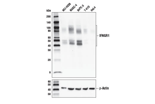
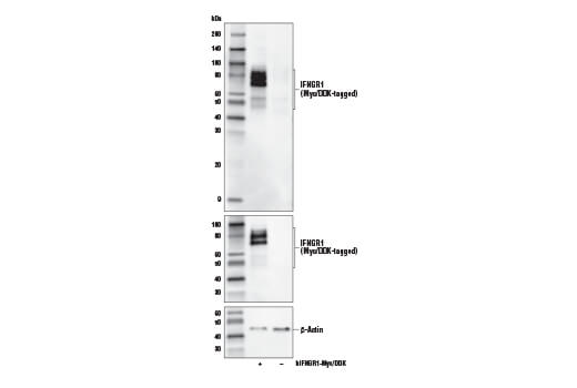
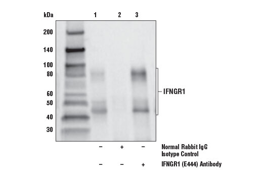
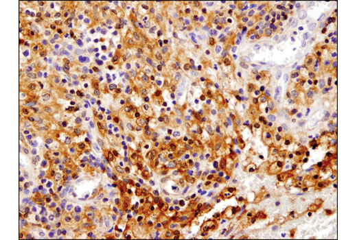
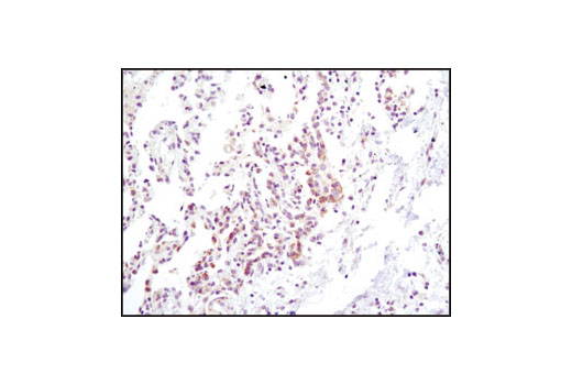
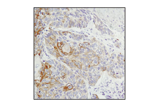
| IFNGR1 (E444) Antibody | 10405 | 20 µl | 45-90 kDa | Rabbit |
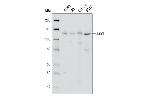
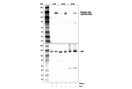
| Jak1 (6G4) Rabbit mAb | 3344 | 20 µl | 130 kDa | Rabbit IgG |
| Phospho-Jak1(Tyr1034/1035) (D7N4Z) Rabbit mAb | 74129 | 20 µl | 130 kDa | Rabbit IgG |
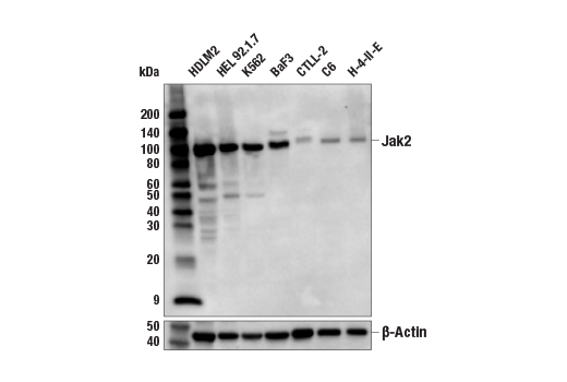
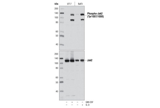
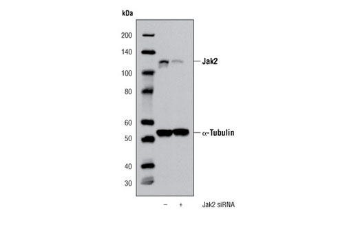
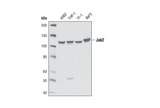
| Jak2 (D2E12) XP® Rabbit mAb | 3230 | 20 µl | 125 kDa | Rabbit IgG |
| Phospho-Jak2 (Tyr1007/1008) (C80C3) Rabbit mAb | 3776 | 20 µl | 125 kDa | Rabbit IgG |
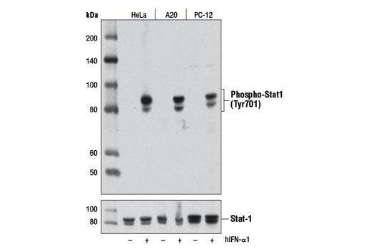
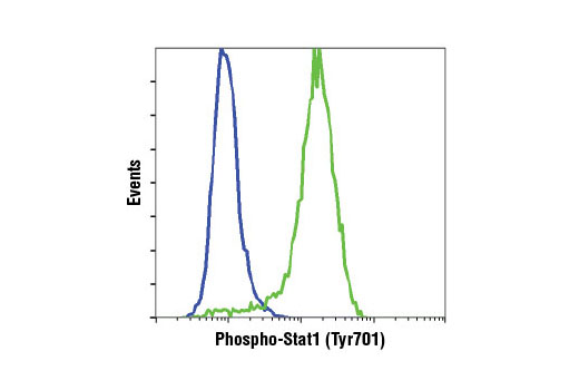
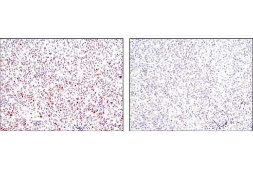
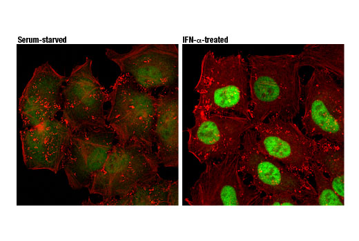
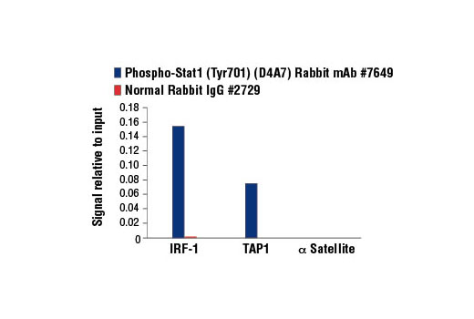
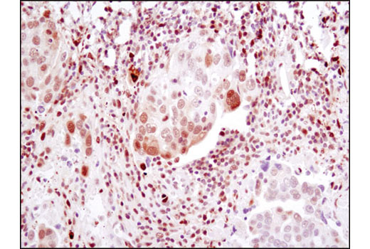
| Phospho-Stat1 (Tyr701) (D4A7) Rabbit mAb | 7649 | 20 µl | 84, 91 kDa | Rabbit IgG |
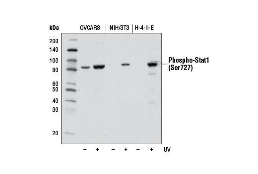
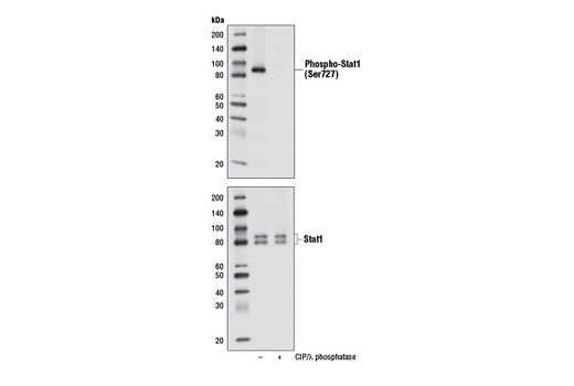
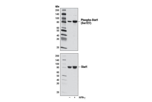
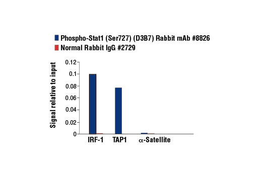
| Phospho-Stat1 (Ser727) (D3B7) Rabbit mAb | 8826 | 20 µl | 91 kDa | Rabbit IgG |
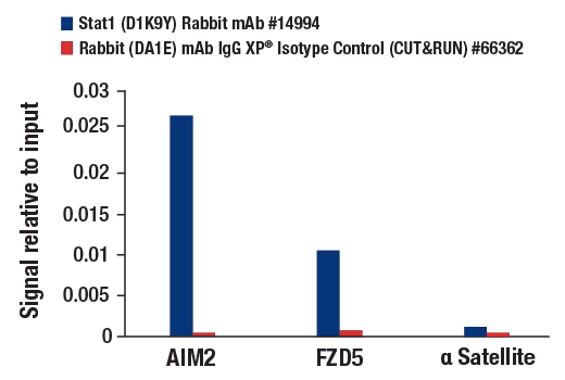
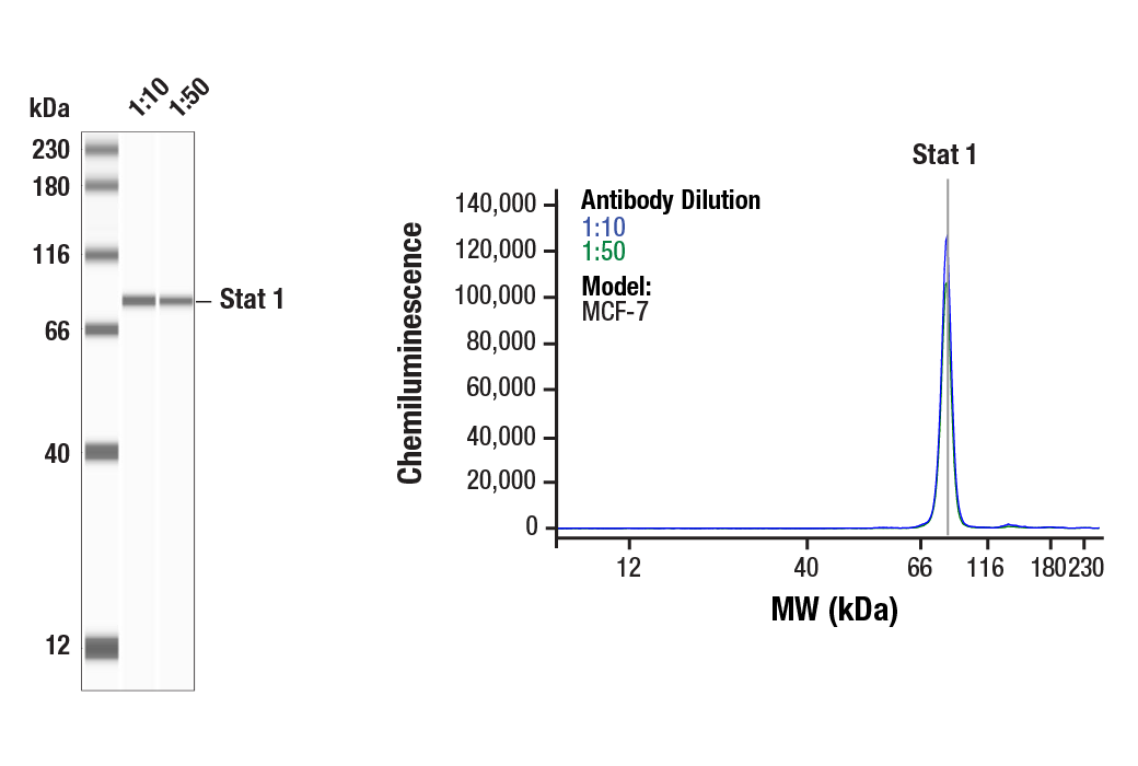
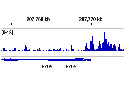
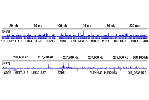
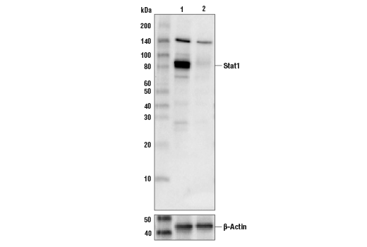
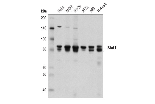
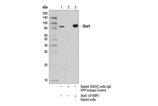
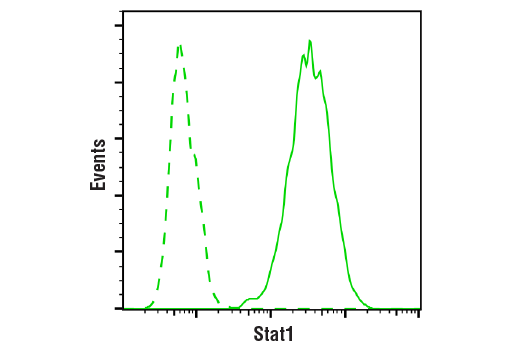
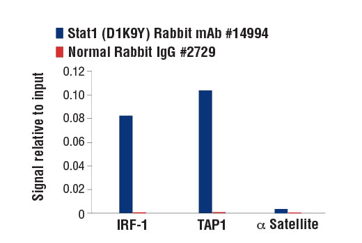
| Stat1 (D1K9Y) Rabbit mAb | 14994 | 20 µl | 84, 91 kDa | Rabbit IgG |
| Anti-rabbit IgG, HRP-linked Antibody | 7074 | 100 µl | Goat |
Please visit cellsignal.com for individual component applications, species cross-reactivity, dilutions, protocols, and additional product information.
Description
The IFN-γ Signaling Pathway Antibody Sampler Kit provides an economical means of detecting the activation of the IFN-γ signaling pathway using phospho-specific and control antibodies. The kit includes enough antibodies to perform two western blot experiments with each primary antibody.
Storage
Background
Originally discovered in the late 1950s for their antiviral activity, interferons (IFNs) have since been assigned diverse roles in many physiological and pathological processes. There are three families of IFNs: types I, II, and III. In humans, type I contains IFN-α (13 different subtypes), IFN-β (also known as IFN-β1), IFN-ε, IFN-κ, and IFN-ω. They bind to a receptor complex containing IFNAR1 and IFNAR2, which is broadly expressed on most cells. IFN-γ is the sole member of type II IFN. It signals through a receptor complex consisting of IFNγR1 and IFNγR2, which is also expressed on most cell types. Type III IFN, also known as interferon lambdas (IFN-λs), have four members in humans: IFN-λ1 (IL29), IFN-λ2 (IL28A), IFN-λ3 (IL28B), and IFN-λ4. IFN-λs signal through a heterodimeric receptor comprised of IFNλR1 and IL-10R2. While IL-10R2 is broadly expressed and shared by the IL-10 family cytokines, IFNλR1 expression is restricted to epithelial cells, neuronal cells, and subsets of myeloid cells (1-3). Engagement of all IFNs with their receptors initiates downstream signaling events, mainly, activation of the Jak–Stat signaling cascade. For type I and III IFNs, Jak1 and Tyk2 are phosphorylated and activated, leading to subsequent phosphorylation of Stat1 and Stat2. Phosphorylated Stat1 and Stat2 are released from the receptor complex and, together with IRF-9, they form so-called ISGF3 (interferon-stimulated gene factor 3) transcriptional complex. ISGF3 translocates to the nucleus, binds to the interferon-stimulated response element (ISRE) to initiate the transcription of a wide array of interferon-stimulated genes (ISGs) (4,5). On the other hand, IFN-γ induces phosphorylation and activation of Jak1 and Jak2, which subsequently phosphorylate Stat1. Phosphorylated Stat1 dimerizes, translocates to the nucleus, and binds to γ-interferon-activated site (GAS) to initiate the transcription of ISGs (6,7).
- Schneider, W.M. et al. (2014) Annu Rev Immunol 32, 513-45.
- Hemann, E.A. et al. (2017) Front Immunol 8, 1707.
- Walter, M.R. (2020) Front Immunol 11, 606489.
- Hervas-Stubbs, S. et al. (2011) Clin Cancer Res 17, 2619-27.
- Mesev, E.V. et al. (2019) Nat Microbiol 4, 914-924.
- Green, D.S. et al. (2017) J Biol Chem 292, 13925-13933.
- Ivashkiv, L.B. (2018) Nat Rev Immunol 18, 545-558.
Background References
Trademarks and Patents
使用に関する制限
法的な権限を与えられたCSTの担当者が署名した書面によって別途明示的に合意された場合を除き、 CST、その関連会社または代理店が提供する製品には以下の条件が適用されます。お客様が定める条件でここに定められた条件に含まれるものを超えるもの、 または、ここに定められた条件と異なるものは、法的な権限を与えられたCSTの担当者が別途書面にて受諾した場合を除き、拒絶され、 いかなる効力も効果も有しません。
研究専用 (For Research Use Only) またはこれに類似する表示がされた製品は、 いかなる目的についても FDA または外国もしくは国内のその他の規制機関により承認、認可または許可を受けていません。 お客様は製品を診断もしくは治療目的で使用してはならず、また、製品に表示された内容に違反する方法で使用してはなりません。 CST が販売または使用許諾する製品は、エンドユーザーであるお客様に対し、使途を研究および開発のみに限定して提供されるものです。 診断、予防もしくは治療目的で製品を使用することまたは製品を再販売 (単独であるか他の製品等の一部であるかを問いません) もしくはその他の商業的利用の目的で購入することについては、CST から別途許諾を得る必要があります。 お客様は以下の事項を遵守しなければなりません。(a) CST の製品 (単独であるか他の資材と一緒であるかを問いません) を販売、使用許諾、貸与、寄付もしくはその他の態様で第三者に譲渡したり使用させたりしてはなりません。また、商用の製品を製造するために CST の製品を使用してはなりません。(b) 複製、改変、リバースエンジニアリング、逆コンパイル、 分解または他の方法により製品の構造または技術を解明しようとしてはなりません。また、 CST の製品またはサービスと競合する製品またはサービスを開発する目的で CST の製品を使用してはなりません。(c) CST の製品の商標、商号、ロゴ、特許または著作権に関する通知または表示を除去したり改変したりしてはなりません。(d) CST の製品をCST 製品販売条件(CST’s Product Terms of Sale) および該当する書面のみに従って使用しなければなりません。(e) CST の製品に関連してお客様が使用する第三者の製品またはサービスに関する使用許諾条件、 サービス提供条件またはこれに類する合意事項を遵守しなければなりません。