WB, IP, IHC-P, FC-L
H
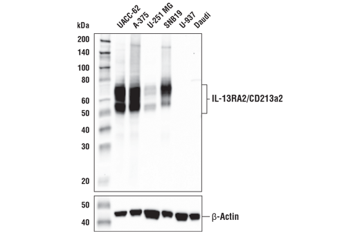
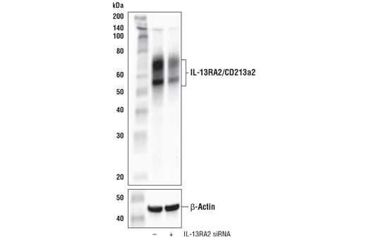
Endogenous
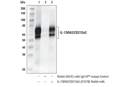
55, 65
Rabbit IgG
#Q14627
3598
Product Information
Product Usage Information
| Application | Dilution |
|---|---|
| Western Blotting | 1:1000 |
| Immunoprecipitation | 1:50 |
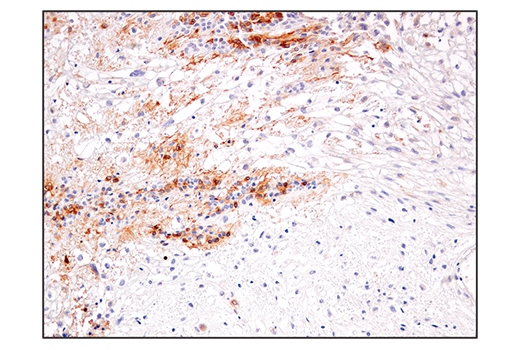
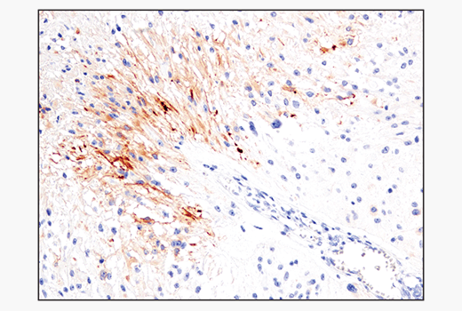
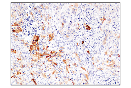
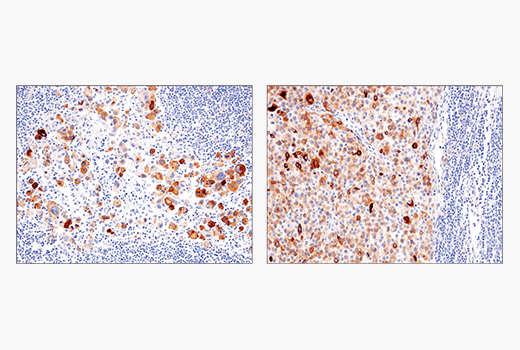
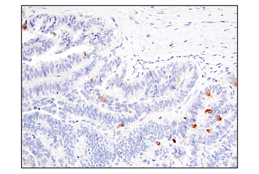
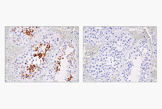
| Immunohistochemistry (Paraffin) | 1:100 - 1:400 |
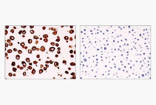
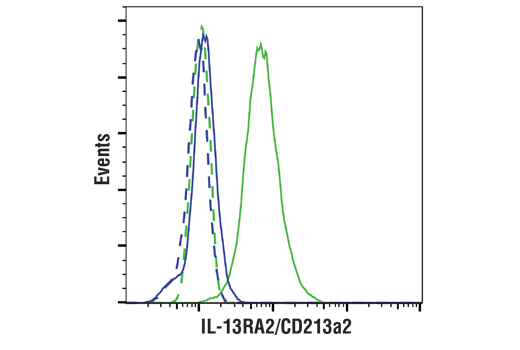
| Flow Cytometry (Live) | 1:50 |
Storage
For a carrier free (BSA and azide free) version of this product see product #68588.
Specificity / Sensitivity
Species Reactivity:
Human
Source / Purification
Monoclonal antibody is produced by immunizing animals with a synthetic peptide corresponding to residues near the amino terminus of human IL-13RA2/CD213a2 protein.
Background
Cancer/testis antigens (CTAs) are a family of more than 100 proteins whose normal expression is largely restricted to immune privileged germ cells of the testis, ovary, and trophoblast cells of the placenta. Although most normal somatic tissues are void of CTA expression, due to epigenetic silencing of gene expression, their expression is upregulated in a wide variety of human solid and liquid tumors (1,2). As such, CTAs have garnered much attention as attractive targets for a variety of immunotherapy-based approaches to selectively attack tumors (3).
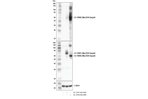
IL-13 receptor alpha-2 (IL-13RA2/CD213a2) is a type-I monomeric transmembrane glycoprotein and component of the IL-4/IL-13 receptor system. Research studies have shown that IL-13RA2 controls IL-13 signaling by serving as a high affinity decoy receptor for IL-13, but not IL-4, in a variety of cellular contexts (4,5). Some studies, however, have suggested that signaling through IL-13RA2 expressed on immunosuppressive myeloid cells facilitates tumor immune evasion through the production of TGF-β (6,7).
IL-13RA2 displays a highly restricted expression pattern in normal human tissues, which positions this receptor as a potentially novel therapeutic target for multiple types of solid tumors (8-10). Indeed, research studies have shown that IL-13RA2 is highly over expressed in glioblastomas (11), which has prompted investigation of different IL-13RA2-directed immunotherapies to treat this cancer (12-14).
- Caballero, O.L. and Chen, Y.T. (2009) Cancer Sci 100, 2014-21.
- De Smet, C. et al. (1999) Mol Cell Biol 19, 7327-35.
- Gjerstorff, M.F. et al. (2015) Oncotarget 6, 15772-87.
- Yoshikawa, M. et al. (2003) Biochem Biophys Res Commun 312, 1248-55.
- Bernard, J. et al. (2001) Lab Invest 81, 1223-31.
- Fichtner-Feigl, S. et al. (2006) Nat Med 12, 99-106.
- Fichtner-Feigl, S. et al. (2008) Cancer Res 68, 3467-75.
- Debinski, W. and Gibo, D.M. (2000) Mol Med 6, 440-9.
- Taguchi, A. et al. (2014) Cancer Res 74, 4694-705.
- Beard, R.E. et al. (2013) Clin Cancer Res 19, 4941-50.
- Jarboe, J.S. et al. (2007) Cancer Res 67, 7983-6.
- Brown, C.E. et al. (2015) Clin Cancer Res 21, 4062-72.
- Brown, C.E. et al. (2012) Clin Cancer Res 18, 2199-209.
- Mintz, A. et al. (2008) Cancer Biother Radiopharm 23, 581-9.
Species Reactivity
Species reactivity is determined by testing in at least one approved application (e.g., western blot).
Western Blot Buffer
IMPORTANT: For western blots, incubate membrane with diluted primary antibody in 5% w/v BSA, 1X TBS, 0.1% Tween® 20 at 4°C with gentle shaking, overnight.
Applications Key
WB: Western Blotting IP: Immunoprecipitation IHC-P: Immunohistochemistry (Paraffin) FC-L: Flow Cytometry (Live)
Cross-Reactivity Key
H: human M: mouse R: rat Hm: hamster Mk: monkey Vir: virus Mi: mink C: chicken Dm: D. melanogaster X: Xenopus Z: zebrafish B: bovine Dg: dog Pg: pig Sc: S. cerevisiae Ce: C. elegans Hr: horse GP: Guinea Pig Rab: rabbit All: all species expected
Trademarks and Patents
使用に関する制限
法的な権限を与えられたCSTの担当者が署名した書面によって別途明示的に合意された場合を除き、 CST、その関連会社または代理店が提供する製品には以下の条件が適用されます。お客様が定める条件でここに定められた条件に含まれるものを超えるもの、 または、ここに定められた条件と異なるものは、法的な権限を与えられたCSTの担当者が別途書面にて受諾した場合を除き、拒絶され、 いかなる効力も効果も有しません。
研究専用 (For Research Use Only) またはこれに類似する表示がされた製品は、 いかなる目的についても FDA または外国もしくは国内のその他の規制機関により承認、認可または許可を受けていません。 お客様は製品を診断もしくは治療目的で使用してはならず、また、製品に表示された内容に違反する方法で使用してはなりません。 CST が販売または使用許諾する製品は、エンドユーザーであるお客様に対し、使途を研究および開発のみに限定して提供されるものです。 診断、予防もしくは治療目的で製品を使用することまたは製品を再販売 (単独であるか他の製品等の一部であるかを問いません) もしくはその他の商業的利用の目的で購入することについては、CST から別途許諾を得る必要があります。 お客様は以下の事項を遵守しなければなりません。(a) CST の製品 (単独であるか他の資材と一緒であるかを問いません) を販売、使用許諾、貸与、寄付もしくはその他の態様で第三者に譲渡したり使用させたりしてはなりません。また、商用の製品を製造するために CST の製品を使用してはなりません。(b) 複製、改変、リバースエンジニアリング、逆コンパイル、 分解または他の方法により製品の構造または技術を解明しようとしてはなりません。また、 CST の製品またはサービスと競合する製品またはサービスを開発する目的で CST の製品を使用してはなりません。(c) CST の製品の商標、商号、ロゴ、特許または著作権に関する通知または表示を除去したり改変したりしてはなりません。(d) CST の製品をCST 製品販売条件(CST’s Product Terms of Sale) および該当する書面のみに従って使用しなければなりません。(e) CST の製品に関連してお客様が使用する第三者の製品またはサービスに関する使用許諾条件、 サービス提供条件またはこれに類する合意事項を遵守しなければなりません。