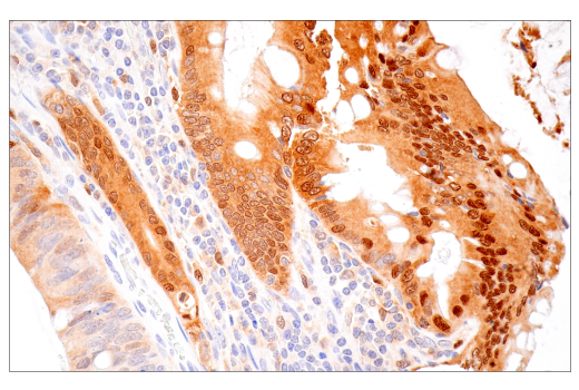
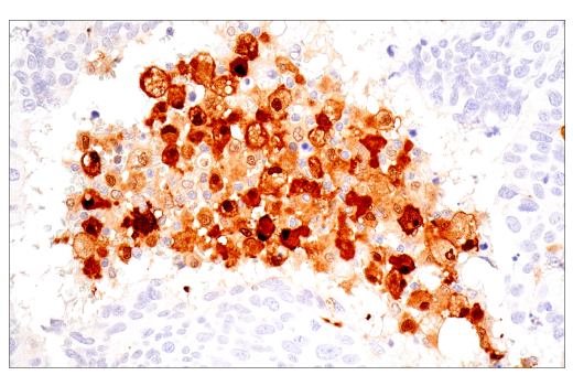
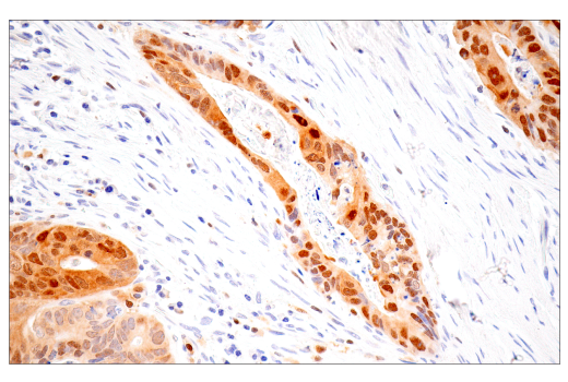
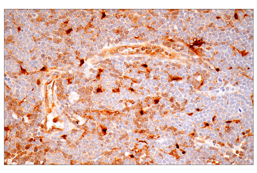
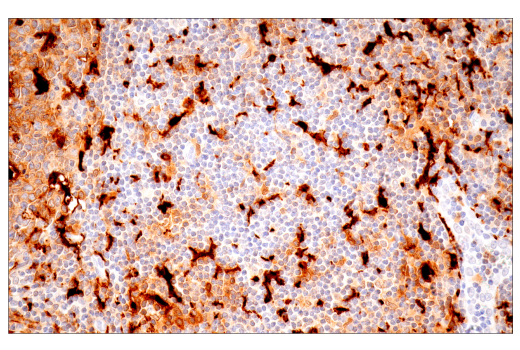
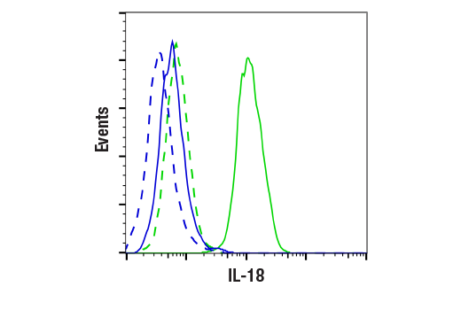
Revision 1
Cell Signaling Technology

Orders: 877-616-CELL (2355) [email protected]

Support: 877-678-TECH (8324)

Web: [email protected] cellsignal.com

3 Trask LaneDanversMassachusetts01923USA
For Research Use Only. Not for Use in Diagnostic Procedures.
Applications:

WB, IHC-P, FC-FP

REACTIVITY:

H

SENSITIVITY:

Endogenous

MW (kDa):

22

Source/Isotype:

Rabbit IgG

UniProt ID:

#Q14116

Entrez-Gene Id:

3606

Product Information

Product Usage Information

This product is the carrier free version of product #67775. All data were generated using the same antibody clone in the standard formulation which contains BSA and glycerol.

This formulation is ideal for use with technologies requiring specialized or custom antibody labeling, including fluorophores, metals, lanthanides, and oligonucleotides. It is not recommended for ChIP, ChIP-seq, CUT&RUN or CUT&Tag assays. If you require a carrier free formulation for chromatin profiling, please contact us. Optimal dilutions/concentrations should be determined by the end user.

Formulation

Supplied in 1X PBS, BSA and Azide Free.

For standard formulation of this product see product #67775

Storage

Store at -20°C. This product will freeze at -20°C so it is recommended to aliquot into single-use vials to avoid multiple freeze/thaw cycles. A slight precipitate may be present and can be dissolved by gently vortexing. This will not interfere with antibody performance.

Specificity / Sensitivity
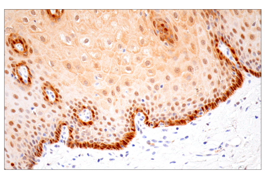
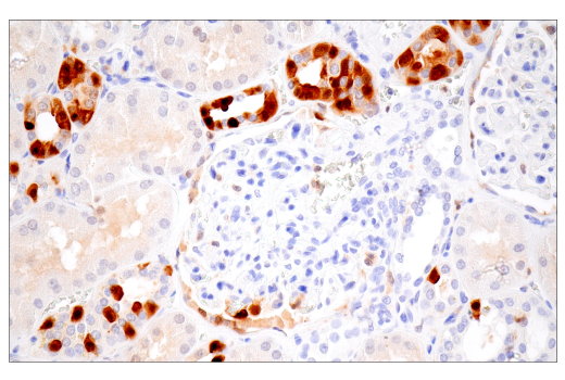
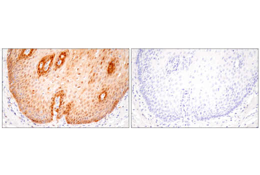
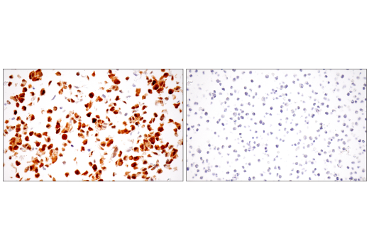
IL-18 (E3G8R) Rabbit mAb (BSA and Azide Free) recognizes endogenous levels of total IL-18 protein.

Species Reactivity:

Human

Source / Purification

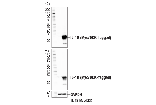
Monoclonal antibody is produced by immunizing animals with human IL-18 recombinant protein.

Background

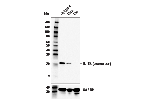
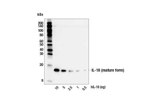
Interleukin-18 (IL-18), originally known as interferon-gamma inducing factor (IGIF), is part of the IL-superfamily of cytokines (1,2). This proinflammatory cytokine is synthesized as an inactive precursor which requires cleavage by Caspase-1 to become an active, mature molecule, which is secreted by monocytes and macrophages (3,4,6). It induces IFNγ production in T-helper-1 (Th1) cells and natural killer (NK) cells, and can regulate innate immunity via both Th1 and Th2 responses (4,5). Elevated IL-18 levels are associated with autoimmune disease, but may be balanced by binding to IL-18 binding protein (IL-18BP), which along with IL-18 neutralizing antibodies, are being examined in clinical trials (6,7).

  1. Okamura, H. et al. (1995) Nature 378, 88-91.
  2. Ushio, S. et al. (1996) J Immunol 156, 4274-9.
  3. Ghayur, T. et al. (1997) Nature 386, 619-23.
  4. Dinarello, C.A. (1999) Methods 19, 121-32.
  5. Nakanishi, K. et al. (2001) Annu Rev Immunol 19, 423-74.
  6. Novick, D. et al. (1999) Immunity 10, 127-36.
  7. Dinarello, C.A. et al. (2013) Front Immunol 4, 289.

Species Reactivity

Species reactivity is determined by testing in at least one approved application (e.g., western blot).

Applications Key

WB: Western Blotting IHC-P: Immunohistochemistry (Paraffin) FC-FP: Flow Cytometry (Fixed/Permeabilized)

Cross-Reactivity Key

H: human M: mouse R: rat Hm: hamster Mk: monkey Vir: virus Mi: mink C: chicken Dm: D. melanogaster X: Xenopus Z: zebrafish B: bovine Dg: dog Pg: pig Sc: S. cerevisiae Ce: C. elegans Hr: horse GP: Guinea Pig Rab: rabbit All: all species expected

Trademarks and Patents

Cell Signaling Technology is a trademark of Cell Signaling Technology, Inc.
All other trademarks are the property of their respective owners. Visit cellsignal.com/trademarks for more information.

使用に関する制限

法的な権限を与えられたCSTの担当者が署名した書面によって別途明示的に合意された場合を除き、 CST、その関連会社または代理店が提供する製品には以下の条件が適用されます。お客様が定める条件でここに定められた条件に含まれるものを超えるもの、 または、ここに定められた条件と異なるものは、法的な権限を与えられたCSTの担当者が別途書面にて受諾した場合を除き、拒絶され、 いかなる効力も効果も有しません。

研究専用 (For Research Use Only) またはこれに類似する表示がされた製品は、 いかなる目的についても FDA または外国もしくは国内のその他の規制機関により承認、認可または許可を受けていません。 お客様は製品を診断もしくは治療目的で使用してはならず、また、製品に表示された内容に違反する方法で使用してはなりません。 CST が販売または使用許諾する製品は、エンドユーザーであるお客様に対し、使途を研究および開発のみに限定して提供されるものです。 診断、予防もしくは治療目的で製品を使用することまたは製品を再販売 (単独であるか他の製品等の一部であるかを問いません) もしくはその他の商業的利用の目的で購入することについては、CST から別途許諾を得る必要があります。 お客様は以下の事項を遵守しなければなりません。(a) CST の製品 (単独であるか他の資材と一緒であるかを問いません) を販売、使用許諾、貸与、寄付もしくはその他の態様で第三者に譲渡したり使用させたりしてはなりません。また、商用の製品を製造するために CST の製品を使用してはなりません。(b) 複製、改変、リバースエンジニアリング、逆コンパイル、 分解または他の方法により製品の構造または技術を解明しようとしてはなりません。また、 CST の製品またはサービスと競合する製品またはサービスを開発する目的で CST の製品を使用してはなりません。(c) CST の製品の商標、商号、ロゴ、特許または著作権に関する通知または表示を除去したり改変したりしてはなりません。(d) CST の製品をCST 製品販売条件(CST’s Product Terms of Sale) および該当する書面のみに従って使用しなければなりません。(e) CST の製品に関連してお客様が使用する第三者の製品またはサービスに関する使用許諾条件、 サービス提供条件またはこれに類する合意事項を遵守しなければなりません。

Revision 1
#33484

IL-18 (E3G8R) Rabbit mAb (BSA and Azide Free)

Western Blotting Image 1: IL-18 (E3G8R) Rabbit mAb (BSA and Azide Free) Expand Image
Western blot analysis of extracts from OVCAR-8, HeLa, and Raji cells using IL-18 (E3G8R) Rabbit mAb (upper) or GAPDH (D16H11) XP® Rabbit mAb #5174 (lower). Negative expression of IL-18 protein in Raji cells is consistent with the predicted expression pattern. Data were generated using the standard formulation of this product.
Western Blotting Image 2: IL-18 (E3G8R) Rabbit mAb (BSA and Azide Free) Expand Image
Western blot analysis of recombinant human IL-18 protein (hIL-18) at indicated levels using IL-18 (E3G8R) Rabbit mAb. Data were generated using the standard formulation of this product.
Western Blotting Image 3: IL-18 (E3G8R) Rabbit mAb (BSA and Azide Free) Expand Image
Western blot analysis of extracts from 293T cells, mock transfected (-) or transfected with a construct expressing Myc/DDK-tagged full-length human IL-18 protein (hIL-18-Myc/DDK; +), using IL-18 (E3G8R) Rabbit mAb (upper), Myc-Tag (71D10) Rabbit mAb #2278 (middle), or GAPDH (D16H11) XP® Rabbit mAb #5174 (lower). Data were generated using the standard formulation of this product.
Immunohistochemistry Image 1: IL-18 (E3G8R) Rabbit mAb (BSA and Azide Free) Expand Image
Immunohistochemical analysis of paraffin-embedded human Hodgkin lymphoma using IL-18 (E3G8R) Rabbit mAb. Data were generated using the standard formulation of this product.
Immunohistochemistry Image 2: IL-18 (E3G8R) Rabbit mAb (BSA and Azide Free) Expand Image
Immunohistochemical analysis of paraffin-embedded human colon carcinoma using IL-18 (E3G8R) Rabbit mAb. Data were generated using the standard formulation of this product.
Immunohistochemistry Image 3: IL-18 (E3G8R) Rabbit mAb (BSA and Azide Free) Expand Image
Immunohistochemical analysis of paraffin-embedded human urothelial carcinoma using IL-18 (E3G8R) Rabbit mAb. Data were generated using the standard formulation of this product.
Immunohistochemistry Image 4: IL-18 (E3G8R) Rabbit mAb (BSA and Azide Free) Expand Image
Immunohistochemical analysis of paraffin-embedded human adenocarcinoma of distal esophagus using IL-18 (E3G8R) Rabbit mAb. Data were generated using the standard formulation of this product.
Immunohistochemistry Image 5: IL-18 (E3G8R) Rabbit mAb (BSA and Azide Free) Expand Image
Immunohistochemical analysis of paraffin-embedded human mantle cell lymphoma using IL-18 (E3G8R) Rabbit mAb. Data were generated using the standard formulation of this product.
Immunohistochemistry Image 6: IL-18 (E3G8R) Rabbit mAb (BSA and Azide Free) Expand Image
Immunohistochemical analysis of paraffin-embedded human tonsil using IL-18 (E3G8R) Rabbit mAb. Data were generated using the standard formulation of this product.
Immunohistochemistry Image 7: IL-18 (E3G8R) Rabbit mAb (BSA and Azide Free) Expand Image
Immunohistochemical analysis of paraffin-embedded human appendix using IL-18 (E3G8R) Rabbit mAb. Data were generated using the standard formulation of this product.
Immunohistochemistry Image 8: IL-18 (E3G8R) Rabbit mAb (BSA and Azide Free) Expand Image
Immunohistochemical analysis of paraffin-embedded normal human thymus using IL-18 (E3G8R) Rabbit mAb. Data were generated using the standard formulation of this product.
Immunohistochemistry Image 9: IL-18 (E3G8R) Rabbit mAb (BSA and Azide Free) Expand Image
Immunohistochemical analysis of paraffin-embedded normal human skin using IL-18 (E3G8R) Rabbit mAb. Data were generated using the standard formulation of this product.
Immunohistochemistry Image 10: IL-18 (E3G8R) Rabbit mAb (BSA and Azide Free) Expand Image
Immunohistochemical analysis of paraffin-embedded normal human liver using IL-18 (E3G8R) Rabbit mAb. Data were generated using the standard formulation of this product.
Immunohistochemistry Image 11: IL-18 (E3G8R) Rabbit mAb (BSA and Azide Free) Expand Image
Immunohistochemical analysis of paraffin-embedded normal human esophagus using IL-18 (E3G8R) Rabbit mAb. Data were generated using the standard formulation of this product.
Immunohistochemistry Image 12: IL-18 (E3G8R) Rabbit mAb (BSA and Azide Free) Expand Image
Immunohistochemical analysis of paraffin-embedded normal human kidney using IL-18 (E3G8R) Rabbit mAb. Data were generated using the standard formulation of this product.
Immunohistochemistry Image 13: IL-18 (E3G8R) Rabbit mAb (BSA and Azide Free) Expand Image
Immunohistochemical analysis of paraffin-embedded normal human esophagus using IL-18 (E3G8R) Rabbit mAb (left) compared to concentration-matched Rabbit (DA1E) mAb IgG XP® Isotype Control #3900 (right). Data were generated using the standard formulation of this product.
Immunohistochemistry Image 14: IL-18 (E3G8R) Rabbit mAb (BSA and Azide Free) Expand Image
Immunohistochemical analysis of paraffin-embedded OVCAR-8 cell pellet (left, positive) or Raji cell pellet (right, negative) using IL-18 (E3G8R) Rabbit mAb. Data were generated using the standard formulation of this product.
Flow Cytometry Image 1: IL-18 (E3G8R) Rabbit mAb (BSA and Azide Free) Expand Image
Flow cytometric analysis of Raji cells (blue, negative) and OVCAR-8 cells (green, positive) using IL-18 (E3G8R) Rabbit mAb (solid lines) or concentration-matched Rabbit (DA1E) mAb IgG XP® Isotype Control #3900 (dashed lines). Anti-rabbit IgG (H+L), F(ab')2 Fragment (Alexa Fluor® 488 Conjugate) #4412 was used as a secondary antibody. Data were generated using the standard formulation of this product.