| Product Includes | Product # | Quantity | Mol. Wt | Isotype/Source |
|---|---|---|---|---|
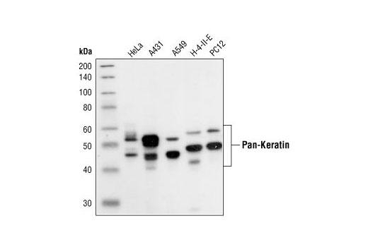
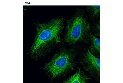
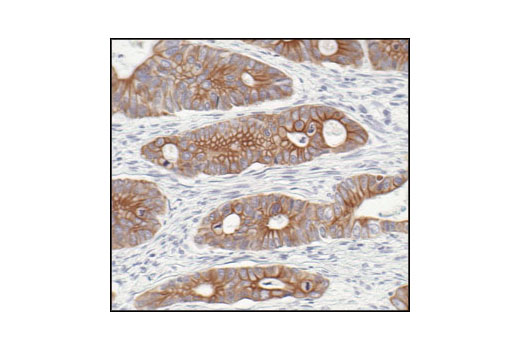
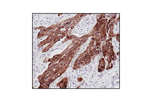
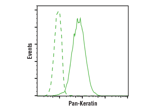
| Pan-Keratin (C11) Mouse mAb | 4545 | 40 µl | 46-58 kDa | Mouse IgG1 |
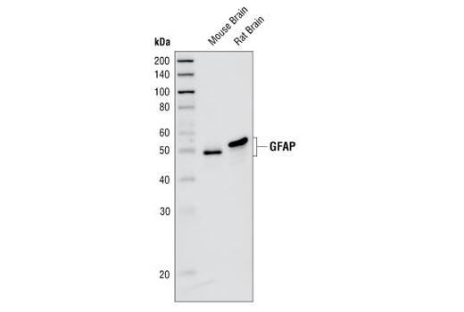
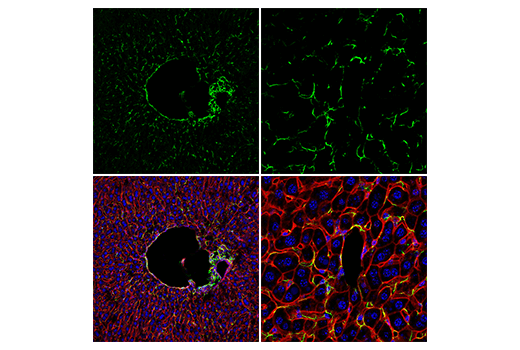
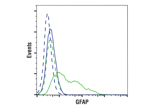
| GFAP (GA5) Mouse mAb | 3670 | 40 µl | 50 kDa | Mouse IgG1 |
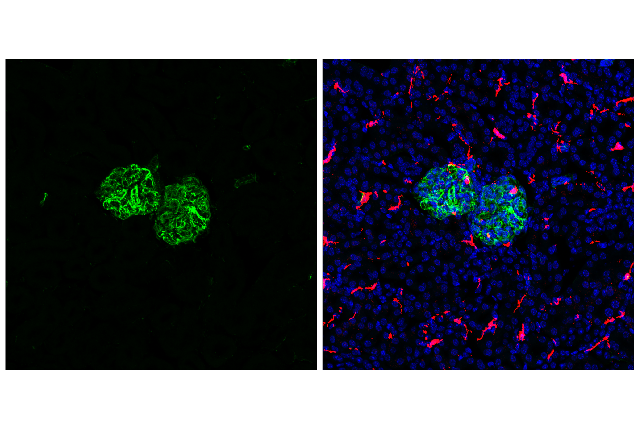
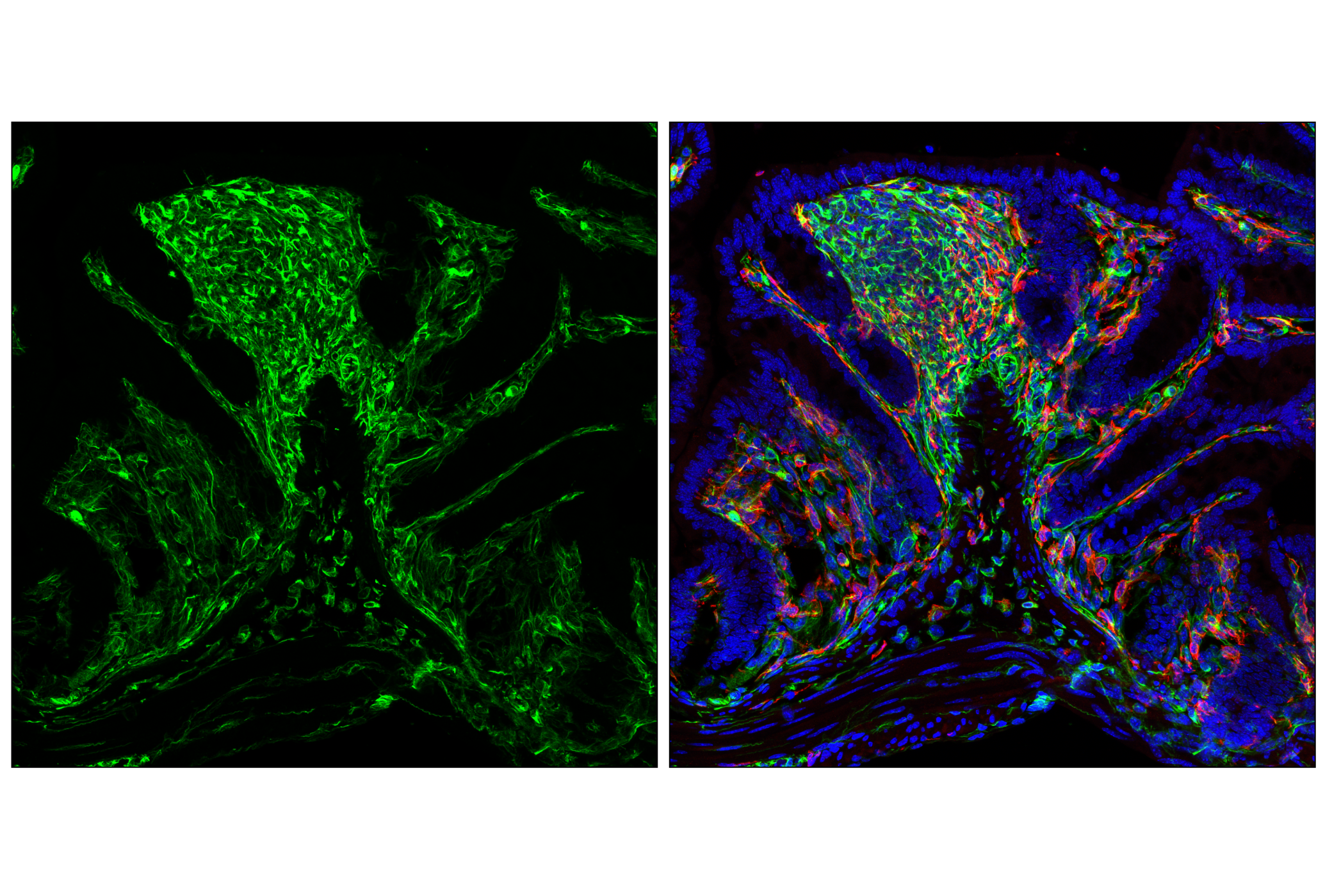
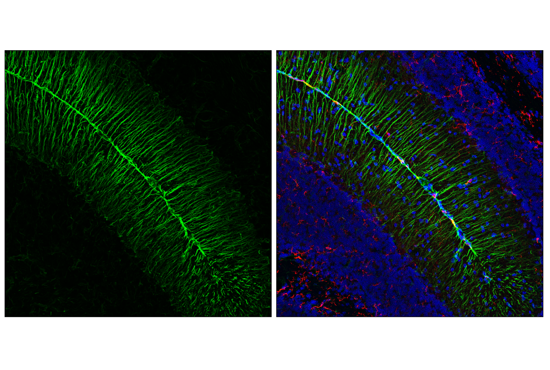
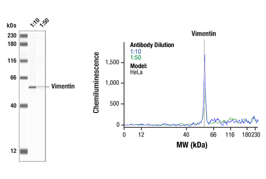
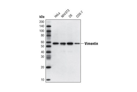
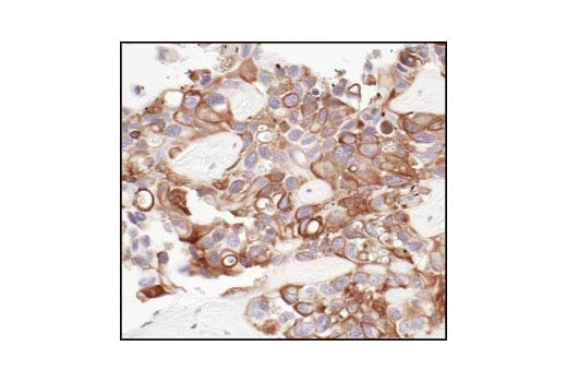
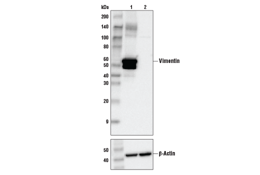
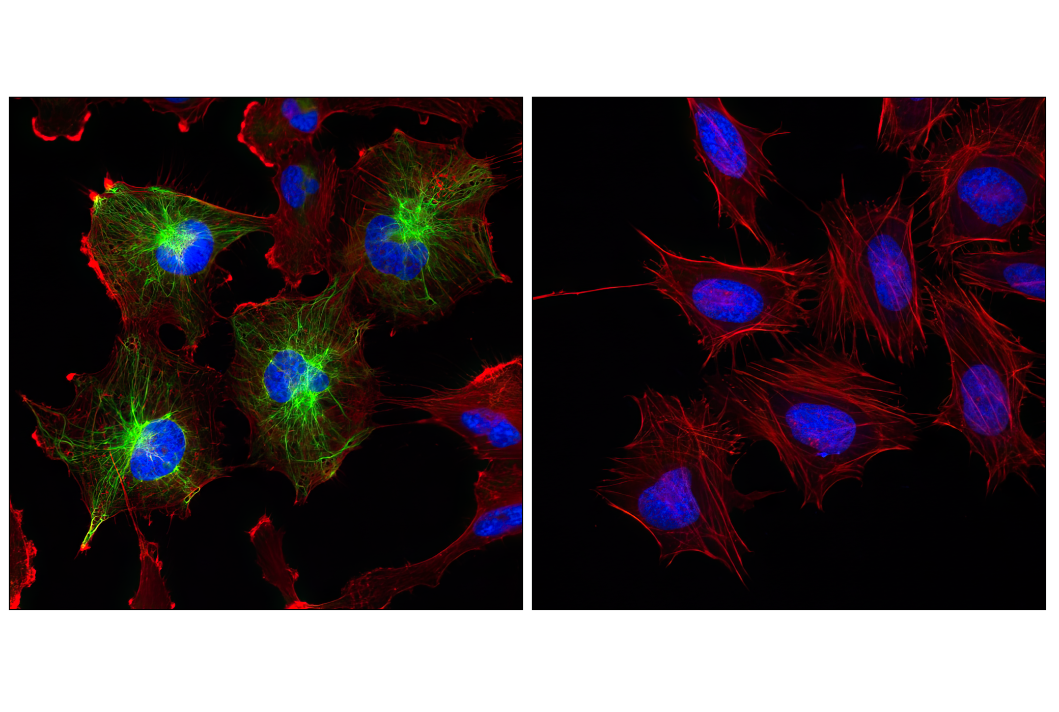
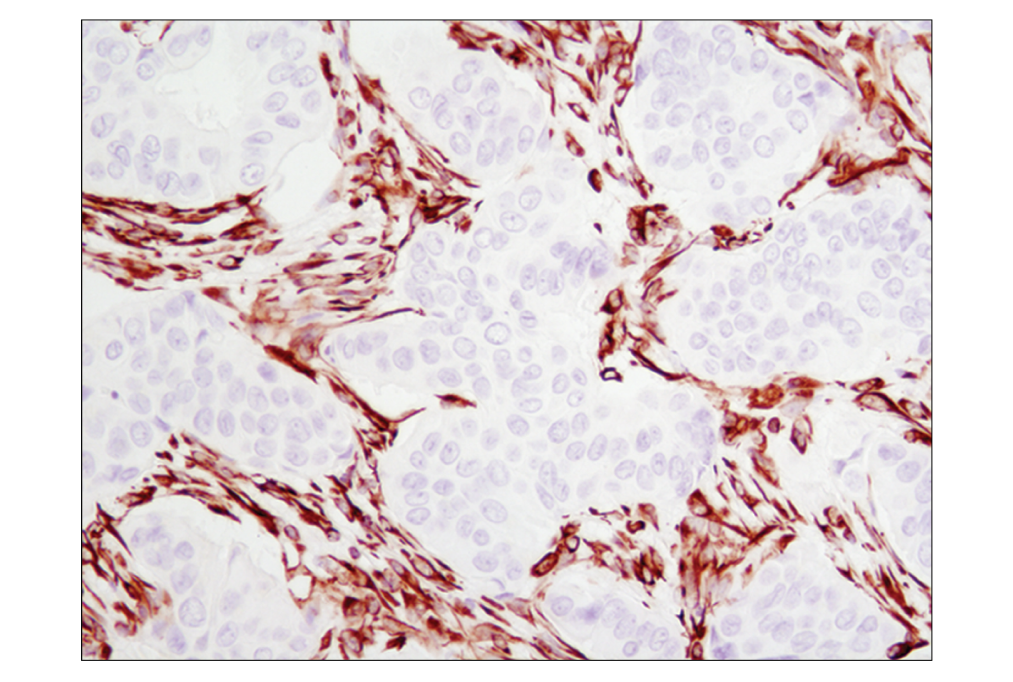
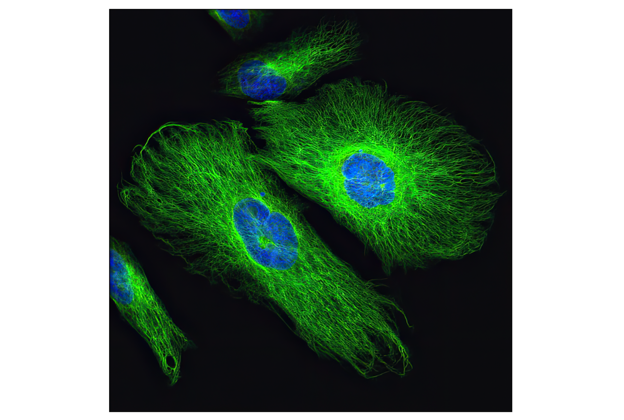
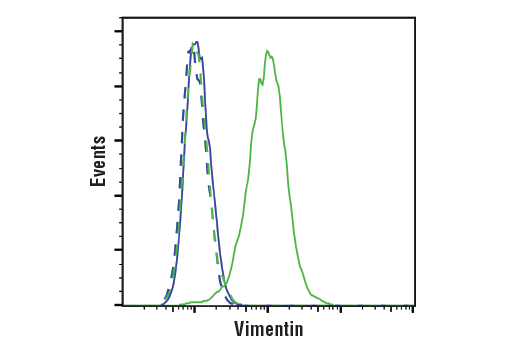
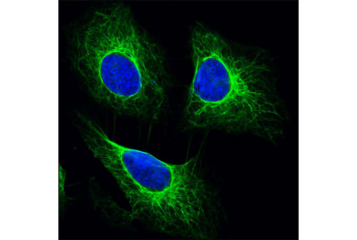
| Vimentin (D21H3) XP® Rabbit mAb | 5741 | 40 µl | 57 kDa | Rabbit IgG |
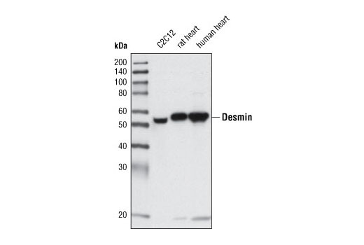
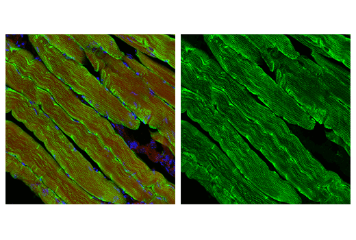
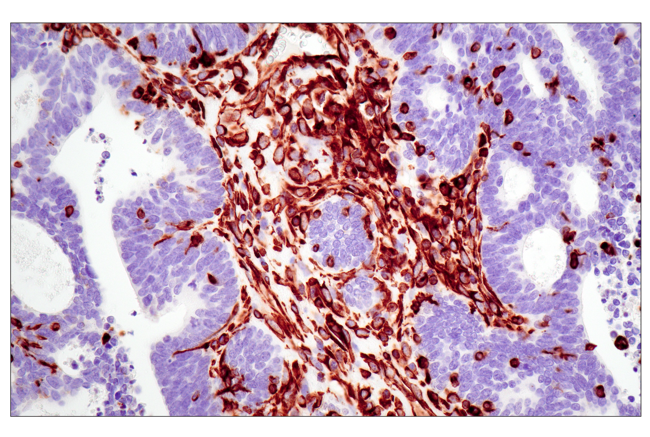
| Desmin (D93F5) XP® Rabbit mAb | 5332 | 40 µl | 53 kDa | Rabbit IgG |
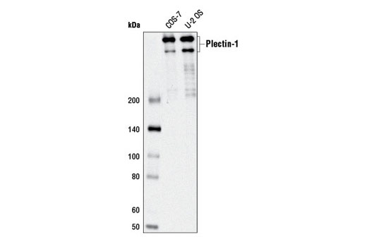
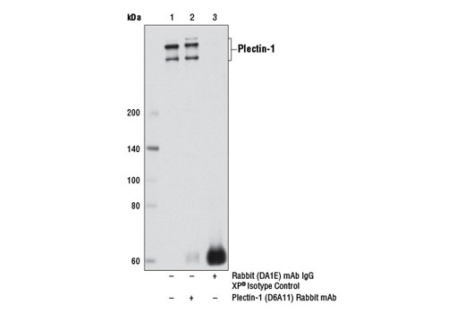
| Plectin-1 (D6A11) Rabbit mAb | 12254 | 40 µl | 400-500 kDa | Rabbit IgG |
| Anti-rabbit IgG, HRP-linked Antibody | 7074 | 100 µl | Goat | |
| Anti-mouse IgG, HRP-linked Antibody | 7076 | 100 µl | Horse |
Please visit cellsignal.com for individual component applications, species cross-reactivity, dilutions, protocols, and additional product information.
Description
The Intermediate Filaments Antibody Sampler Kit provides an economical means to evaluate the presence and status of intermediate filaments. The kit includes enough primary and secondary antibody to perform four Western blot experiments per antibody.
Storage
Background
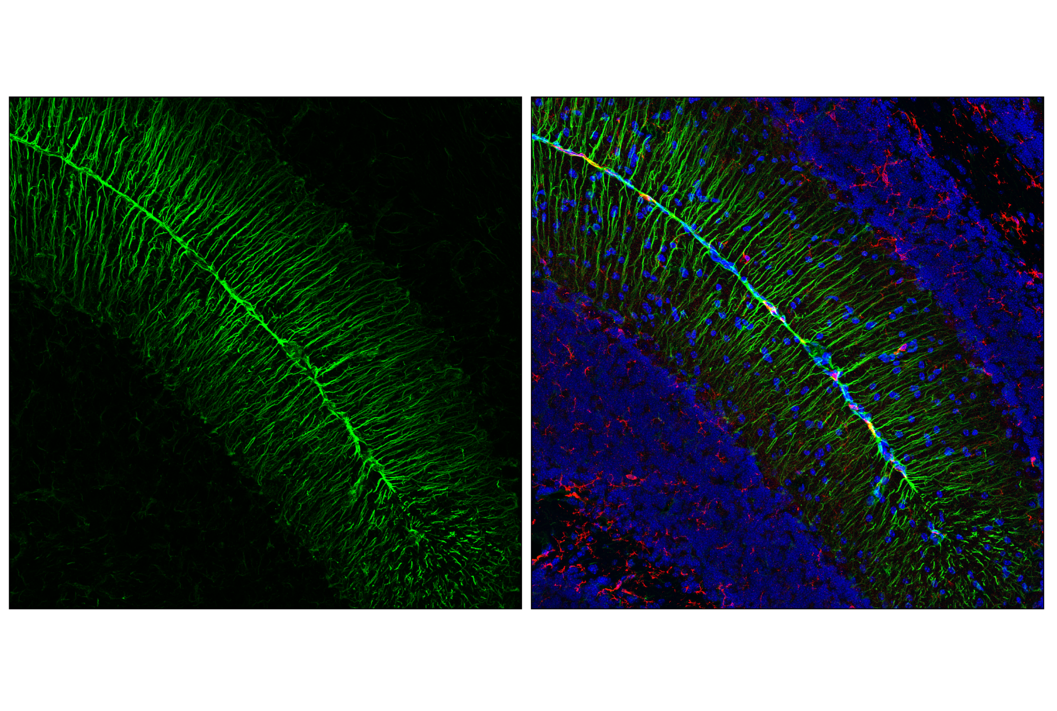
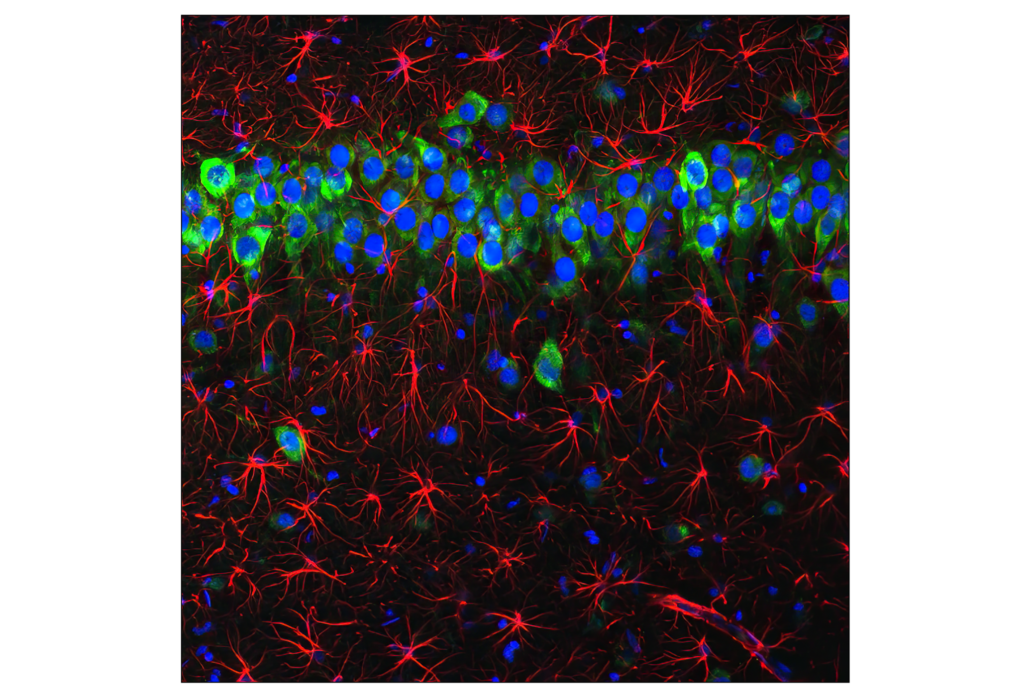
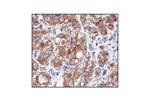
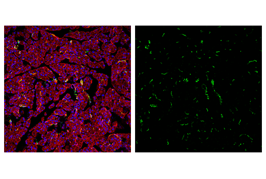
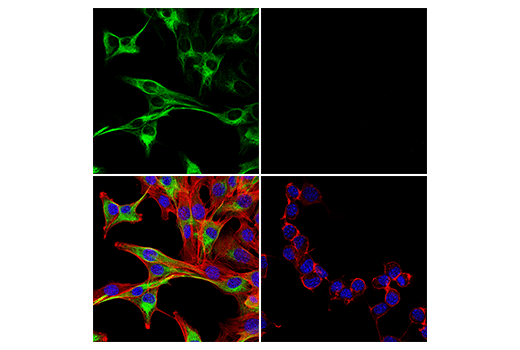
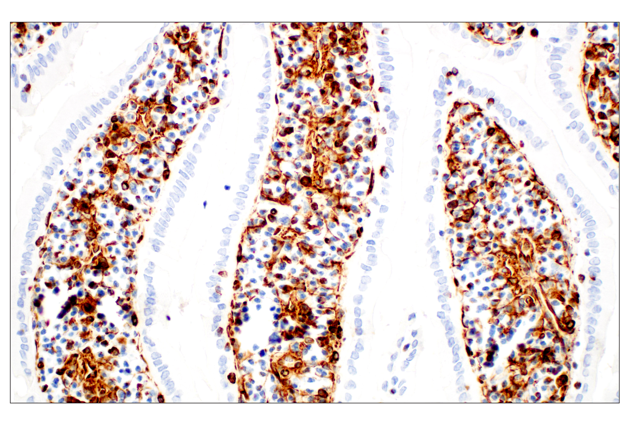
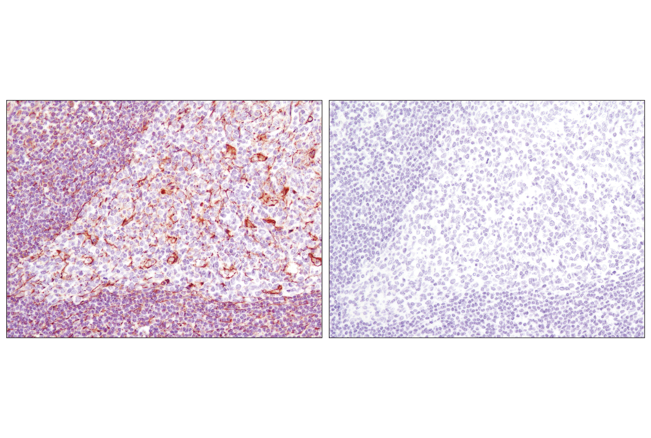
The cytoskeleton consists of three types of cytosolic fibers: microfilaments (actin filaments), intermediate filaments and microtubules. Major types of intermediate filaments are distinguished and expressed in particular cell types: cytokeratins (epithelial cells), glial fibrillary acidic protein, GFAP (glial cells), desmin (skeletal, visceral and certain vascular smooth muscle cells), vimentin (mesenchyme origin) and neurofilaments (neurons). GFAP and vimentin form intermediate filaments in astroglial cells and modulate their motility and shape (1). In particular, vimentin filaments are present at early developmental stages, while GFAP filaments are characteristic of differentiated and mature brain astrocytes. Thus, GFAP is commonly used as a marker for intracranial and intraspinal tumors arising from astrocytes (2). Vimentin is present in sarcomas, but not carcinomas, and its expression is examined in conjunction with that of other markers to distinguish between the two (3).
Desmin is a myogenic marker expressed in early development that forms a network of filaments that extends across the myofibril and surrounds Z discs. The desmin cytoskeleton provides a connection between myofibrils, organelles and the cytoskeleton (4). Desmin knockout mice develop cardiomyopathy as well as skeletal and smooth muscle defects (5). In humans, desmin related myopathies might be caused by mutations in the corresponding desmin gene or in proteins with which desmin interacts, including αB-crystallin and synemin. Disorganized desmin filaments and the accumulation of protein aggregates comprised predominantly of desmin characterize desmin-related myopathies (reviewed in 6,7).
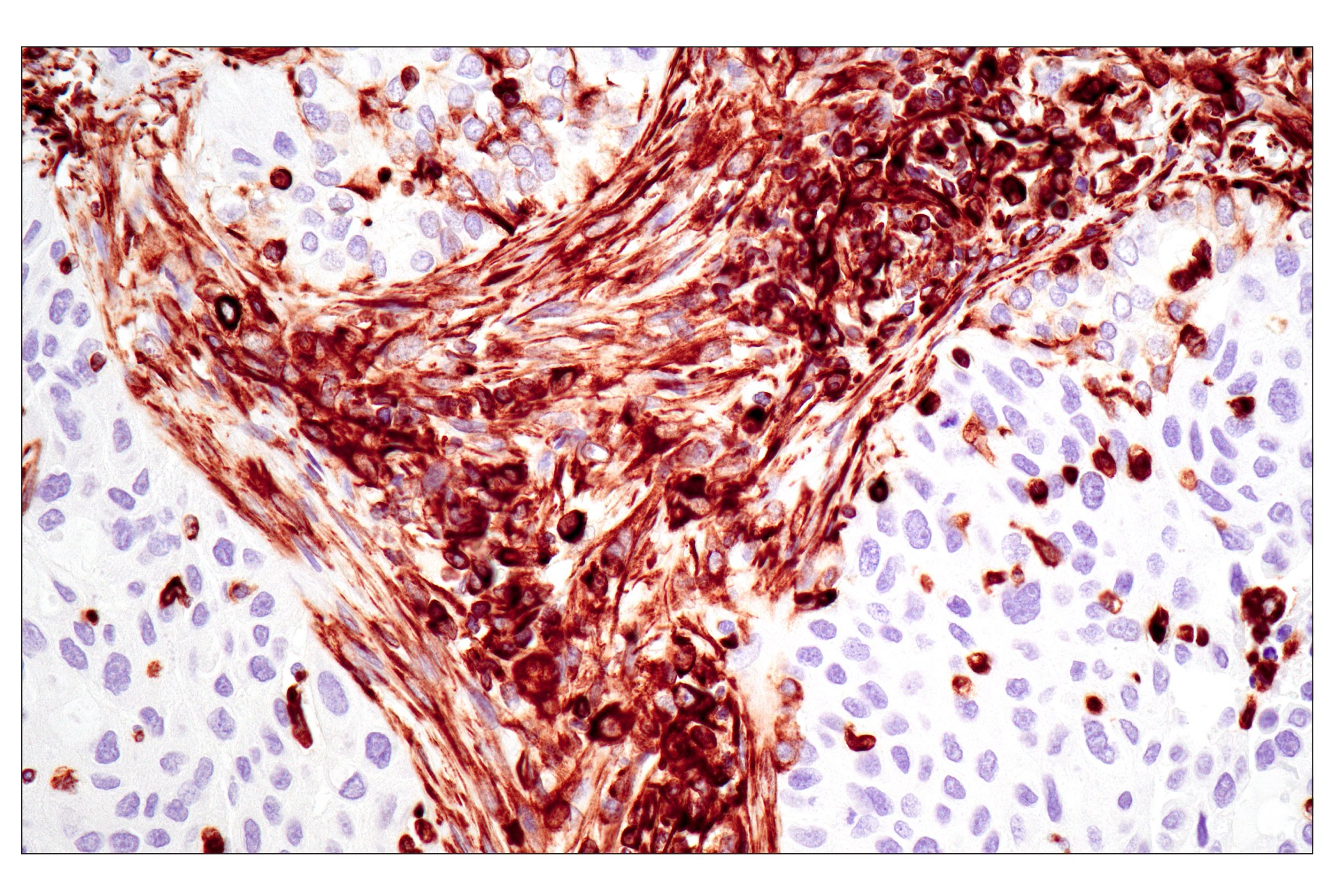
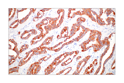
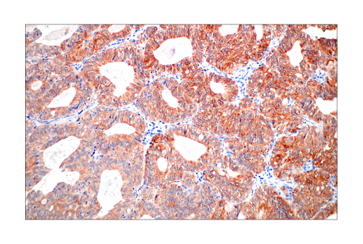
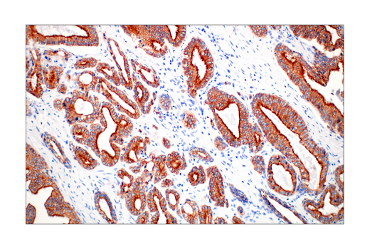
Keratins assemble into filaments, forming heterodimers of an acidic keratin (or type I keratin, keratins 9 to 23) and a basic keratin (or type II keratin, keratins 1 to 8) (8,9). Keratin isoforms demonstrate tissue- and differentiation-specific profiles, which make them useful as biomarkers (8). Mutations in keratin genes are associated with skin disorders, liver and pancreatic diseases, and inflammatory intestinal diseases (10-13).
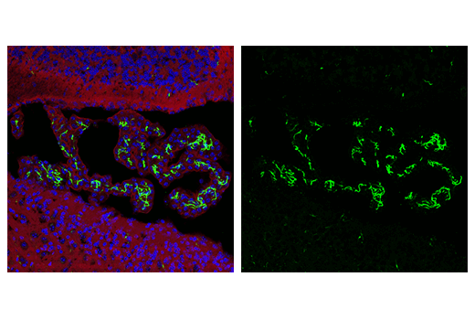
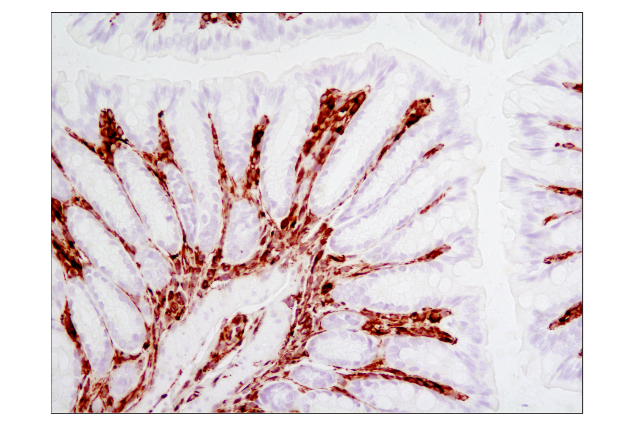
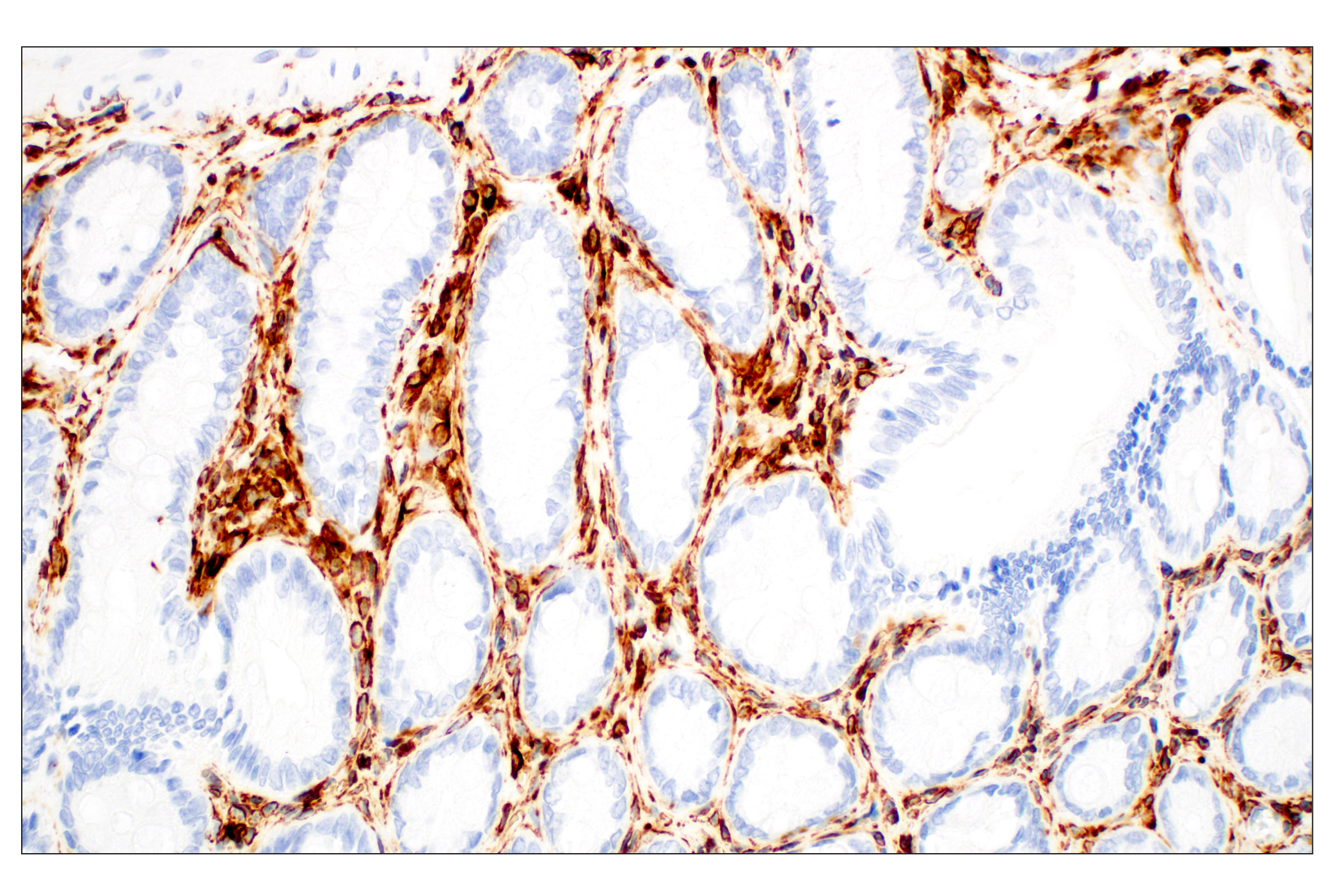
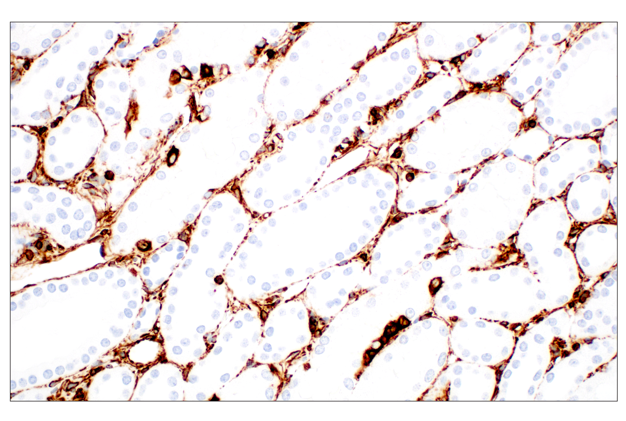
Plectin is a large, widely expressed protein that crosslinks the intermediate filament and actin cytoskeleton, mechanically stabilizing cells and tissues. Plectin also plays a role in the regulation of actin dynamics and acts as a scaffold for signaling molecules (14). It is important in the stabilization of hemidesmosomes, crosslinking them to the intermediate filament network. Plectin has been shown to be involved in several signaling cascades. It signals to PKC by binding to and sequestering RACK1, the receptor for activated C kinase 1 (15,16). Plectin is also involved in the regulation of cytokeratin architecture and cell stress response (16), signaling through the chemokine receptor CXCR4 (17), regulation of AMP-activated protein kinase (AMPK) activity and signaling in mouse myotubes (18).
- Eng, L.F. et al. (2000) Neurochem Res 25, 1439-51.
- Goebel, H.H. et al. (1987) Acta Histochem Suppl 34, 81-93.
- Leader, M. et al. (1987) Histopathology 11, 63-72.
- Capetanaki, Y. et al. (2007) Exp Cell Res 313, 2063-76.
- Li, Z. et al. (1996) Dev Biol 175, 362-6.
- Paulin, D. and Li, Z. (2004) Exp Cell Res 301, 1-7.
- Paulin, D. et al. (2004) J Pathol 204, 418-27.
- Moll, R. et al. (1982) Cell 31, 11-24.
- Chang, L. and Goldman, R.D. (2004) Nat Rev Mol Cell Biol 5, 601-13.
- Ramaekers, F.C. and Bosman, F.T. (2004) J Pathol 204, 351-4.
- Lane, E.B. and McLean, W.H. (2004) J Pathol 204, 355-66.
- Zatloukal, K. et al. (2004) J Pathol 204, 367-76.
- Owens, D.W. and Lane, E.B. (2004) J Pathol 204, 377-85.
- Wiche, G. (1998) J Cell Sci 111 ( Pt 17), 2477-86.
- Osmanagic-Myers, S. and Wiche, G. (2004) J Biol Chem 279, 18701-10.
- Osmanagic-Myers, S. et al. (2006) J Cell Biol 174, 557-68.
- Ding, Y. et al. (2008) Exp Cell Res 314, 590-602.
- Gregor, M. et al. (2006) J Cell Sci 119, 1864-75.
Background References
Trademarks and Patents
使用に関する制限
法的な権限を与えられたCSTの担当者が署名した書面によって別途明示的に合意された場合を除き、 CST、その関連会社または代理店が提供する製品には以下の条件が適用されます。お客様が定める条件でここに定められた条件に含まれるものを超えるもの、 または、ここに定められた条件と異なるものは、法的な権限を与えられたCSTの担当者が別途書面にて受諾した場合を除き、拒絶され、 いかなる効力も効果も有しません。
研究専用 (For Research Use Only) またはこれに類似する表示がされた製品は、 いかなる目的についても FDA または外国もしくは国内のその他の規制機関により承認、認可または許可を受けていません。 お客様は製品を診断もしくは治療目的で使用してはならず、また、製品に表示された内容に違反する方法で使用してはなりません。 CST が販売または使用許諾する製品は、エンドユーザーであるお客様に対し、使途を研究および開発のみに限定して提供されるものです。 診断、予防もしくは治療目的で製品を使用することまたは製品を再販売 (単独であるか他の製品等の一部であるかを問いません) もしくはその他の商業的利用の目的で購入することについては、CST から別途許諾を得る必要があります。 お客様は以下の事項を遵守しなければなりません。(a) CST の製品 (単独であるか他の資材と一緒であるかを問いません) を販売、使用許諾、貸与、寄付もしくはその他の態様で第三者に譲渡したり使用させたりしてはなりません。また、商用の製品を製造するために CST の製品を使用してはなりません。(b) 複製、改変、リバースエンジニアリング、逆コンパイル、 分解または他の方法により製品の構造または技術を解明しようとしてはなりません。また、 CST の製品またはサービスと競合する製品またはサービスを開発する目的で CST の製品を使用してはなりません。(c) CST の製品の商標、商号、ロゴ、特許または著作権に関する通知または表示を除去したり改変したりしてはなりません。(d) CST の製品をCST 製品販売条件(CST’s Product Terms of Sale) および該当する書面のみに従って使用しなければなりません。(e) CST の製品に関連してお客様が使用する第三者の製品またはサービスに関する使用許諾条件、 サービス提供条件またはこれに類する合意事項を遵守しなければなりません。