| Product Includes | Product # | Quantity | Mol. Wt | Isotype/Source |
|---|---|---|---|---|
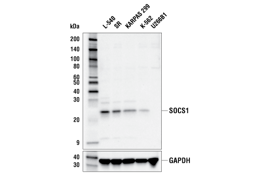
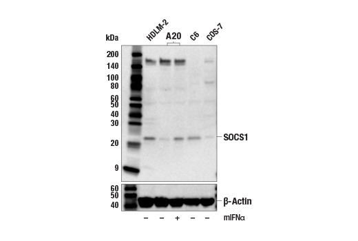
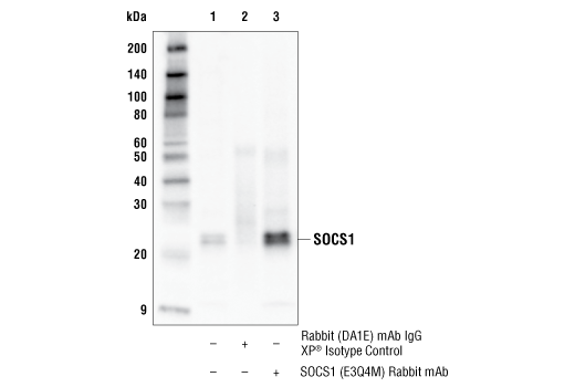
| SOCS1 (E3Q4M) Rabbit mAb | 55313 | 20 µl | 23 kDa | Rabbit IgG |
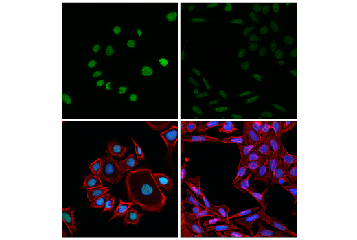
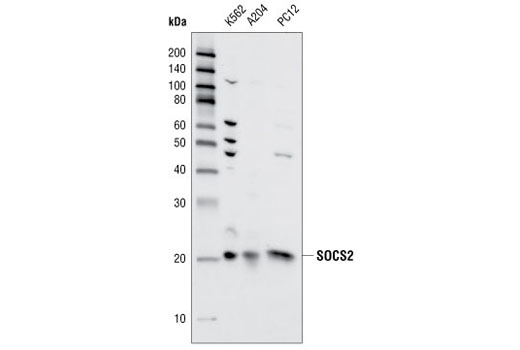
| SOCS2 Antibody | 2779 | 20 µl | 22 kDa | Rabbit |
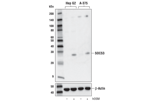
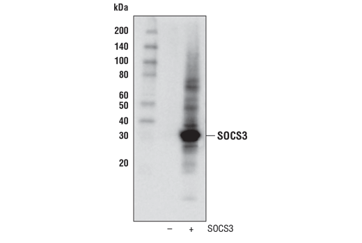
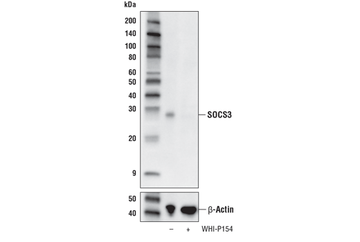
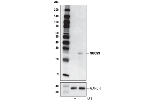
| SOCS3 (D6E1T) Rabbit mAb | 52113 | 20 µl | 28 kDa | Rabbit IgG |
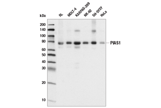
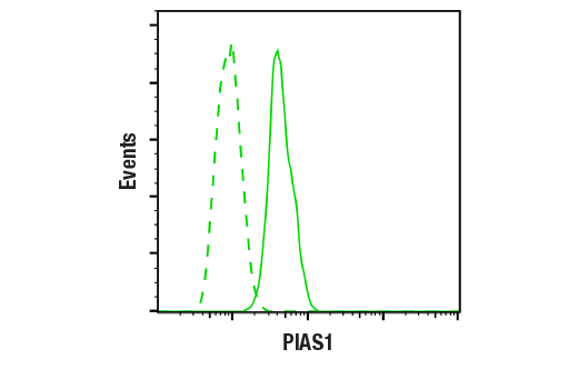
| PIAS1 (D33A7) XP® Rabbit mAb | 3550 | 20 µl | 76 kDa | Rabbit IgG |
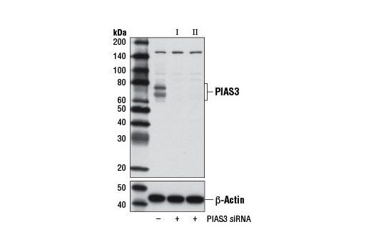
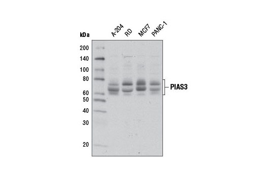
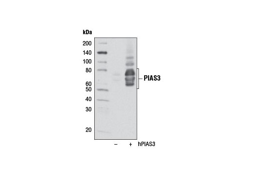
| PIAS3 (D5F9) XP® Rabbit mAb | 9042 | 20 µl | 65-75 kDa | Rabbit IgG |
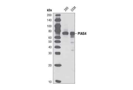
| PIAS4 (D2F12) Rabbit mAb | 4392 | 20 µl | 75 kDa | Rabbit IgG |
| Anti-rabbit IgG, HRP-linked Antibody | 7074 | 100 µl | Goat |
Please visit cellsignal.com for individual component applications, species cross-reactivity, dilutions, protocols, and additional product information.
Description
The Jak/Stat Pathway Inhibitors Antibody Sampler Kit provides an economical means to examine several inhibitors of Jak/Stat signaling, including PIAS1, PIAS3, PIAS4, SOCS1, SOCS2, and SOCS3. The kit contains enough primary antibody to perform two western blot experiments with each primary antibody.
Storage
Background
Jak (Janus Kinase) and Stat (signal transducer and activator of transcription) proteins are utilized by receptors for a wide varity of ligands including cytokines, hormones, growth factors, and neurotransmitters (1). Jaks and Stats play important roles in oncogenesis, tumor progression, angiogenesis, cell motility, immune responses, and stem cell differentiation (2-5). Therefore, regulation of Jak/Stat signaling is crucial to prevent aberrant signaling which can lead to disease progression. Two methods for regulating Jak/Stat signaling involve SOCS and PIAS proteins (6,7).The SOCS (suppressor or cytokine signaling) family members are negative regulators of cytokine signal transduction that inhibit the Jak/Stat pathway and consist of 8 known members, including the originally identified protein CIS1 (cytokine-inducible SH2-containing protein) and SOCS1-SOCS7. Each SOCS family member contains a central SH2 domain and a conserved carboxy-terminal motif designated as the SOCS box. These proteins are important regulators of cytokine signaling, proliferation, differentiation, and immune responses (8-10). SOCS proteins are involved in regulating over 30 cytokines, including interleukins, growth hormone (GH), interferons, leptin, and leukemia inhibitory factor (7). SOCS1, also known as JAB (Janus Kinase binding protein) and SSI-1 (Stat-induced Stat inhibitor-1), shares the most homology with SOCS3 and both are highly induced by cytokines (7,11). Both SOCS1 and SOCS3 directly inhibit Jak activity; SOCS1 inhibits Jak through an interaction involving a phospohotyrosine located in the kinase activation loop; SOCS3 inhibits Jak via its SH2 domain (12,13). In addition to inhibiting Jak/Stat signaling, the SOCS box of SOCS1 and SOCS3 can trigger ubiquitin-mediated degradation of proteins within and outside the Jak/Stat pathway (14,15). SOCS2 is also incduced upon cytokine stimulation and the activity of SOCS2 has been predominately linked to GH and insulin-like growth fac
- Darnell, J.E. et al. (1994) Science 264, 1415-21.
- Bromberg, J.F. et al. (1999) Cell 98, 295-303.
- Dentelli, P. et al. (1999) J Immunol 163, 2151-9.
- Cattaneo, E. et al. (1999) Trends Neurosci 22, 365-9.
- Su, L. et al. (1999) J Biol Chem 274, 31770-4.
- Shuai, K. (2006) Cell Res 16, 196-202.
- Croker, B.A. et al. (2008) Semin Cell Dev Biol 19, 414-22.
- Alexander, W.S. et al. (1999) J Leukoc Biol 66, 588-92.
- Chen, X.P. et al. (2000) Immunity 13, 287-90.
- Hilton, D.J. et al. (1998) Proc Natl Acad Sci U S A 95, 114-9.
- Starr, R. et al. (1997) Nature 387, 917-21.
- Yasukawa, H. et al. (1999) EMBO J 18, 1309-20.
- Sasaki, A. et al. (1999) Genes Cells 4, 339-51.
- Kamizono, S. et al. (2001) J Biol Chem 276, 12530-8.
- Rui, L. et al. (2002) J Biol Chem 277, 42394-8.
- Dey, B.R. et al. (1998) J Biol Chem 273, 24095-101.
- Liu, B. et al. (1998) Proc Natl Acad Sci U S A 95, 10626-31.
- Chung, C.D. et al. (1997) Science 278, 1803-5.
- Arora, T. et al. (2003) J Biol Chem 278, 21327-30.
- Nishida, T. et al. (2007) Biochem J 405, 481-8.
- Zhou, S. et al. (2008) J Biol Chem 283, 20137-48.
Background References
Trademarks and Patents
使用に関する制限
法的な権限を与えられたCSTの担当者が署名した書面によって別途明示的に合意された場合を除き、 CST、その関連会社または代理店が提供する製品には以下の条件が適用されます。お客様が定める条件でここに定められた条件に含まれるものを超えるもの、 または、ここに定められた条件と異なるものは、法的な権限を与えられたCSTの担当者が別途書面にて受諾した場合を除き、拒絶され、 いかなる効力も効果も有しません。
研究専用 (For Research Use Only) またはこれに類似する表示がされた製品は、 いかなる目的についても FDA または外国もしくは国内のその他の規制機関により承認、認可または許可を受けていません。 お客様は製品を診断もしくは治療目的で使用してはならず、また、製品に表示された内容に違反する方法で使用してはなりません。 CST が販売または使用許諾する製品は、エンドユーザーであるお客様に対し、使途を研究および開発のみに限定して提供されるものです。 診断、予防もしくは治療目的で製品を使用することまたは製品を再販売 (単独であるか他の製品等の一部であるかを問いません) もしくはその他の商業的利用の目的で購入することについては、CST から別途許諾を得る必要があります。 お客様は以下の事項を遵守しなければなりません。(a) CST の製品 (単独であるか他の資材と一緒であるかを問いません) を販売、使用許諾、貸与、寄付もしくはその他の態様で第三者に譲渡したり使用させたりしてはなりません。また、商用の製品を製造するために CST の製品を使用してはなりません。(b) 複製、改変、リバースエンジニアリング、逆コンパイル、 分解または他の方法により製品の構造または技術を解明しようとしてはなりません。また、 CST の製品またはサービスと競合する製品またはサービスを開発する目的で CST の製品を使用してはなりません。(c) CST の製品の商標、商号、ロゴ、特許または著作権に関する通知または表示を除去したり改変したりしてはなりません。(d) CST の製品をCST 製品販売条件(CST’s Product Terms of Sale) および該当する書面のみに従って使用しなければなりません。(e) CST の製品に関連してお客様が使用する第三者の製品またはサービスに関する使用許諾条件、 サービス提供条件またはこれに類する合意事項を遵守しなければなりません。