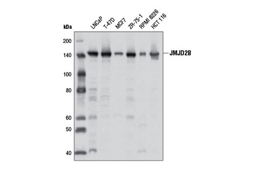
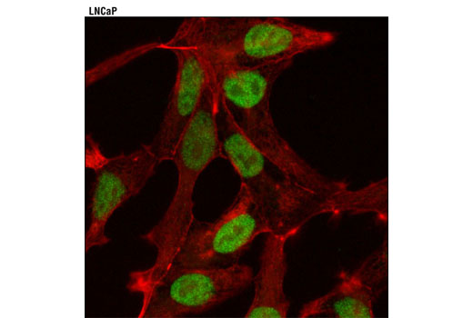
WB, IP, IF-IC
H Mk
Endogenous
150
Rabbit IgG
#O94953
23030
Product Information
Product Usage Information
| Application | Dilution |
|---|---|
| Western Blotting | 1:1000 |
| Immunoprecipitation | 1:50 |
| Immunofluorescence (Immunocytochemistry) | 1:800 |
Storage
Specificity / Sensitivity
Species Reactivity:
Human, Monkey
Source / Purification
Monoclonal antibody is produced by immunizing animals with a synthetic peptide corresponding to residues near the carboxy terminus of human JMJD2B protein.
Background
The methylation state of lysine residues in histone proteins is a major determinant of the formation of active and inactive regions of the genome and is crucial for proper programming of the genome during development (1,2). Jumonji C (JmjC) domain-containing proteins represent the largest class of potential histone demethylase proteins (3). The JmjC domain can catalyze the demethylation of mono-, di-, and tri-methyl lysine residues via an oxidative reaction that requires iron and α-ketoglutarate (3). Based on homology, both humans and mice contain at least 30 such proteins, which can be divided into 7 separate families (3). The jumonji domain-containing protein 2 (JMJD2) family, also known as the JmjC domain-containing histone demethylation protein 3 (JHDM3) family, contains four members: JMJD2A/JHDM3A, JMJD2B/JHDM3B, JMJD2C/JHDM3C, and JMJD2D/JHDM3D. In addition to the JmjC domain, these proteins also contain JmjN, PHD, and tudor domains, the latter of which has been shown to bind to methylated histone H3 at Lys4 and Lys9, and methylated histone H4 at Lys20 (4,5). JMJD2 proteins have been shown to demethylate di- and tri-methyl histone H3 at Lys9 and Lys36 and function as both activators and repressors of transcription (6-11). JMJD2A, JMJD2C, and JMJD2D function as coactivators of the androgen receptor in prostate tumor cells (7). In contrast, JMJD2A also associates with Rb and NCoR corepressor complexes and is necessary for transcriptional repression of target genes (8,9). JMJD2B antagonizes histone H3 Lys9 tri-methylation at pericentric heterochromatin (10). JMJD2C, also known as GASC1, is amplified in squamous cell carcinomas and metastatic lung carcinoma and inhibition of JMJD2C expression decreases cell proliferation (11,12). JMJD2C has also been identified as a downstream target of Oct-4 and is critical for the regulation of self-renewal in embryonic stem cells (13).
Recent studies have demonstrated that JMJD2B is physically associated with and an integral component of the mixed-lineage leukemia (MLL) 2 H3K4 methyltransferase complex. JMJD2B also interacts with estrogen receptor α (ERα) and members of a chromatin remodeling complex, SWI/SNF-B. It is likely that JMJD2B removes repressive histone marks at ERα binding sites, which may also generate docking sites for enzymes and transcription factors that remodel chromatin in order to facilitate ERα-mediated transcription. Of note, JMJD2B is expressed in a high percentage of human breast tumors and its expression positively correlates with ERα expression. Researchers have shown that JMJD2B is a transcriptional target of ERα and may participate in a feed-forward regulatory loop involved in driving estrogen responsive breast tumor formation (14,15).
- Kubicek, S. et al. (2006) Ernst Schering Res Found Workshop , 1-27.
- Lin, W. and Dent, S.Y. (2006) Curr Opin Genet Dev 16, 137-42.
- Klose, R.J. et al. (2006) Nat Rev Genet 7, 715-27.
- Chen, Z. et al. (2007) Proc Natl Acad Sci USA 104, 10818-23.
- Lee, J. et al. (2008) Nat Struct Mol Biol 15, 109-11.
- Whetstine, J.R. et al. (2006) Cell 125, 467-81.
- Shin, S. and Janknecht, R. (2007) Biochem Biophys Res Commun 359, 742-6.
- Gray, S.G. et al. (2005) J Biol Chem 280, 28507-18.
- Zhang, D. et al. (2005) Mol Cell Biol 25, 6404-14.
- Fodor, B.D. et al. (2006) Genes Dev 20, 1557-62.
- Cloos, P.A. et al. (2006) Nature 442, 307-11.
- Italiano, A. et al. (2006) Cancer Genet Cytogenet 167, 122-30.
- Loh, Y.H. et al. (2007) Genes Dev 21, 2545-57.
- Shi, L. et al. (2011) Proc Natl Acad Sci USA 108, 7541-6.
- Kawazu, M. et al. (2011) PLoS One 6, e17830.
Species Reactivity
Species reactivity is determined by testing in at least one approved application (e.g., western blot).
Western Blot Buffer
IMPORTANT: For western blots, incubate membrane with diluted primary antibody in 5% w/v nonfat dry milk, 1X TBS, 0.1% Tween® 20 at 4°C with gentle shaking, overnight.
Applications Key
WB: Western Blotting IP: Immunoprecipitation IF-IC: Immunofluorescence (Immunocytochemistry)
Cross-Reactivity Key
H: human M: mouse R: rat Hm: hamster Mk: monkey Vir: virus Mi: mink C: chicken Dm: D. melanogaster X: Xenopus Z: zebrafish B: bovine Dg: dog Pg: pig Sc: S. cerevisiae Ce: C. elegans Hr: horse GP: Guinea Pig Rab: rabbit All: all species expected
Trademarks and Patents
使用に関する制限
法的な権限を与えられたCSTの担当者が署名した書面によって別途明示的に合意された場合を除き、 CST、その関連会社または代理店が提供する製品には以下の条件が適用されます。お客様が定める条件でここに定められた条件に含まれるものを超えるもの、 または、ここに定められた条件と異なるものは、法的な権限を与えられたCSTの担当者が別途書面にて受諾した場合を除き、拒絶され、 いかなる効力も効果も有しません。
研究専用 (For Research Use Only) またはこれに類似する表示がされた製品は、 いかなる目的についても FDA または外国もしくは国内のその他の規制機関により承認、認可または許可を受けていません。 お客様は製品を診断もしくは治療目的で使用してはならず、また、製品に表示された内容に違反する方法で使用してはなりません。 CST が販売または使用許諾する製品は、エンドユーザーであるお客様に対し、使途を研究および開発のみに限定して提供されるものです。 診断、予防もしくは治療目的で製品を使用することまたは製品を再販売 (単独であるか他の製品等の一部であるかを問いません) もしくはその他の商業的利用の目的で購入することについては、CST から別途許諾を得る必要があります。 お客様は以下の事項を遵守しなければなりません。(a) CST の製品 (単独であるか他の資材と一緒であるかを問いません) を販売、使用許諾、貸与、寄付もしくはその他の態様で第三者に譲渡したり使用させたりしてはなりません。また、商用の製品を製造するために CST の製品を使用してはなりません。(b) 複製、改変、リバースエンジニアリング、逆コンパイル、 分解または他の方法により製品の構造または技術を解明しようとしてはなりません。また、 CST の製品またはサービスと競合する製品またはサービスを開発する目的で CST の製品を使用してはなりません。(c) CST の製品の商標、商号、ロゴ、特許または著作権に関する通知または表示を除去したり改変したりしてはなりません。(d) CST の製品をCST 製品販売条件(CST’s Product Terms of Sale) および該当する書面のみに従って使用しなければなりません。(e) CST の製品に関連してお客様が使用する第三者の製品またはサービスに関する使用許諾条件、 サービス提供条件またはこれに類する合意事項を遵守しなければなりません。