WB, IP, ChIP
H
Endogenous
62
Rabbit IgG
#P12524
4610
Product Information
Product Usage Information
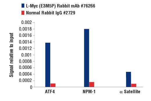
For optimal ChIP results, use 10 μl of antibody and 10 μg of chromatin (approximately 4 × 10⁶ cells) per IP. This antibody has been validated using SimpleChIP® Enzymatic Chromatin IP Kits.
| Application | Dilution |
|---|---|
| Western Blotting | 1:1000 |
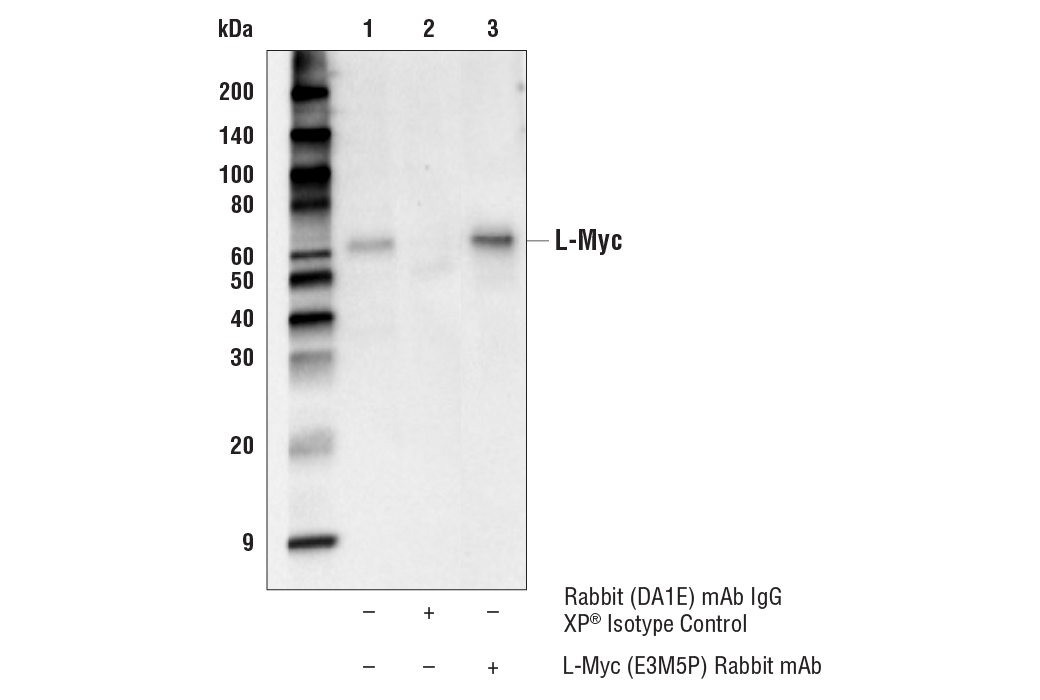
| Immunoprecipitation | 1:200 |
| Chromatin IP | 1:50 |
Storage
Specificity / Sensitivity
Species Reactivity:
Human
Source / Purification
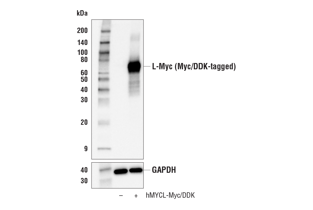
Monoclonal antibody is produced by immunizing animals with recombinant protein specific to the carboxy terminus of human L-Myc protein. The epitope has been mapped to residues surrounding Thr190.
Background
Members of the Myc/Max/Mad network function as transcriptional regulators with roles in various aspects of cell behavior, including proliferation, differentiation, and apoptosis (1). These proteins share a common basic-helix-loop-helix leucine zipper (bHLH-ZIP) motif required for dimerization and DNA-binding. Max was originally discovered based on its ability to associate with c-Myc and found to be required for the ability of Myc to bind DNA and activate transcription (2). Subsequently, Max has been viewed as a central component of the transcriptional network, forming homodimers as well as heterodimers with other members of the Myc and Mad families (1). The association between Max and either Myc or Mad can have opposing effects on transcriptional regulation and cell behavior (1). The Mad family consists of four related proteins; Mad1, Mad2 (Mxi1), Mad3, and Mad4, and the more distantly related members of the bHLH-ZIP family, Mnt and Mga. Like Myc, the Mad proteins are tightly regulated with short half-lives. In general, Mad family members interfere with Myc-mediated processes, such as proliferation, transformation, and prevention of apoptosis by inhibiting transcription (3,4).
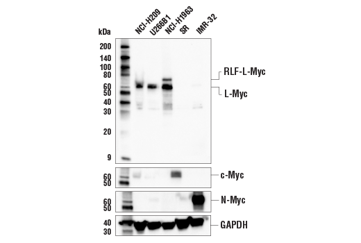
The Myc family is comprised of c-Myc, N-Myc, and L-Myc with often distinct patterns of expression during development as well as cancer (5). Amplification of each of the family members in cancer is frequently mutually exclusive with c-Myc being the most widely studied and most commonly amplified. N-Myc amplification, on the other hand, is found predominantly in neuroblastomas, and L-Myc amplification has been described in small cell lung cancer (SCLC) (6). Amplification of L-Myc in SCLC may result from chromosomal rearrangement between the rlf and MYCL genes (7). L-Myc expression by dendritic cells may be required for optimal T cell priming (8).
- Baudino, T.A. and Cleveland, J.L. (2001) Mol Cell Biol 21, 691-702.
- Blackwood, E.M. and Eisenman, R.N. (1991) Science 251, 1211-7.
- Henriksson, M. and Lüscher, B. (1996) Adv Cancer Res 68, 109-82.
- Grandori, C. et al. (2000) Annu Rev Cell Dev Biol 16, 653-99.
- Nesbit, C.E. et al. (1999) Oncogene 18, 3004-16.
- Nau, M.M. et al. Nature 318, 69-73.
- Mäkelä, T.P. et al. (1991) EMBO J 10, 1331-5.
- KC, W. et al. (2014) Nature 507, 243-7.
Species Reactivity
Species reactivity is determined by testing in at least one approved application (e.g., western blot).
Western Blot Buffer
IMPORTANT: For western blots, incubate membrane with diluted primary antibody in 5% w/v BSA, 1X TBS, 0.1% Tween® 20 at 4°C with gentle shaking, overnight.
Applications Key
WB: Western Blotting IP: Immunoprecipitation ChIP: Chromatin IP
Cross-Reactivity Key
H: human M: mouse R: rat Hm: hamster Mk: monkey Vir: virus Mi: mink C: chicken Dm: D. melanogaster X: Xenopus Z: zebrafish B: bovine Dg: dog Pg: pig Sc: S. cerevisiae Ce: C. elegans Hr: horse GP: Guinea Pig Rab: rabbit All: all species expected
Trademarks and Patents
使用に関する制限
法的な権限を与えられたCSTの担当者が署名した書面によって別途明示的に合意された場合を除き、 CST、その関連会社または代理店が提供する製品には以下の条件が適用されます。お客様が定める条件でここに定められた条件に含まれるものを超えるもの、 または、ここに定められた条件と異なるものは、法的な権限を与えられたCSTの担当者が別途書面にて受諾した場合を除き、拒絶され、 いかなる効力も効果も有しません。
研究専用 (For Research Use Only) またはこれに類似する表示がされた製品は、 いかなる目的についても FDA または外国もしくは国内のその他の規制機関により承認、認可または許可を受けていません。 お客様は製品を診断もしくは治療目的で使用してはならず、また、製品に表示された内容に違反する方法で使用してはなりません。 CST が販売または使用許諾する製品は、エンドユーザーであるお客様に対し、使途を研究および開発のみに限定して提供されるものです。 診断、予防もしくは治療目的で製品を使用することまたは製品を再販売 (単独であるか他の製品等の一部であるかを問いません) もしくはその他の商業的利用の目的で購入することについては、CST から別途許諾を得る必要があります。 お客様は以下の事項を遵守しなければなりません。(a) CST の製品 (単独であるか他の資材と一緒であるかを問いません) を販売、使用許諾、貸与、寄付もしくはその他の態様で第三者に譲渡したり使用させたりしてはなりません。また、商用の製品を製造するために CST の製品を使用してはなりません。(b) 複製、改変、リバースエンジニアリング、逆コンパイル、 分解または他の方法により製品の構造または技術を解明しようとしてはなりません。また、 CST の製品またはサービスと競合する製品またはサービスを開発する目的で CST の製品を使用してはなりません。(c) CST の製品の商標、商号、ロゴ、特許または著作権に関する通知または表示を除去したり改変したりしてはなりません。(d) CST の製品をCST 製品販売条件(CST’s Product Terms of Sale) および該当する書面のみに従って使用しなければなりません。(e) CST の製品に関連してお客様が使用する第三者の製品またはサービスに関する使用許諾条件、 サービス提供条件またはこれに類する合意事項を遵守しなければなりません。