WB, IF-IC, FC-FP, ChIP, ChIP-seq
H
Endogenous
18
Rabbit IgG
#P25791
4005
Product Information
Product Usage Information
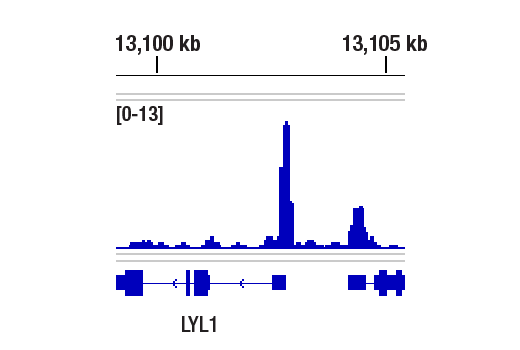
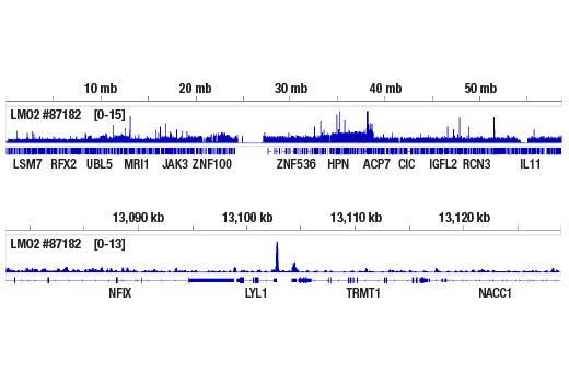
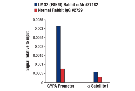
For optimal ChIP and ChIP-seq results, use 5 μL of antibody and 10 μg of chromatin (approximately 4 x 10^6 cells) per IP. This antibody has been validated using SimpleChIP® Enzymatic Chromatin IP Kits.
| Application | Dilution |
|---|---|
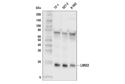
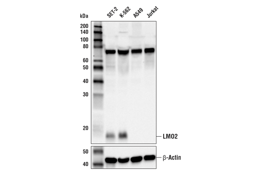
| Western Blotting | 1:1000 |
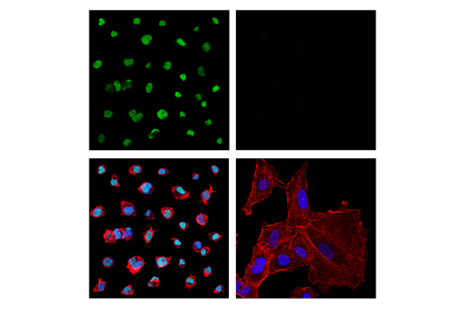
| Immunofluorescence (Immunocytochemistry) | 1:1600 - 1:6400 |
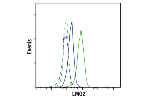
| Flow Cytometry (Fixed/Permeabilized) | 1:400 - 1:1600 |
| Chromatin IP | 1:100 |
| Chromatin IP-seq | 1:100 |
Storage
Specificity / Sensitivity
Species Reactivity:
Human
Source / Purification
Monoclonal antibody is produced by immunizing animals with a synthetic peptide corresponding to residues near the amino terminus of human LMO2 protein.
Background
LIM-domain-only protein 2 (LMO2) is a transcriptional regulator that was first identified as a proto-oncogene activated by chromosomal translocations in T-cell acute lymphocytic leukemia (T-ALL) (1). Since then, LMO2 has been found to be a master regulator essential for erythroid development, as evidenced by homozygous LMO2 knockouts resulting in embryonic lethality in mice (2). LMO2 is an intrinsically unstructured protein that does not bind DNA directly, but rather acts as a scaffolding protein that recruits various transcription factors via its tandem cysteine-rich LIM domains to form a multi-protein DNA binding complex (3,4). The LMO2 complex plays a major role in hematopoiesis and was originally shown to consist of the transcription factors TAL1, E47, and GATA-1 in erythroid lineage cells, but variations of this complex may contain alternate transcription factors including LYL1, TAL2, GATA-2, and GATA-3 (5-9). The LMO2 complex also requires interaction with LIM-domain binding protein 1 (LDB1), which is necessary for LMO2 protein stability (10-11). In addition to hematopoietic tissue, LMO2 is also expressed in embryonic brain tissue, where it associates with BEX2 and the transcription factor NSCL-2 (12). Aberrant LMO2 expression is observed in several types of hematopoietic cancers, including large diffuse B-cell lymphoma (DLBCL), B-cell acute leukemia, (B-ALL), acute myeloid leukemia (AML), and T-ALL (13-16). LMO2-mediated T-ALL is primarily caused by the translocation t(11;14)(p13;q11) with the TCRD/A gene from chromosome 14q11, or the t(7;11)(q35;p13) translocation involving TCRB from 7q35 (1). In addition to hematopoietic cancers, overexpression of LMO2 in prostate stromal cells also facilitates prostate cancer progression by inducing expression of Interleukin-11 (IL-11), which stimulates STAT3 signaling in these cells (17).
- Boehm, T. et al. (1991) Proc Natl Acad Sci U S A 88, 4367-71.
- Warren, A.J. et al. (1994) Cell 78, 45-57.
- Chambers, J. and Rabbitts, T.H. (2015) Open Biol 5, 150062.
- Lécuyer, E. et al. (2007) J Biol Chem 282, 33649-58.
- Valge-Archer, V.E. et al. (1994) Proc Natl Acad Sci U S A 91, 8617-21.
- Wadman, I. et al. (1994) EMBO J 13, 4831-9.
- Wadman, I.A. et al. (1997) EMBO J 16, 3145-57.
- Osada, H. et al. (1995) Proc Natl Acad Sci U S A 92, 9585-9.
- Ono, Y. et al. (1998) Mol Cell Biol 18, 6939-50.
- Agulnick, A.D. et al. (1996) Nature 384, 270-2.
- Layer, J.H. et al. (2016) Mol Cell Biol 36, 488-506.
- Han, C. et al. (2005) Nucleic Acids Res 33, 6555-65.
- Natkunam, Y. et al. (2007) Blood 109, 1636-42.
- de Boer, J. et al. (2011) Leukemia 25, 321-30.
- Atay, M.H. et al. (2013) Histopathology 63, 293-4.
- Patel, J.L. et al. (2014) Histopathology 64, 226-33.
- Jiang, C.Y. et al. (2016) Oncotarget 7, 26247-58.
Species Reactivity
Species reactivity is determined by testing in at least one approved application (e.g., western blot).
Western Blot Buffer
IMPORTANT: For western blots, incubate membrane with diluted primary antibody in 5% w/v BSA, 1X TBS, 0.1% Tween® 20 at 4°C with gentle shaking, overnight.
Applications Key
WB: Western Blotting IF-IC: Immunofluorescence (Immunocytochemistry) FC-FP: Flow Cytometry (Fixed/Permeabilized) ChIP: Chromatin IP ChIP-seq: Chromatin IP-seq
Cross-Reactivity Key
H: human M: mouse R: rat Hm: hamster Mk: monkey Vir: virus Mi: mink C: chicken Dm: D. melanogaster X: Xenopus Z: zebrafish B: bovine Dg: dog Pg: pig Sc: S. cerevisiae Ce: C. elegans Hr: horse GP: Guinea Pig Rab: rabbit All: all species expected
Trademarks and Patents
使用に関する制限
法的な権限を与えられたCSTの担当者が署名した書面によって別途明示的に合意された場合を除き、 CST、その関連会社または代理店が提供する製品には以下の条件が適用されます。お客様が定める条件でここに定められた条件に含まれるものを超えるもの、 または、ここに定められた条件と異なるものは、法的な権限を与えられたCSTの担当者が別途書面にて受諾した場合を除き、拒絶され、 いかなる効力も効果も有しません。
研究専用 (For Research Use Only) またはこれに類似する表示がされた製品は、 いかなる目的についても FDA または外国もしくは国内のその他の規制機関により承認、認可または許可を受けていません。 お客様は製品を診断もしくは治療目的で使用してはならず、また、製品に表示された内容に違反する方法で使用してはなりません。 CST が販売または使用許諾する製品は、エンドユーザーであるお客様に対し、使途を研究および開発のみに限定して提供されるものです。 診断、予防もしくは治療目的で製品を使用することまたは製品を再販売 (単独であるか他の製品等の一部であるかを問いません) もしくはその他の商業的利用の目的で購入することについては、CST から別途許諾を得る必要があります。 お客様は以下の事項を遵守しなければなりません。(a) CST の製品 (単独であるか他の資材と一緒であるかを問いません) を販売、使用許諾、貸与、寄付もしくはその他の態様で第三者に譲渡したり使用させたりしてはなりません。また、商用の製品を製造するために CST の製品を使用してはなりません。(b) 複製、改変、リバースエンジニアリング、逆コンパイル、 分解または他の方法により製品の構造または技術を解明しようとしてはなりません。また、 CST の製品またはサービスと競合する製品またはサービスを開発する目的で CST の製品を使用してはなりません。(c) CST の製品の商標、商号、ロゴ、特許または著作権に関する通知または表示を除去したり改変したりしてはなりません。(d) CST の製品をCST 製品販売条件(CST’s Product Terms of Sale) および該当する書面のみに従って使用しなければなりません。(e) CST の製品に関連してお客様が使用する第三者の製品またはサービスに関する使用許諾条件、 サービス提供条件またはこれに類する合意事項を遵守しなければなりません。