| Product Includes | Product # | Quantity | Mol. Wt | Isotype/Source |
|---|---|---|---|---|
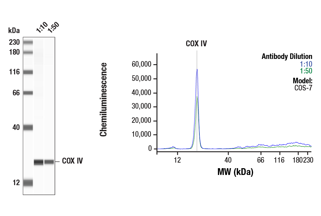
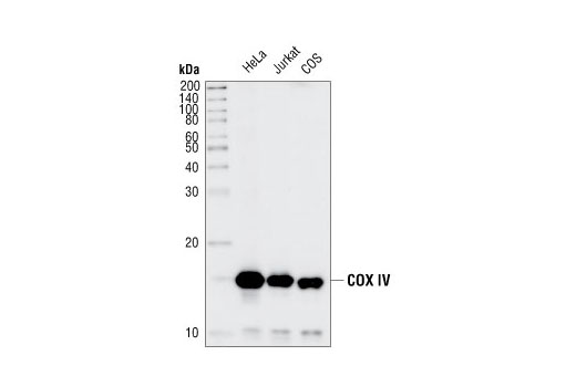
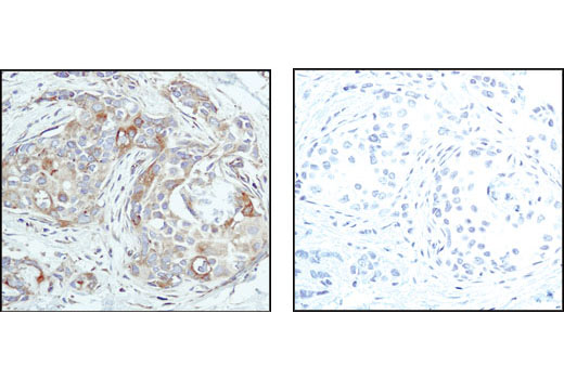
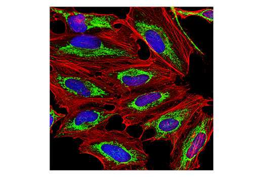
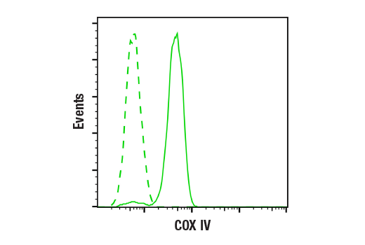
| COX IV (3E11) Rabbit mAb | 4850 | 20 µl | 17 kDa | Rabbit IgG |
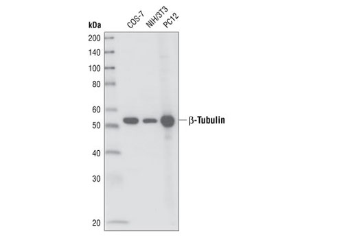
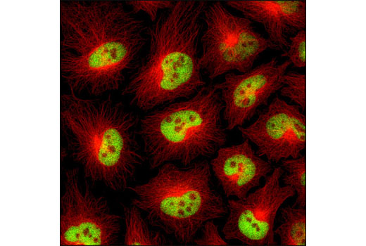
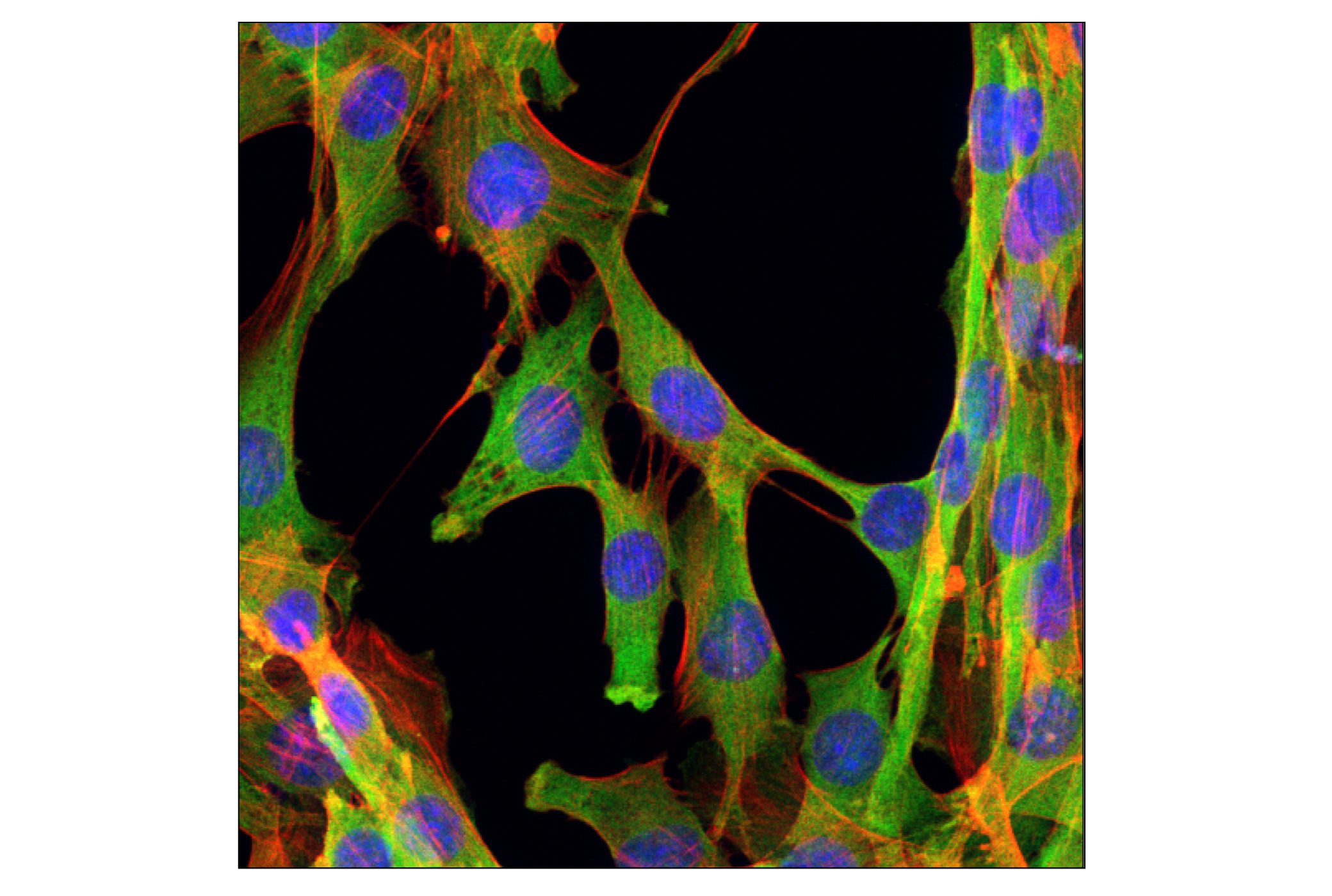
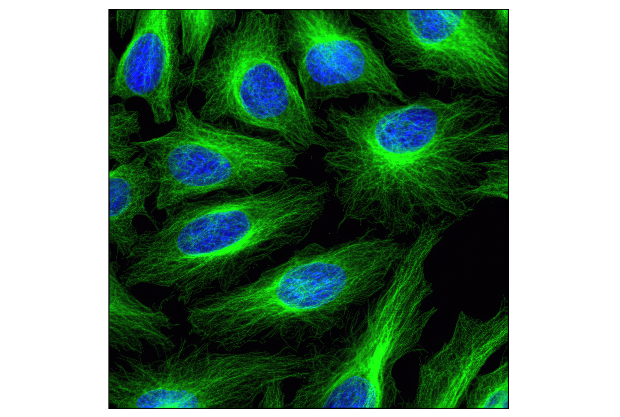
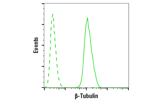
| β-Tubulin (9F3) Rabbit mAb | 2128 | 20 µl | 55 kDa | Rabbit IgG |
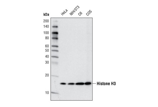
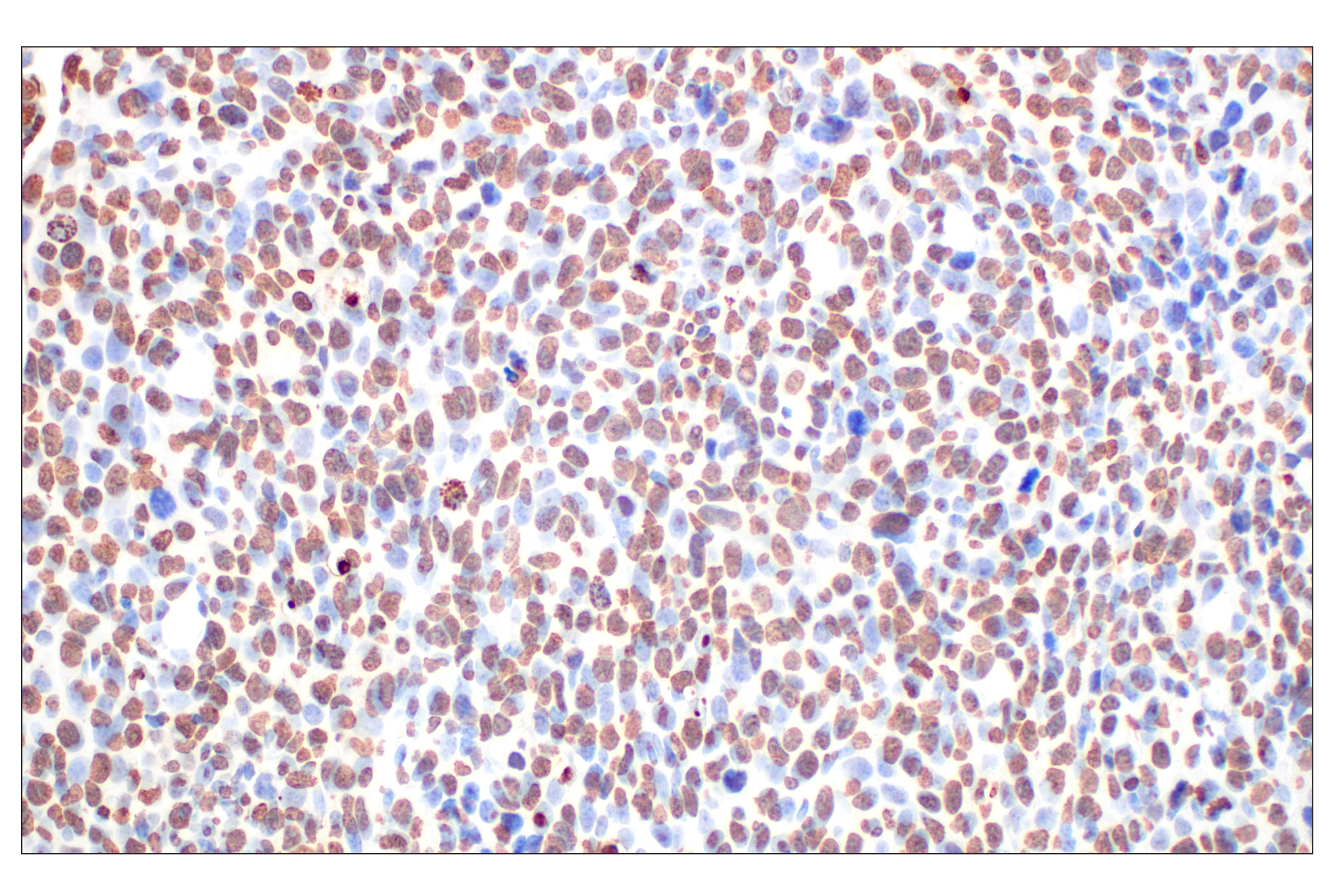
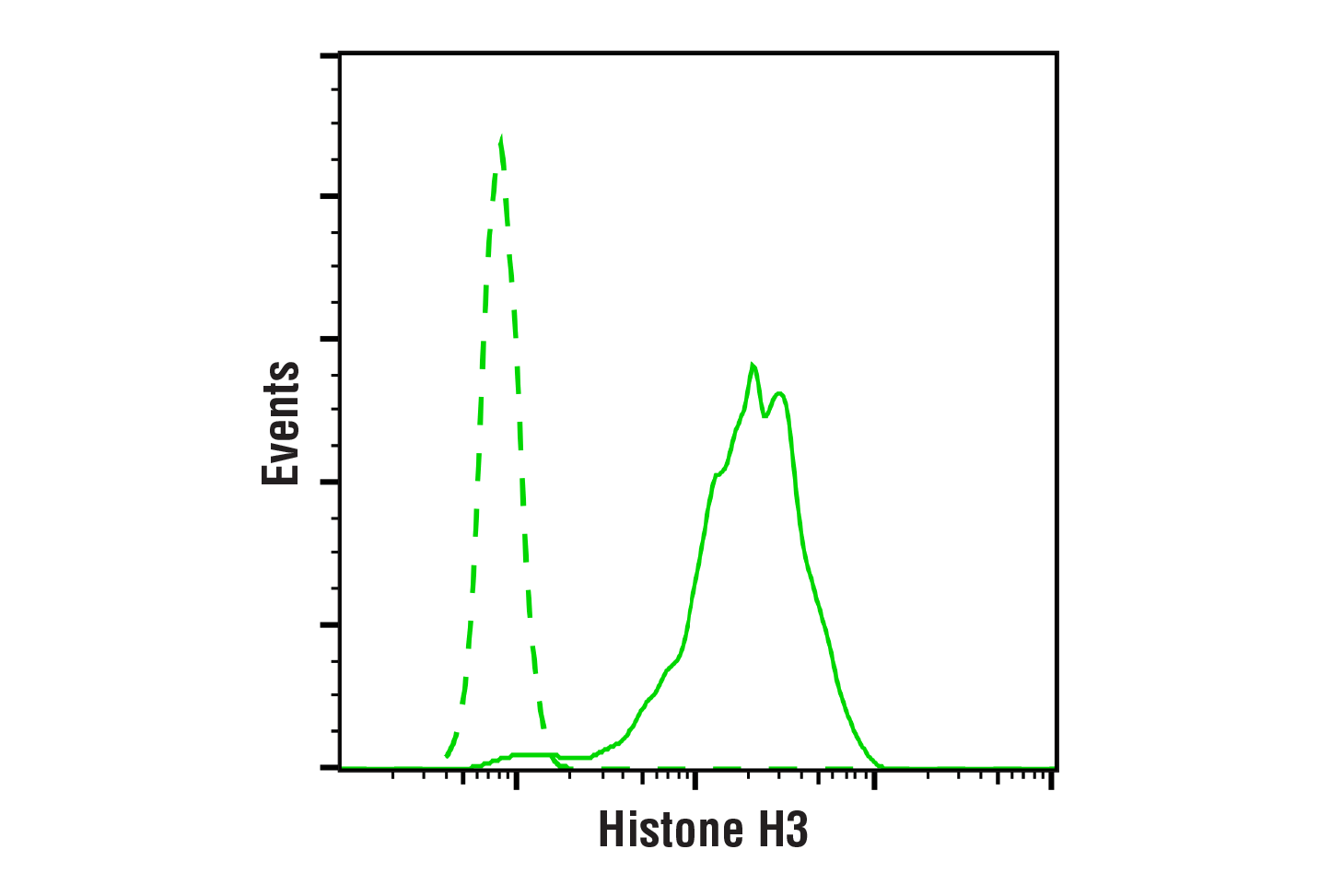
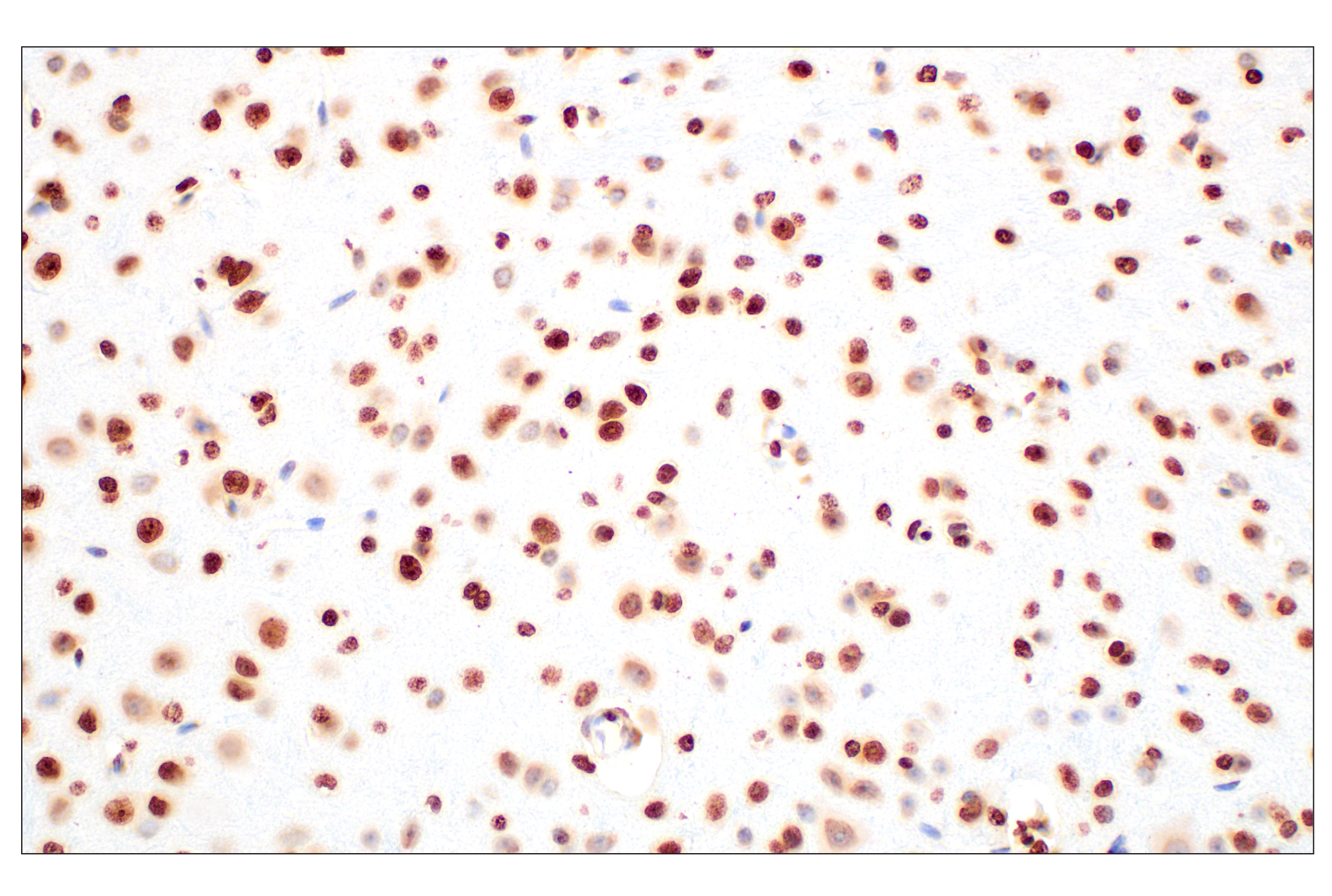
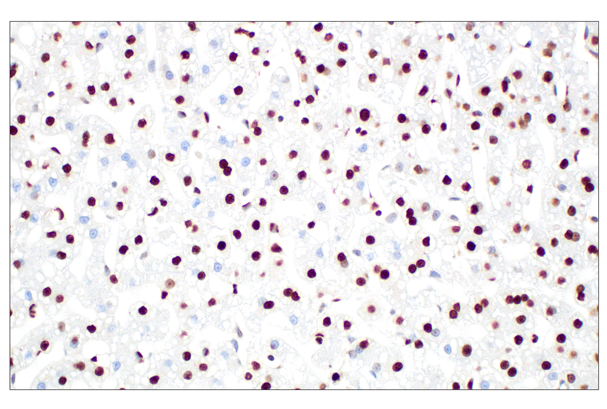
| Histone H3 (D1H2) XP® Rabbit mAb | 4499 | 20 µl | 17 kDa | Rabbit IgG |
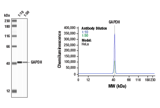
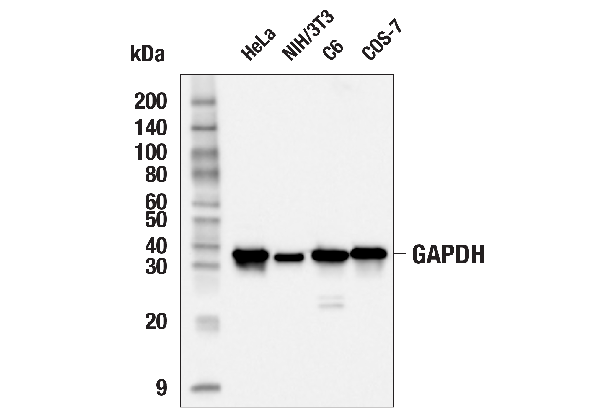
| GAPDH (D16H11) XP® Rabbit mAb | 5174 | 20 µl | 37 kDa | Rabbit IgG |
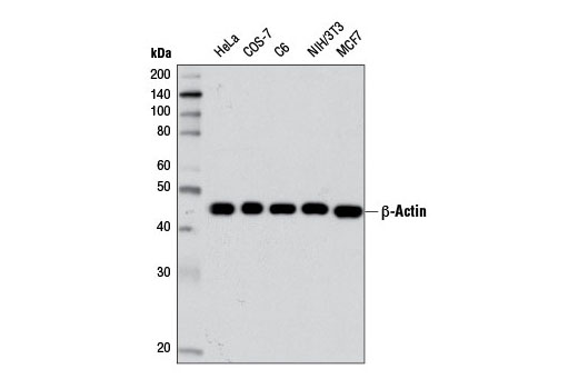
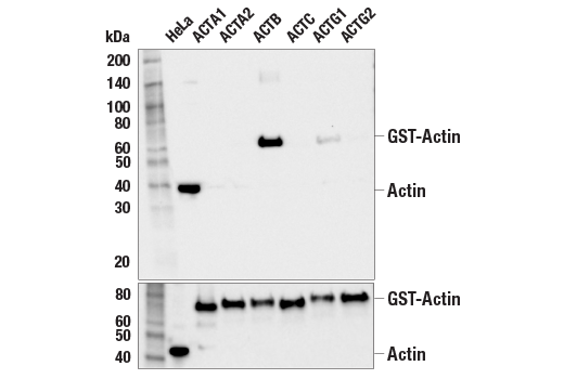
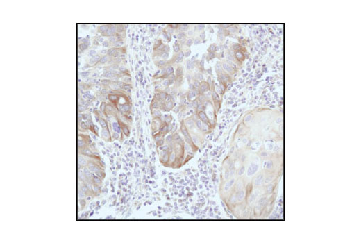
| β-Actin (D6A8) Rabbit mAb | 8457 | 20 µl | 45 kDa | Rabbit IgG |
| Anti-rabbit IgG, HRP-linked Antibody | 7074 | 100 µl | Goat |
Please visit cellsignal.com for individual component applications, species cross-reactivity, dilutions, protocols, and additional product information.
Description
The Loading Control Antibody Sampler Kit contains antibodies to a variety of housekeeping proteins. The kit contains enough primary and secondary antibodies to perform two western blots per primary antibody.
Storage
Background
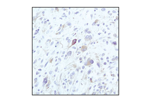
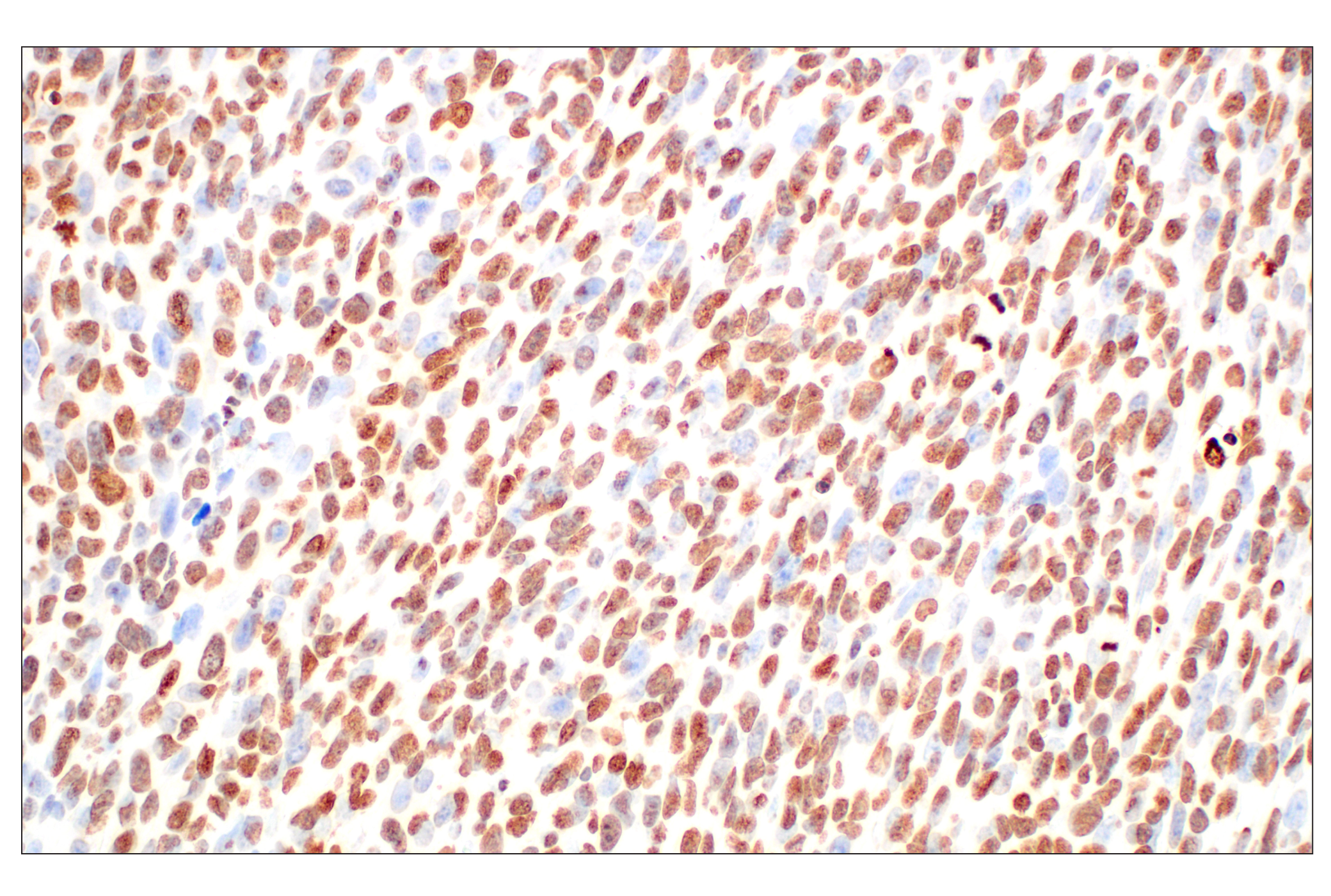
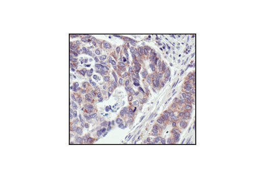
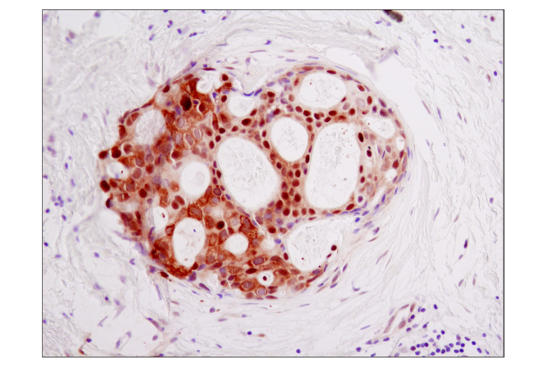
Housekeeping proteins perform numerous basic functions within the cell and are constitutively expressed at high levels in a variety of tissues and cell types. Western blot analysis commonly uses housekeeping proteins such as β-actin, COX IV, GAPDH, histone H3 and the α- and β-tubulins as loading controls. Actin is a ubiquitous protein and a major component of the eukaryotic cytoskeleton. Actin exists mainly as the F-actin fibrous polymer (1). Glyceraldehyde-3-phosphate dehydrogenase (GAPDH) catalyzes the phosphorylation of glyceraldehyde-3-phosphate during glycolysis. Recent work has demonstrated that GAPDH plays roles in apoptosis (2), gene expression (3), and nuclear transport (4). Globular tubulin subunits made up of α- and β-tubulin heterodimers are the building blocks of microtubules, one of three types of cytosolic fibers that comprise the cytoskeleton (5). Histone proteins, including histone H3, make up the primary building block of chromatin known as nucleosomes. Modulation of the chromatin structure plays an important role in the regulation of transcription in eukaryotes (6). Cytochrome c oxidase (COX) is a hetero-oligomeric enzyme consisting of 13 subunits localized to the inner mitochondrial membrane (7-9). It is the terminal enzyme complex in the respiratory chain, catalyzing the reduction of protons across the mitochondrial inner membrane to drive ATP synthesis (10).
- Condeelis, J. (2001) Trends Cell Biol 11, 288-93.
- Hara, M.R. and Snyder, S.H. Cell Mol Neurobiol (2006) 26, 527-38.
- Zheng, L. et al. (2003) Cell 114, 255-66.
- Bae, B.I. et al. (2006) Proc Natl Acad Sci USA 103, 3405-9.
- Westermann, S. and Weber, K. (2003) Nat Rev Mol Cell Biol 4, 938-47.
- Workman, J.L. and Kingston, R.E. (1998) Annu Rev Biochem 67, 545-79.
- Ostermeier, C. et al. (1996) Curr Opin Struct Biol 6, 460-6.
- Capaldi, R.A. et al. (1983) Biochim Biophys Acta 726, 135-48.
- Kadenbach, B. et al. (2000) Free Radic Biol Med 29, 211-21.
- Barrientos, A. et al. (2002) Gene 286, 53-63.
Background References
Trademarks and Patents
使用に関する制限
法的な権限を与えられたCSTの担当者が署名した書面によって別途明示的に合意された場合を除き、 CST、その関連会社または代理店が提供する製品には以下の条件が適用されます。お客様が定める条件でここに定められた条件に含まれるものを超えるもの、 または、ここに定められた条件と異なるものは、法的な権限を与えられたCSTの担当者が別途書面にて受諾した場合を除き、拒絶され、 いかなる効力も効果も有しません。
研究専用 (For Research Use Only) またはこれに類似する表示がされた製品は、 いかなる目的についても FDA または外国もしくは国内のその他の規制機関により承認、認可または許可を受けていません。 お客様は製品を診断もしくは治療目的で使用してはならず、また、製品に表示された内容に違反する方法で使用してはなりません。 CST が販売または使用許諾する製品は、エンドユーザーであるお客様に対し、使途を研究および開発のみに限定して提供されるものです。 診断、予防もしくは治療目的で製品を使用することまたは製品を再販売 (単独であるか他の製品等の一部であるかを問いません) もしくはその他の商業的利用の目的で購入することについては、CST から別途許諾を得る必要があります。 お客様は以下の事項を遵守しなければなりません。(a) CST の製品 (単独であるか他の資材と一緒であるかを問いません) を販売、使用許諾、貸与、寄付もしくはその他の態様で第三者に譲渡したり使用させたりしてはなりません。また、商用の製品を製造するために CST の製品を使用してはなりません。(b) 複製、改変、リバースエンジニアリング、逆コンパイル、 分解または他の方法により製品の構造または技術を解明しようとしてはなりません。また、 CST の製品またはサービスと競合する製品またはサービスを開発する目的で CST の製品を使用してはなりません。(c) CST の製品の商標、商号、ロゴ、特許または著作権に関する通知または表示を除去したり改変したりしてはなりません。(d) CST の製品をCST 製品販売条件(CST’s Product Terms of Sale) および該当する書面のみに従って使用しなければなりません。(e) CST の製品に関連してお客様が使用する第三者の製品またはサービスに関する使用許諾条件、 サービス提供条件またはこれに類する合意事項を遵守しなければなりません。