| Product Includes | Product # | Quantity | Mol. Wt | Isotype/Source |
|---|---|---|---|---|
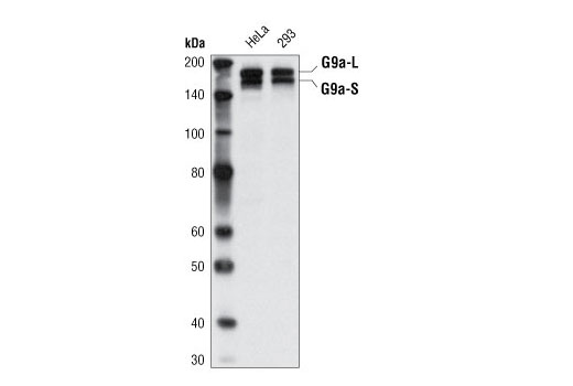
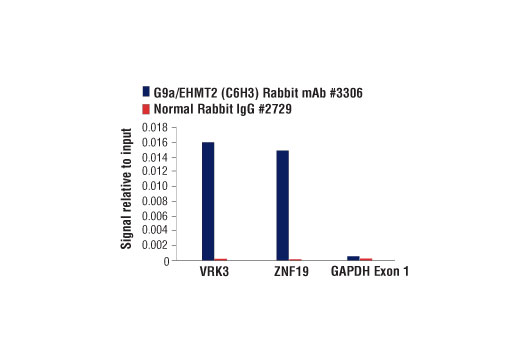
| G9a/EHMT2 (C6H3) Rabbit mAb | 3306 | 20 µl | 160,180 kDa | Rabbit IgG |
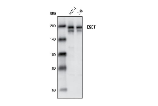
| ESET (C1C12) Rabbit mAb | 2196 | 20 µl | 180 kDa | Rabbit IgG |
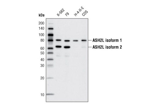
| ASH2L (D93F6) XP® Rabbit mAb | 5019 | 20 µl | 80, 65 kDa | Rabbit IgG |
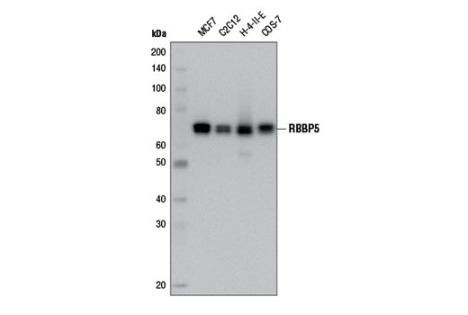
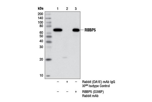
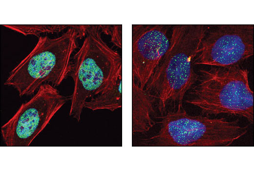
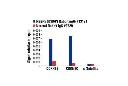
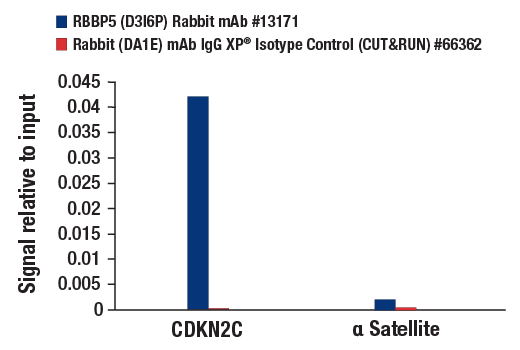
| RBBP5 (D3I6P) Rabbit mAb | 13171 | 20 µl | 70 kDa | Rabbit IgG |
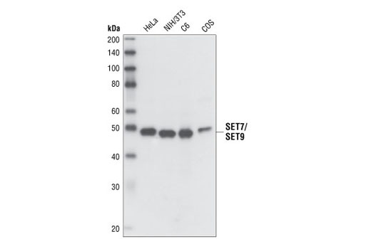
| SET7/SET9 Antibody | 2813 | 20 µl | 48 kDa | Rabbit |
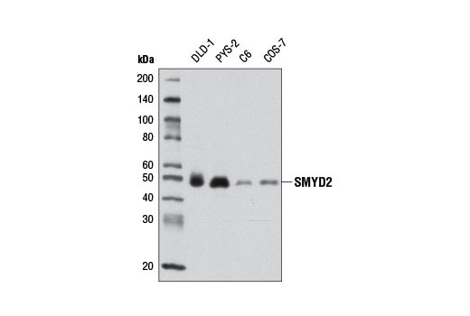
| SMYD2 (D14H7) Rabbit mAb | 9734 | 20 µl | 49 kDa | Rabbit IgG |
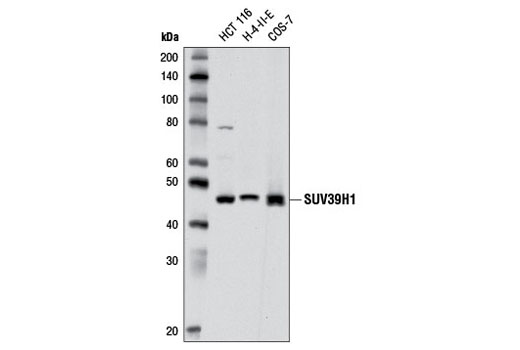
| SUV39H1 (D11B6) Rabbit mAb | 8729 | 20 µl | 48 kDa | Rabbit IgG |
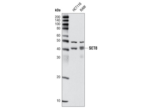
| SET8 (C18B7) Rabbit mAb | 2996 | 20 µl | 43 kDa | Rabbit IgG |
| Anti-rabbit IgG, HRP-linked Antibody | 7074 | 100 µl | Goat |
Please visit cellsignal.com for individual component applications, species cross-reactivity, dilutions, protocols, and additional product information.
Description
The Lysine Methyltransferase Antibody Sampler Kit provides a fast and economical means to evaluate endogenous levels of lysine methyltransferases. The kit contains enough primary antibody to perform two western blot experiments per primary antibody.
Storage
Background
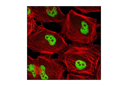
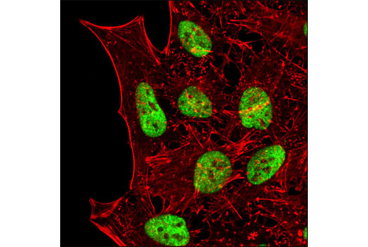
SET domain-containing proteins are potential histone methyltransferases (HMTases), which are classified into subgroups by their putative substrate specificities. Histone H3 Lys9 (H3-K9) methyltransferase group genes include Suv39h1, Suv39h2, G9a, G9a related protein (GLP) and SETDB1/ESET (1). The H3-K9 methylation mark plays an important role as a binding site for the chromo-containing protein, resulting in chromatin compaction and heterochromatin generation (2). Histone H3-K4 methylation is exclusively associated with actively transcribed genes (2). The first H3-K4 methylase complex, COMPASS, was identified in the yeast S. cerevisiae and consists of Set1/KMT2 and seven other polypeptides, Cps60-Cps15 (2). Set1/KMT2 functions within COMPASS and is capable of mono-, di-, and trimethylating H3-K4 (2). There are several Set1 related proteins in mammals including WDR5, RBBP5, ASH2L, CXXC1, and DPY30 (2,3). SET7/SET9 is a member of the SET domain-containing family that can specifically methylate H3-K4, Lys189 of the TAF10, a member of the TFIID transcription factor complex, and Lys372 of the p53 tumor suppressor protein (4-6). SET domain-containing lysine methyltransferase 8 (SET8), also known as PR/SET domain-containing protein 7 (PR/SET7), is a single-subunit enzyme that mono-methylates histone H4-K20, preferably on nucleosomal substrates (7-9). SET and MYND domain-containing protein 2 (SMYD2), also known as lysine methyltransferase protein 3C (KMT3C), functions to repress transcription by interacting with the Sin3A repressor complex and methylating H3-K36 (10). SMYD2 also methylates H3-K4 through interaction with HSP90α, and methylates p53 at Lys370 to repress p53-mediated transcriptional activation and apoptosis (11,12).
- Tachibana, M. et al. (2005) Genes Dev 19, 815-26.
- Shilatifard, A. (2008) Curr Opin Cell Biol 20, 341-8.
- Lee, J.H. et al. (2007) J Biol Chem 282, 13419-28.
- Nishioka, K. et al. (2002) Genes Dev 16, 479-89.
- Kouskouti, A. et al. (2004) Mol Cell 14, 175-82.
- Chuikov, S. et al. (2004) Nature 432, 353-60.
- Fang, J. et al. (2002) Curr Biol 12, 1086-99.
- Xiao, B. et al. (2005) Genes Dev 19, 1444-54.
- Couture, J.F. et al. (2005) Genes Dev 19, 1455-65.
- Brown, M.A. et al. (2006) Mol Cancer 5, 26.
- Abu-Farha, M. et al. (2008) Mol Cell Proteomics 7, 560-72.
- Huang, J. et al. (2006) Nature 444, 629-32.
Background References
Trademarks and Patents
使用に関する制限
法的な権限を与えられたCSTの担当者が署名した書面によって別途明示的に合意された場合を除き、 CST、その関連会社または代理店が提供する製品には以下の条件が適用されます。お客様が定める条件でここに定められた条件に含まれるものを超えるもの、 または、ここに定められた条件と異なるものは、法的な権限を与えられたCSTの担当者が別途書面にて受諾した場合を除き、拒絶され、 いかなる効力も効果も有しません。
研究専用 (For Research Use Only) またはこれに類似する表示がされた製品は、 いかなる目的についても FDA または外国もしくは国内のその他の規制機関により承認、認可または許可を受けていません。 お客様は製品を診断もしくは治療目的で使用してはならず、また、製品に表示された内容に違反する方法で使用してはなりません。 CST が販売または使用許諾する製品は、エンドユーザーであるお客様に対し、使途を研究および開発のみに限定して提供されるものです。 診断、予防もしくは治療目的で製品を使用することまたは製品を再販売 (単独であるか他の製品等の一部であるかを問いません) もしくはその他の商業的利用の目的で購入することについては、CST から別途許諾を得る必要があります。 お客様は以下の事項を遵守しなければなりません。(a) CST の製品 (単独であるか他の資材と一緒であるかを問いません) を販売、使用許諾、貸与、寄付もしくはその他の態様で第三者に譲渡したり使用させたりしてはなりません。また、商用の製品を製造するために CST の製品を使用してはなりません。(b) 複製、改変、リバースエンジニアリング、逆コンパイル、 分解または他の方法により製品の構造または技術を解明しようとしてはなりません。また、 CST の製品またはサービスと競合する製品またはサービスを開発する目的で CST の製品を使用してはなりません。(c) CST の製品の商標、商号、ロゴ、特許または著作権に関する通知または表示を除去したり改変したりしてはなりません。(d) CST の製品をCST 製品販売条件(CST’s Product Terms of Sale) および該当する書面のみに従って使用しなければなりません。(e) CST の製品に関連してお客様が使用する第三者の製品またはサービスに関する使用許諾条件、 サービス提供条件またはこれに類する合意事項を遵守しなければなりません。