| Product Includes | Product # | Quantity | Mol. Wt | Isotype/Source |
|---|---|---|---|---|
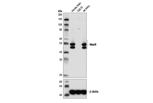
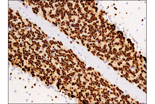
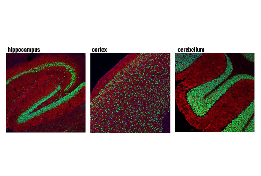
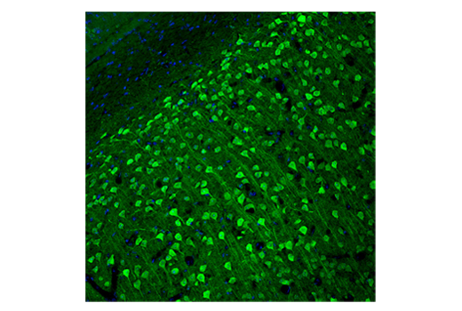
| NeuN (D4G4O) XP® Rabbit mAb | 24307 | 20 µl | 46-55 kDa | Rabbit IgG |
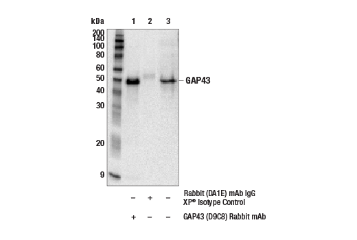
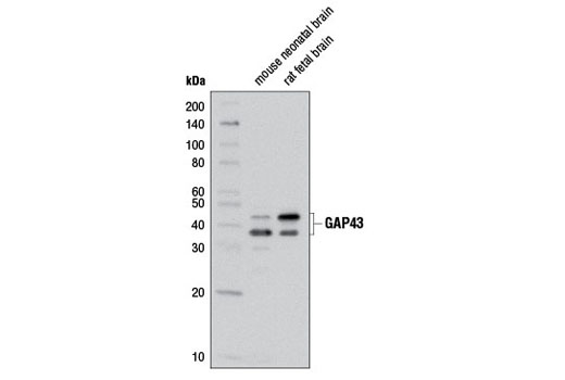
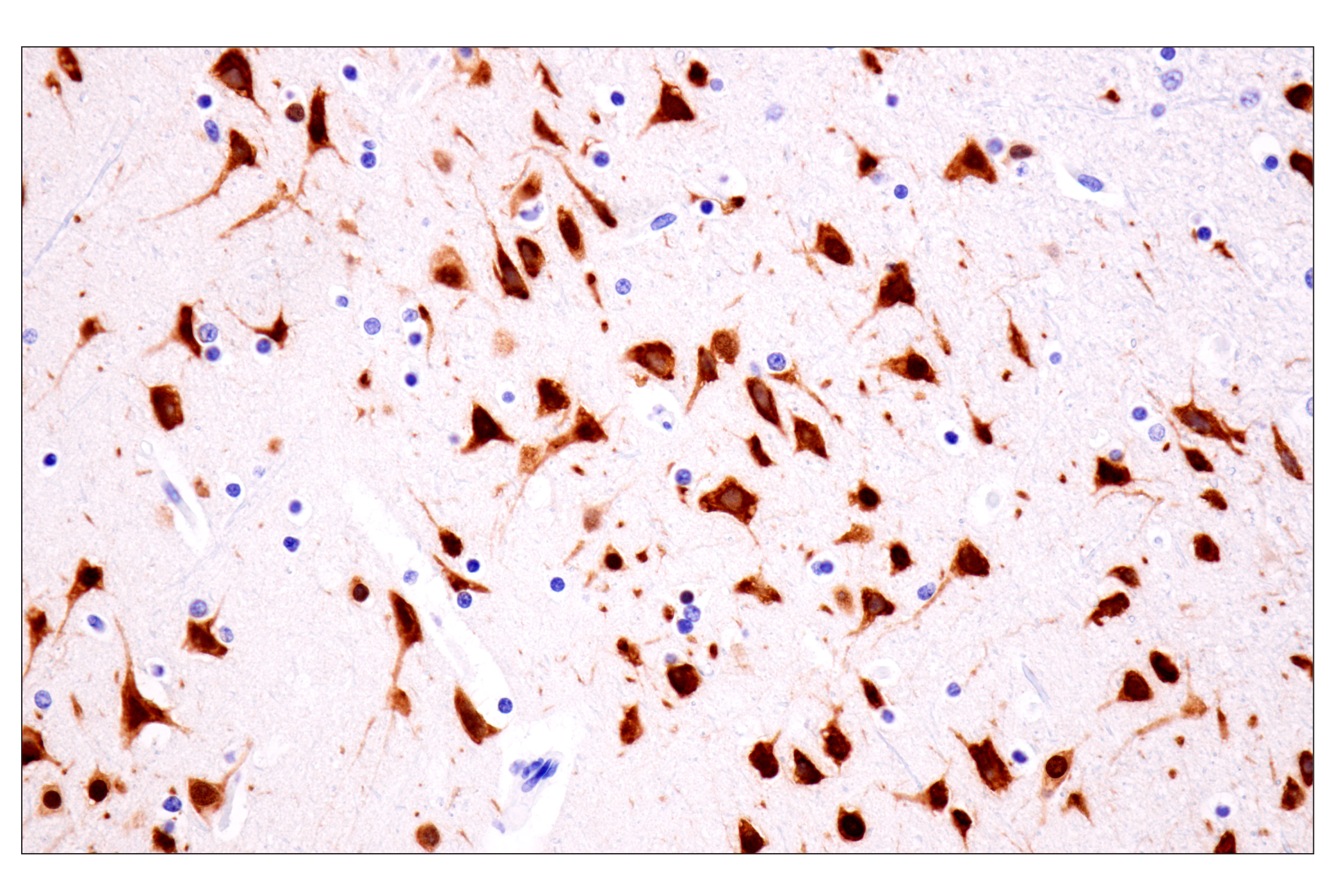
| GAP43 (D9C8) Rabbit mAb | 8945 | 20 µl | 38, 43 kDa | Rabbit IgG |
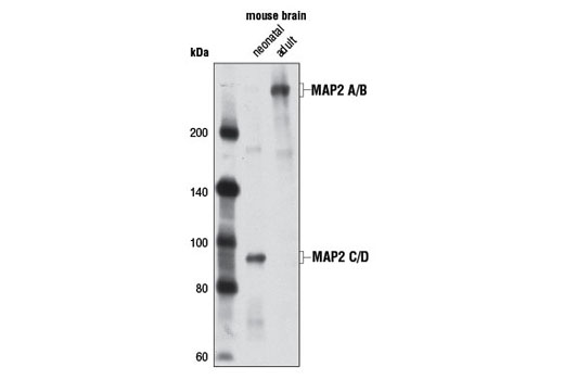
| MAP2 (D5G1) XP® Rabbit mAb | 8707 | 20 µl | 75, 82, 280 kDa | Rabbit IgG |
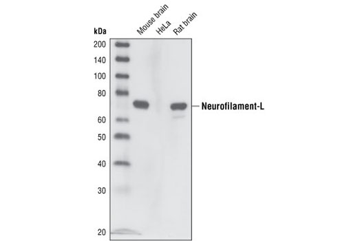
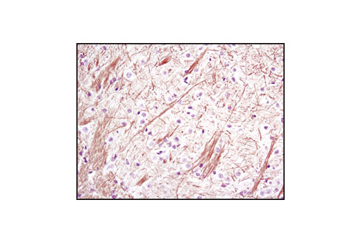
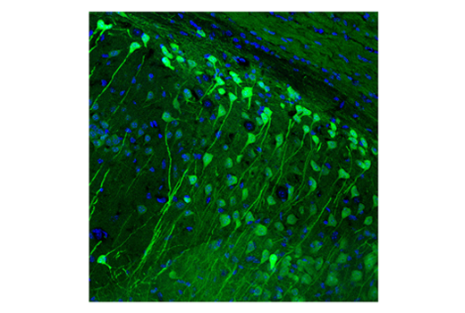
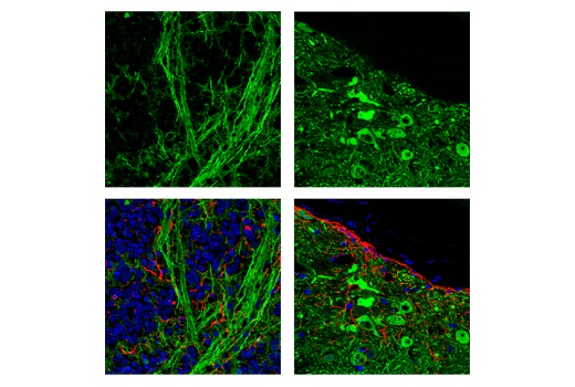
| Neurofilament-L (C28E10) Rabbit mAb | 2837 | 20 µl | 70 kDa | Rabbit IgG |
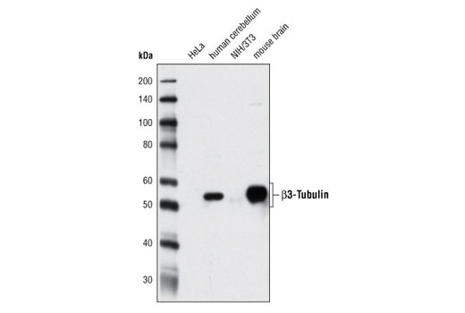
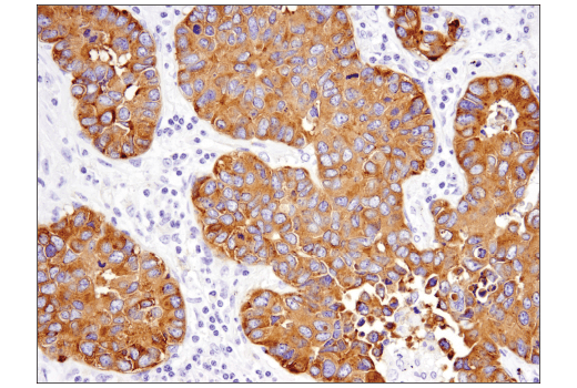
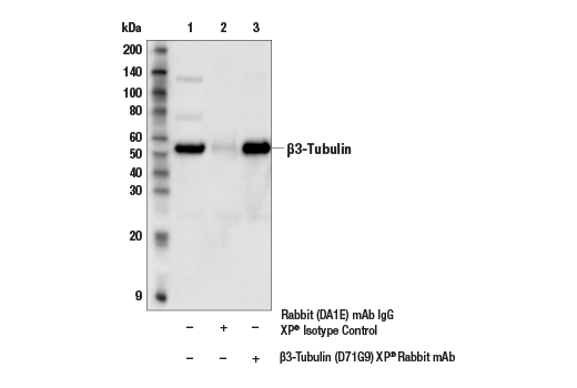
| β3-Tubulin (D71G9) XP® Rabbit mAb | 5568 | 20 µl | 55 kDa | Rabbit IgG |
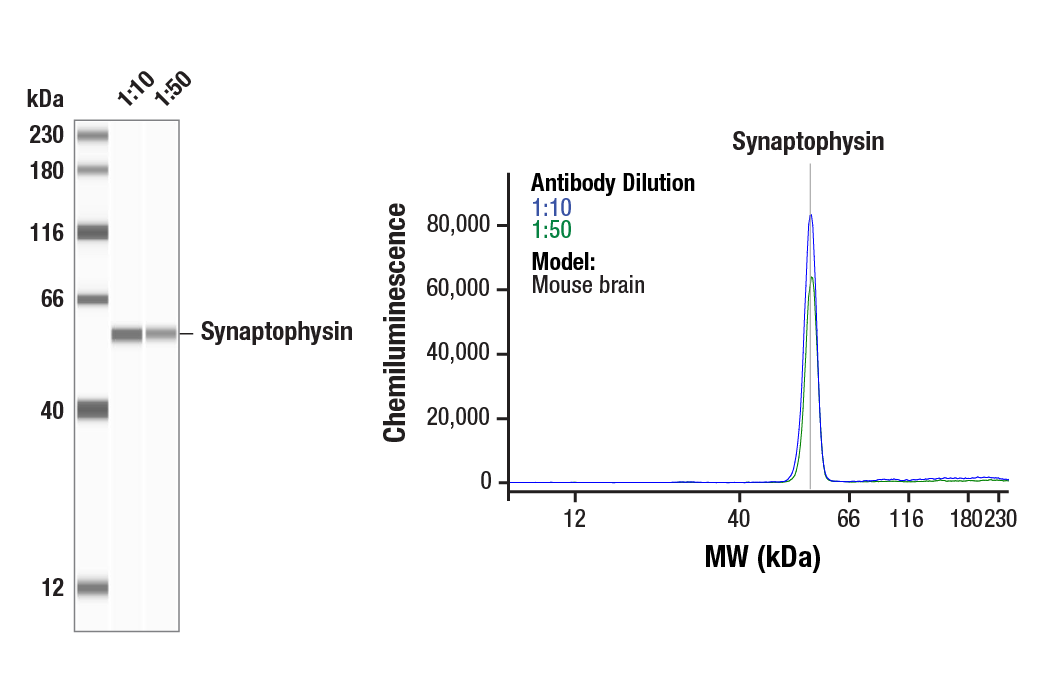
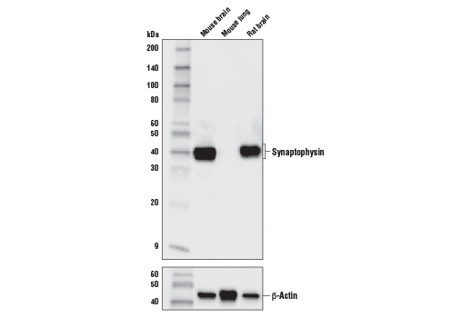
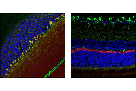
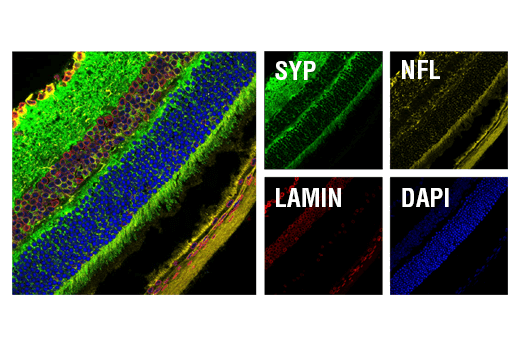
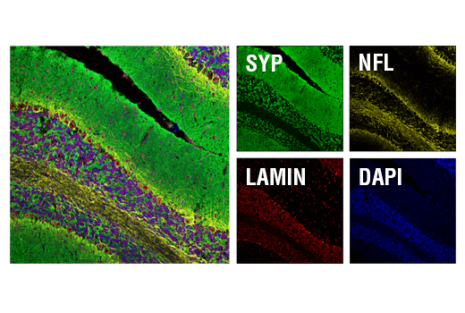
| Synaptophysin (D8F6H) XP® Rabbit mAb | 36406 | 20 µl | 38 kDa | Rabbit IgG |
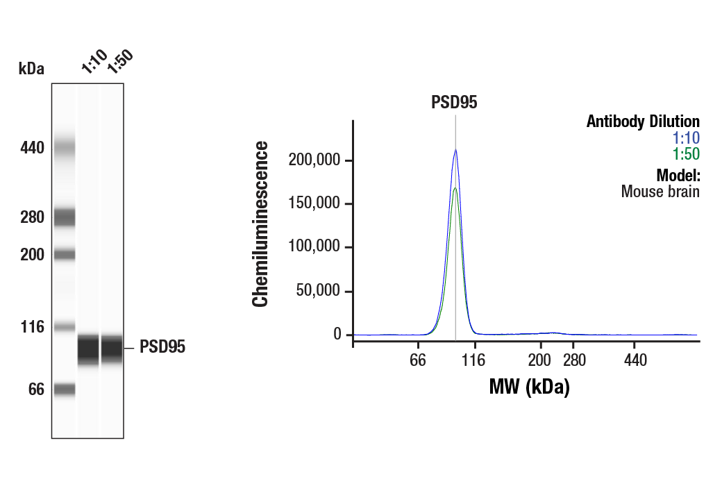
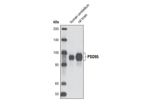
| PSD95 (D27E11) XP® Rabbit mAb | 3450 | 20 µl | 95 kDa | Rabbit IgG |
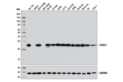
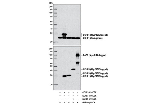
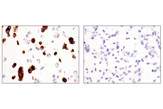
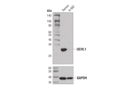
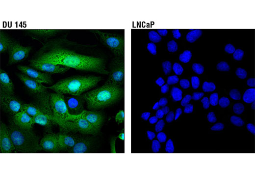
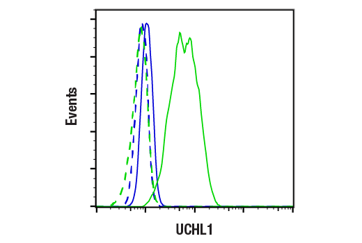
| UCHL1 (D3T2E) XP® Rabbit mAb | 13179 | 20 µl | 27 kDa | Rabbit IgG |
| Anti-rabbit IgG, HRP-linked Antibody | 7074 | 100 µl | Goat |
Please visit cellsignal.com for individual component applications, species cross-reactivity, dilutions, protocols, and additional product information.
Description
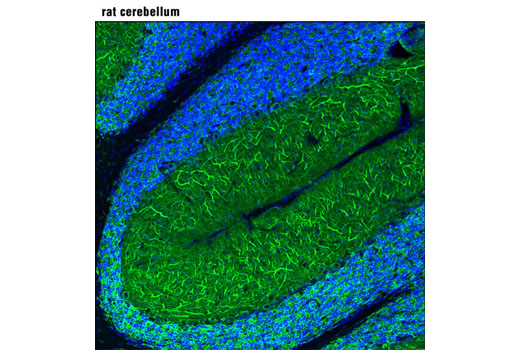
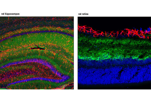
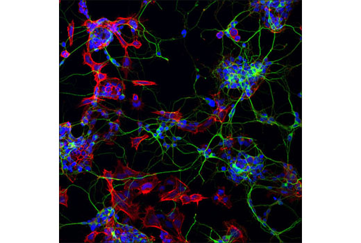
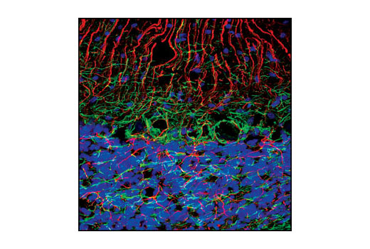
The Mature Neuron Marker Antibody Sampler Kit provides an economical means for detecting mature neuron proteins by western and labeling mature neuronal structures by immunofluorescence (IF). This kit includes enough primary antibodies to perform two western blot experiments or at least forty IF tests per primary antibody.
Storage
Background
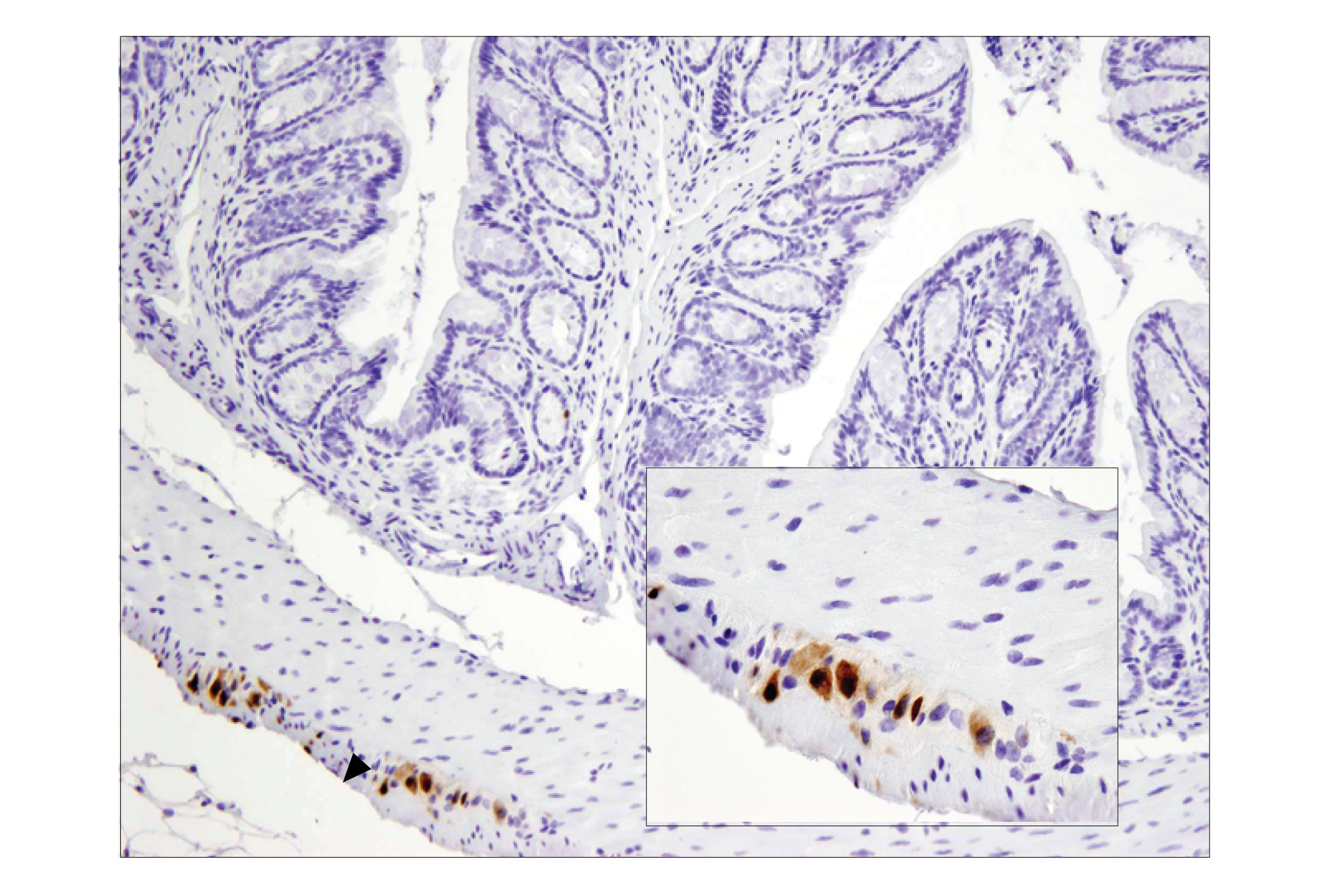
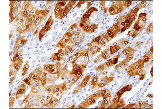
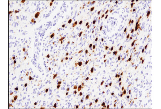
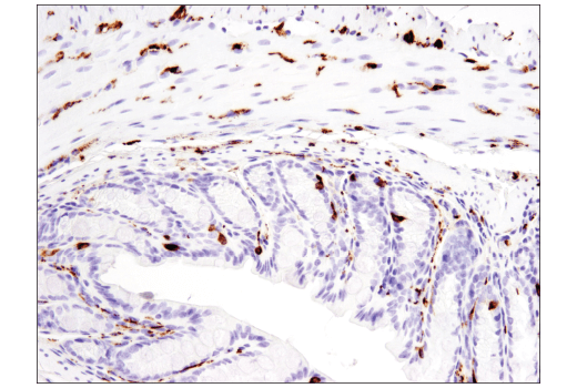
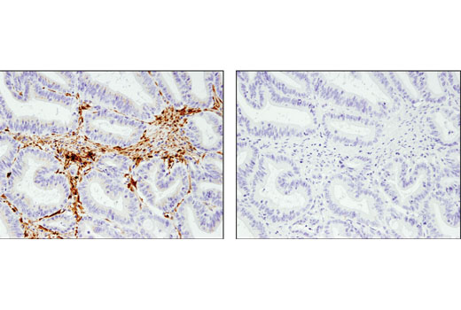
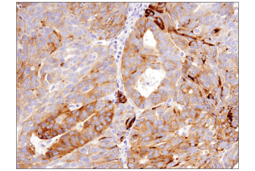
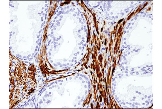
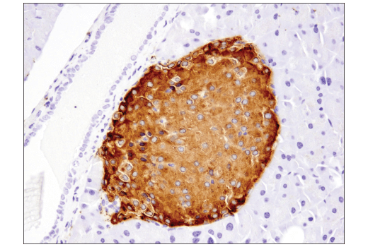
The antibodies in this kit serve to characterize and identify mature neurons. Neural stem cells differentiate into mature post-mitotic neurons that are incapable of cellular division. Several neuron-enriched markers can be used to identify mature neurons. Neuronal nuclei (NeuN, Fox-3, RBFOX3) is a nuclear protein expressed in most post-mitotic neurons of the central and peripheral nervous systems. NeuN is not detected in Purkinje cells, sympathetic ganglion cells, Cajal-Retzius cells, INL retinal cells, inferior olivary, or dentate nucleus neurons (1). This neuronal protein was originally identified by immunoreactivity with a monoclonal antibody also called NeuN. Using MS-analysis, NeuN was later identified as the Fox-3 gene product, which contains an RNA recognition motif and functions as a splicing regulator (2). As neurons mature, they develop elaborate processes like axons and dendrites that are necessary to drive core neuronal functions, including synaptic transmission.
GAP43 is a nervous system specific, growth-associated protein enriched in growth cones and areas of high plasticity (3). GAP43 is integral to growth cone formation, neurite outgrowth, and the development of a functional cerebral cortex (4). The cytoskeleton, which is important in generating neuronal processes, consists of three types of cytosolic fibers: actin microfilaments, intermediate filaments, and microtubules. β3-tubulin is one of six β-tubulin isoforms that make up the building blocks of microtubules (5). Microtubule-associated protein 2 (MAP2) is a neuronal phosphoprotein that regulates the structure and stability of microtubules, neuronal morphogenesis, cytoskeleton dynamics, and organelle trafficking in axons and dendrites (6). MAP2 is preferentially localized to dendrites in cultured neurons (7). Neurofilaments are the major intermediate filaments found in neurons and consist of light (NFL), medium (NFM), and heavy (NFH) subunits (8). Similar in structure to other intermediate filament proteins, neurofilaments have a globular amino-terminal head, a central α-helical rod domain, and a carboxy-terminal tail. A heterotetrameric unit (NFL-NFM and NFL-NFH) forms a protofilament, with eight protofilaments comprising the typical 10 nm intermediate filament (9). Neurofilaments are critical for radial axon growth and determine axon caliber, serving as markers for neuronal axons.
Mature neurons function as cellular mediators of synaptic transmission. Synaptophysin is a neuronal synaptic vesicle glycoprotein (10). Synaptophysin is responsible for targeting synaptobrevin 2/VAMP2 to synaptic vesicles, and is a critical component and marker for the presynaptic fusion complex (11). Postsynaptic Density protein 95 (PSD95) is a member of the membrane-associated guanylate kinase (MAGUK) family of proteins. These family members consist of an amino-terminal variable segment followed by three PDZ domains, an SH3 domain, and an inactive guanylate kinase (GK) domain. PSD95 is a scaffolding protein involved in the assembly and function of mature postsynaptic density complexes (12,13).
Several cellular processes are required to support dynamic functions existing in mature neurons, including protein regulation by protein ubiquitination. Ubiquitin C-terminal hydrolase L1 (UCH-L1) is a deubiquitinating enzyme that is selectively and abundantly expressed in the brain, and its activity is required for normal synaptic function (14).
- Mullen, R.J. et al. (1992) Development 116, 201-11.
- Kim, K.K. et al. (2009) J Biol Chem 284, 31052-61.
- Biewenga, J.E. et al. (1996) Acta Biochim Pol 43, 327-38.
- Aigner, L. and Caroni, P. (1993) J Cell Biol 123, 417-29.
- Jiang, Y.Q. and Oblinger, M.M. (1992) J Cell Sci 103 (Pt 3), 643-51.
- Sánchez, C. et al. (2000) Prog Neurobiol 61, 133-68.
- Caceres, A. et al. (1984) Brain Res 315, 314-8.
- Al-Chalabi, A. and Miller, C.C. (2003) Bioessays 25, 346-55.
- Cohlberg, J.A. et al. (1995) J Biol Chem 270, 9334-9.
- Wiedenmann, B. and Franke, W.W. (1985) Cell 41, 1017-28.
- Bonanomi, D. et al. (2007) Biochem J 404, 525-34.
- Cao, J. et al. (2005) J Cell Biol 168, 117-26.
- Chetkovich, D.M. et al. (2002) J Neurosci 22, 6415-25.
- Gong, B. et al. (2006) Cell 126, 775-88.
Background References
Trademarks and Patents
使用に関する制限
法的な権限を与えられたCSTの担当者が署名した書面によって別途明示的に合意された場合を除き、 CST、その関連会社または代理店が提供する製品には以下の条件が適用されます。お客様が定める条件でここに定められた条件に含まれるものを超えるもの、 または、ここに定められた条件と異なるものは、法的な権限を与えられたCSTの担当者が別途書面にて受諾した場合を除き、拒絶され、 いかなる効力も効果も有しません。
研究専用 (For Research Use Only) またはこれに類似する表示がされた製品は、 いかなる目的についても FDA または外国もしくは国内のその他の規制機関により承認、認可または許可を受けていません。 お客様は製品を診断もしくは治療目的で使用してはならず、また、製品に表示された内容に違反する方法で使用してはなりません。 CST が販売または使用許諾する製品は、エンドユーザーであるお客様に対し、使途を研究および開発のみに限定して提供されるものです。 診断、予防もしくは治療目的で製品を使用することまたは製品を再販売 (単独であるか他の製品等の一部であるかを問いません) もしくはその他の商業的利用の目的で購入することについては、CST から別途許諾を得る必要があります。 お客様は以下の事項を遵守しなければなりません。(a) CST の製品 (単独であるか他の資材と一緒であるかを問いません) を販売、使用許諾、貸与、寄付もしくはその他の態様で第三者に譲渡したり使用させたりしてはなりません。また、商用の製品を製造するために CST の製品を使用してはなりません。(b) 複製、改変、リバースエンジニアリング、逆コンパイル、 分解または他の方法により製品の構造または技術を解明しようとしてはなりません。また、 CST の製品またはサービスと競合する製品またはサービスを開発する目的で CST の製品を使用してはなりません。(c) CST の製品の商標、商号、ロゴ、特許または著作権に関する通知または表示を除去したり改変したりしてはなりません。(d) CST の製品をCST 製品販売条件(CST’s Product Terms of Sale) および該当する書面のみに従って使用しなければなりません。(e) CST の製品に関連してお客様が使用する第三者の製品またはサービスに関する使用許諾条件、 サービス提供条件またはこれに類する合意事項を遵守しなければなりません。