Revision 1

#53312Store at -20C

1 Kit

(4 x 20 microliters)

Cell Signaling Technology

Orders: 877-616-CELL (2355) [email protected]

Support: 877-678-TECH (8324)

Web: [email protected] cellsignal.com

3 Trask LaneDanversMassachusetts01923USA
For Research Use Only. Not for Use in Diagnostic Procedures.
Product Includes Product # Quantity Mol. Wt Isotype/Source
Tri-Methyl-Histone H3 (Lys27) (C36B11) Rabbit mAb 9733 20 µl 17 kDa Rabbit IgG
Di-Methyl-Histone H3 (Lys27) (D18C8) XP® Rabbit mAb 9728 20 µl 17 kDa Rabbit IgG
Mono-Methyl-Histone H3 (Lys27) (D3R8N) Rabbit mAb 84932 20 µl 17 kDa Rabbit IgG
Histone H3 (D1H2) XP® Rabbit mAb 4499 20 µl 17 kDa Rabbit IgG
Anti-rabbit IgG, HRP-linked Antibody 7074 100 µl Goat 

Please visit cellsignal.com for individual component applications, species cross-reactivity, dilutions, protocols, and additional product information.

Description

The Methyl-Histone H3 (Lys27) Antibody Sampler Kit provides an economical means of detecting levels of mono-, di-, and tri-methyl histone H3 Lys27 using methyl-specific and control histone H3 antibodies. The kit contains enough primary antibodies to perform at least two western blot experiments.

Storage

Supplied in 10 mM sodium HEPES (pH 7.5), 150 mM NaCl, 100 µg/ml BSA, 50% glycerol and less than 0.02% sodium azide. Store at –20°C. Do not aliquot the antibodies.

Background

The nucleosome, made up of four core histone proteins (H2A, H2B, H3, and H4), is the primary building block of chromatin. Originally thought to function as a static scaffold for DNA packaging, histones have now been shown to be dynamic proteins, undergoing multiple types of post-translational modifications, including acetylation, phosphorylation, methylation, and ubiquitination (1). Histone methylation is a major determinant for the formation of active and inactive regions of the genome and is crucial for the proper programming of the genome during development (2,3). Arginine methylation of histones H3 (Arg2, 17, 26) and H4 (Arg3) promotes transcriptional activation and is mediated by a family of protein arginine methyltransferases (PRMTs), including the co-activators PRMT1 and CARM1 (PRMT4) (4). In contrast, a more diverse set of histone lysine methyltransferases has been identified, all but one of which contain a conserved catalytic SET domain originally identified in the Drosophila Su(var)3-9, Enhancer of zeste, and Trithorax proteins. Lysine methylation occurs primarily on histones H3 (Lys4, 9, 27, 36, 79) and H4 (Lys20) and has been implicated in both transcriptional activation and silencing (4). Methylation of these lysine residues coordinates the recruitment of chromatin modifying enzymes containing methyl-lysine binding modules such as chromodomains (HP1, PRC1), PHD fingers (BPTF, ING2), tudor domains (53BP1), and WD-40 domains (WDR5) (5-8). The discovery of histone demethylases, such as PADI4, LSD1, JMJD1, JMJD2, and JHDM1, has shown that methylation is a reversible epigenetic marker (9).

Methylation of histone H3 Lys27 is generally associated with transcriptional repression and regulation of stem cell pluripotency and differentiation, through the regulation of facultative heterochromatin. Mono-, di-, and tri-methylation of histone H3 Lys27 are all mediated by polycomb repressor complex 2 (PRC2), which contains either the EZH1 or EZH2 methyltransferase proteins. Tri- and di-methyl-histone H3 Lys27 levels are highest at the promoters of polycomb-repressed genes. In addition, tri- and di-methyl-histone H3 Lys27 is found at the promoters of inactive, but transcriptionally poised genes that also contain the active tri-methyl-histone H3 Lys4 modification. Mono-methyl-histone H3 Lys27 is more widely dispersed and found in the bodies of active genes.

  1. Peterson, C.L. and Laniel, M.A. (2004) Curr Biol 14, R546-51.
  2. Kubicek, S. et al. (2006) Ernst Schering Res Found Workshop, 1-27.
  3. Lin, W. and Dent, S.Y. (2006) Curr Opin Genet Dev 16, 137-42.
  4. Lee, D.Y. et al. (2005) Endocr Rev 26, 147-70.
  5. Daniel, J.A. et al. (2005) Cell Cycle 4, 919-26.
  6. Shi, X. et al. (2006) Nature 442, 96-9.
  7. Wysocka, J. et al. (2006) Nature 442, 86-90.
  8. Wysocka, J. et al. (2005) Cell 121, 859-72.
  9. Trojer, P. and Reinberg, D. (2006) Cell 125, 213-7.

Background References

    Trademarks and Patents

    Cell Signaling Technology is a trademark of Cell Signaling Technology, Inc.
    XP is a registered trademark of Cell Signaling Technology, Inc.
    U.S. Patent No. 7,429,487, foreign equivalents, and child patents deriving therefrom.
    All other trademarks are the property of their respective owners. Visit cellsignal.com/trademarks for more information.

    使用に関する制限

    法的な権限を与えられたCSTの担当者が署名した書面によって別途明示的に合意された場合を除き、 CST、その関連会社または代理店が提供する製品には以下の条件が適用されます。お客様が定める条件でここに定められた条件に含まれるものを超えるもの、 または、ここに定められた条件と異なるものは、法的な権限を与えられたCSTの担当者が別途書面にて受諾した場合を除き、拒絶され、 いかなる効力も効果も有しません。

    研究専用 (For Research Use Only) またはこれに類似する表示がされた製品は、 いかなる目的についても FDA または外国もしくは国内のその他の規制機関により承認、認可または許可を受けていません。 お客様は製品を診断もしくは治療目的で使用してはならず、また、製品に表示された内容に違反する方法で使用してはなりません。 CST が販売または使用許諾する製品は、エンドユーザーであるお客様に対し、使途を研究および開発のみに限定して提供されるものです。 診断、予防もしくは治療目的で製品を使用することまたは製品を再販売 (単独であるか他の製品等の一部であるかを問いません) もしくはその他の商業的利用の目的で購入することについては、CST から別途許諾を得る必要があります。 お客様は以下の事項を遵守しなければなりません。(a) CST の製品 (単独であるか他の資材と一緒であるかを問いません) を販売、使用許諾、貸与、寄付もしくはその他の態様で第三者に譲渡したり使用させたりしてはなりません。また、商用の製品を製造するために CST の製品を使用してはなりません。(b) 複製、改変、リバースエンジニアリング、逆コンパイル、 分解または他の方法により製品の構造または技術を解明しようとしてはなりません。また、 CST の製品またはサービスと競合する製品またはサービスを開発する目的で CST の製品を使用してはなりません。(c) CST の製品の商標、商号、ロゴ、特許または著作権に関する通知または表示を除去したり改変したりしてはなりません。(d) CST の製品をCST 製品販売条件(CST’s Product Terms of Sale) および該当する書面のみに従って使用しなければなりません。(e) CST の製品に関連してお客様が使用する第三者の製品またはサービスに関する使用許諾条件、 サービス提供条件またはこれに類する合意事項を遵守しなければなりません。
    Revision 1
    #53312

    Methyl-Histone H3 (Lys27) Antibody Sampler Kit

    Methyl-Histone H3 (Lys27) Antibody Sampler Kit: Image 1 Expand Image
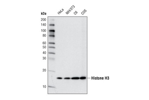
    Western blot analysis of extracts from various cell lines using Histone H3 (D1H2) XP® Rabbit mAb.
    Methyl-Histone H3 (Lys27) Antibody Sampler Kit: Image 2 Expand Image
    Immunohistochemical analysis of paraffin-embedded human breast carcinoma using Histone H3 (D1H2) XP® Rabbit mAb.
    Methyl-Histone H3 (Lys27) Antibody Sampler Kit: Image 3 Expand Image
    After the primary antibody is bound to the target protein, a complex with HRP-linked secondary antibody is formed. The LumiGLO® is added and emits light during enzyme catalyzed decomposition.
    Methyl-Histone H3 (Lys27) Antibody Sampler Kit: Image 4 Expand Image
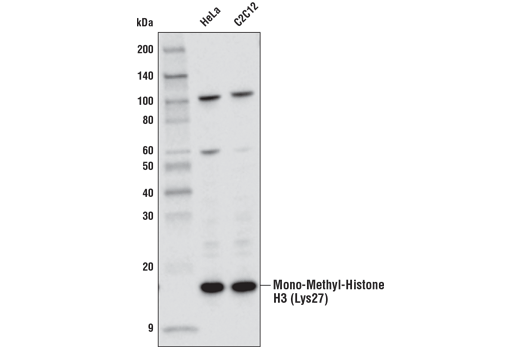
    Western blot analysis of extracts from HeLa and C2C12 cell lines using Mono-Methyl-Histone H3 (Lys27) (D3R8N) Rabbit mAb.
    Methyl-Histone H3 (Lys27) Antibody Sampler Kit: Image 5 Expand Image
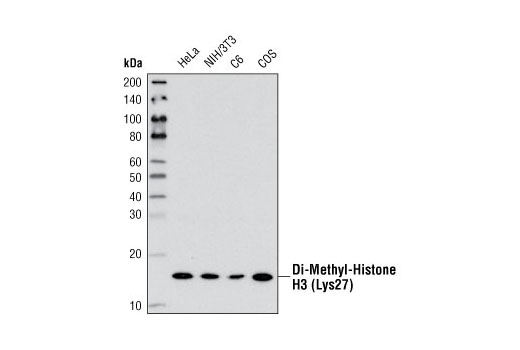
    Western blot analysis of extracts from various cell lines using Di-Methyl-Histone H3 (Lys27) (D18C8) XP® Rabbit mAb.
    Methyl-Histone H3 (Lys27) Antibody Sampler Kit: Image 6 Expand Image
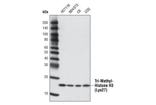
    Western blot analysis of various cell lines using Tri-Methyl-Histone H3 (Lys27) (C36B11) Rabbit mAb.
    Methyl-Histone H3 (Lys27) Antibody Sampler Kit: Image 7 Expand Image
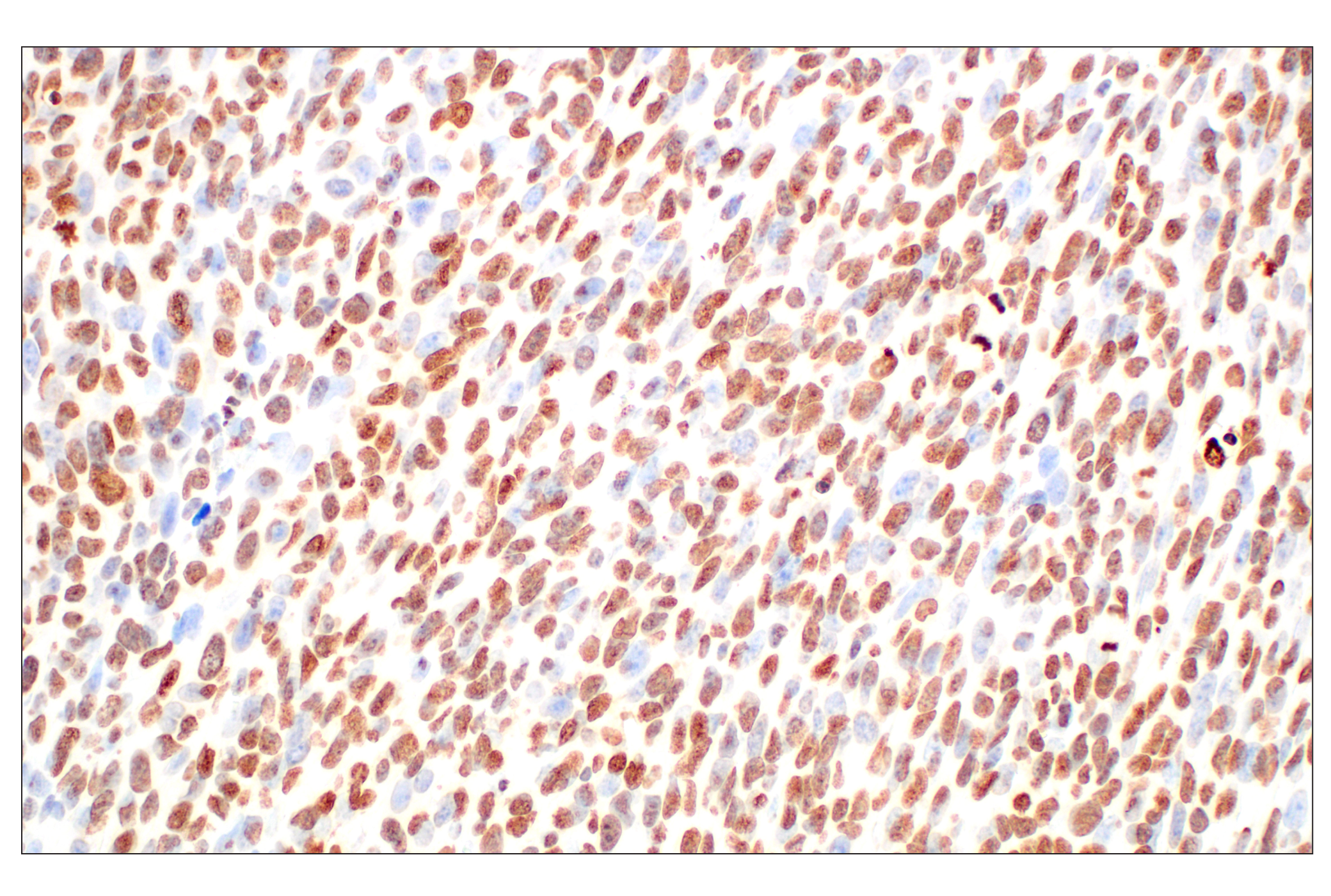
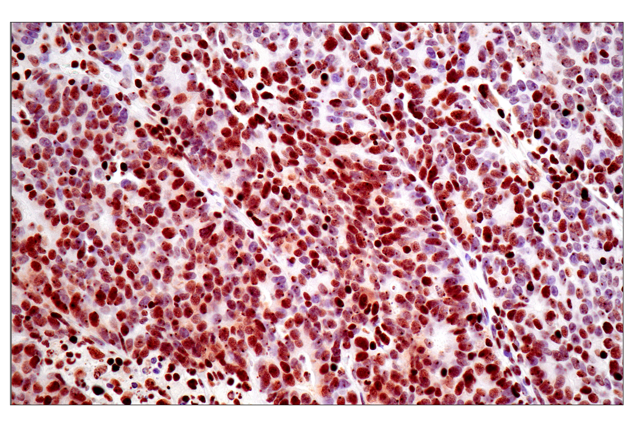
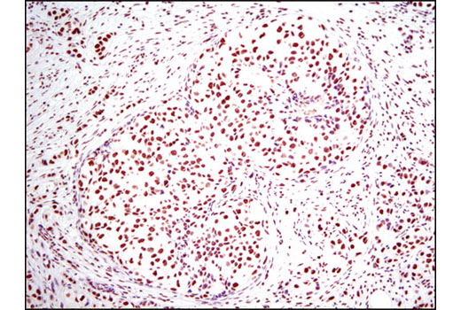
    Immunohistochemical analysis of paraffin-embedded human endometrioid adenocarcinoma using Tri-Methyl-Histone H3 (Lys27) (C36B11) Rabbit mAb performed on the Leica® BOND Rx.
    Methyl-Histone H3 (Lys27) Antibody Sampler Kit: Image 8 Expand Image
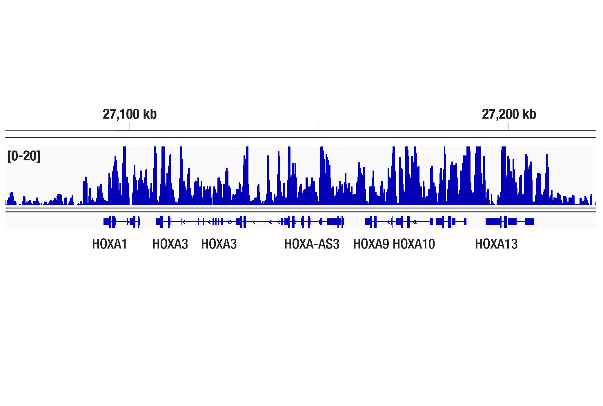
    CUT&Tag was performed with NCCIT cells and Tri-Methyl-Histone H3 (Lys27) (C36B11) Rabbit mAb, using CUT&Tag Assay Kit #77552. DNA library was prepared using CUT&Tag Dual Index Primers and PCR Master Mix for Illumina Systems #47415. The figure shows binding across the HOXA gene cluster.
    Methyl-Histone H3 (Lys27) Antibody Sampler Kit: Image 9 Expand Image
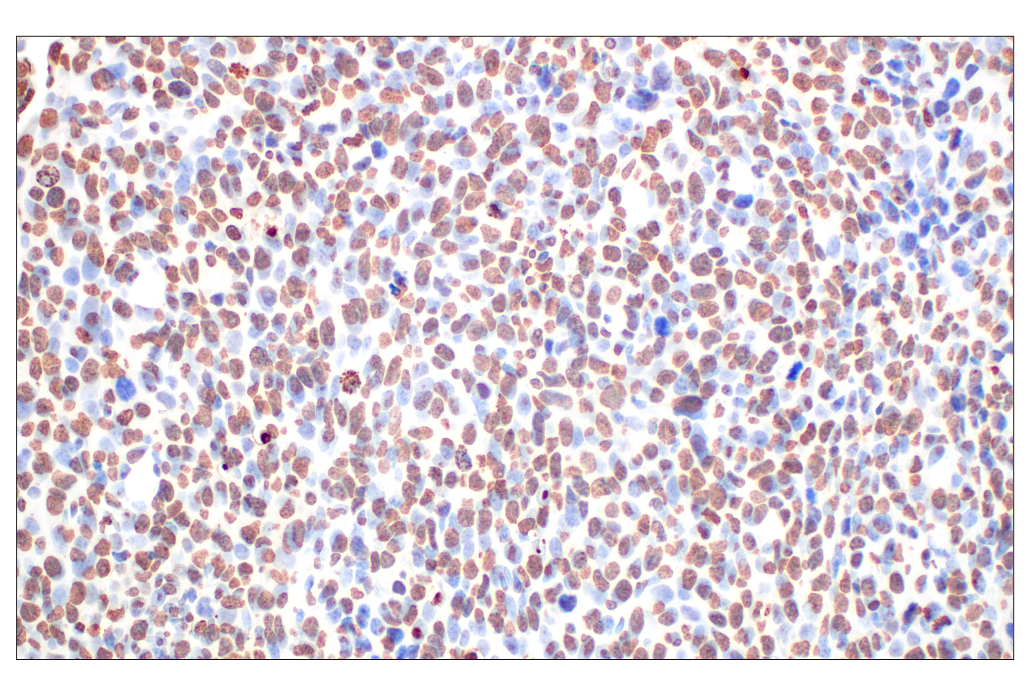
    Immunohistochemical analysis of paraffin-embedded 4T1 syngeneic mammary tumor using Histone H3 (D1H2) XP® Rabbit mAb.
    Methyl-Histone H3 (Lys27) Antibody Sampler Kit: Image 10 Expand Image
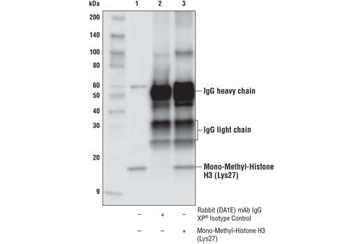
    Immunoprecipitation of mono-methyl-histone H3 Lys27 from HeLa cell extracts. Lane 1 is 10% input, lane 2 is Rabbit (DA1E) mAb IgG XP® Isotype Control #3900, and lane 3 is Mono-Methyl-Histone H3 (Lys27) (D3R8N) Rabbit mAb. Western blot analysis was performed using Mono-Methyl-Histone H3 (Lys27) (D3R8N) Rabbit mAb.
    Methyl-Histone H3 (Lys27) Antibody Sampler Kit: Image 11 Expand Image
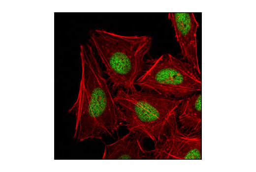
    Confocal immumunofluorescent analysis of HeLa cells using Di-Methyl-Histone H3 (Lys27) (D18C8) XP® Rabbit mAb (green). Actin filaments have been labled with DY-554 phalloidin (red).
    Methyl-Histone H3 (Lys27) Antibody Sampler Kit: Image 12 Expand Image
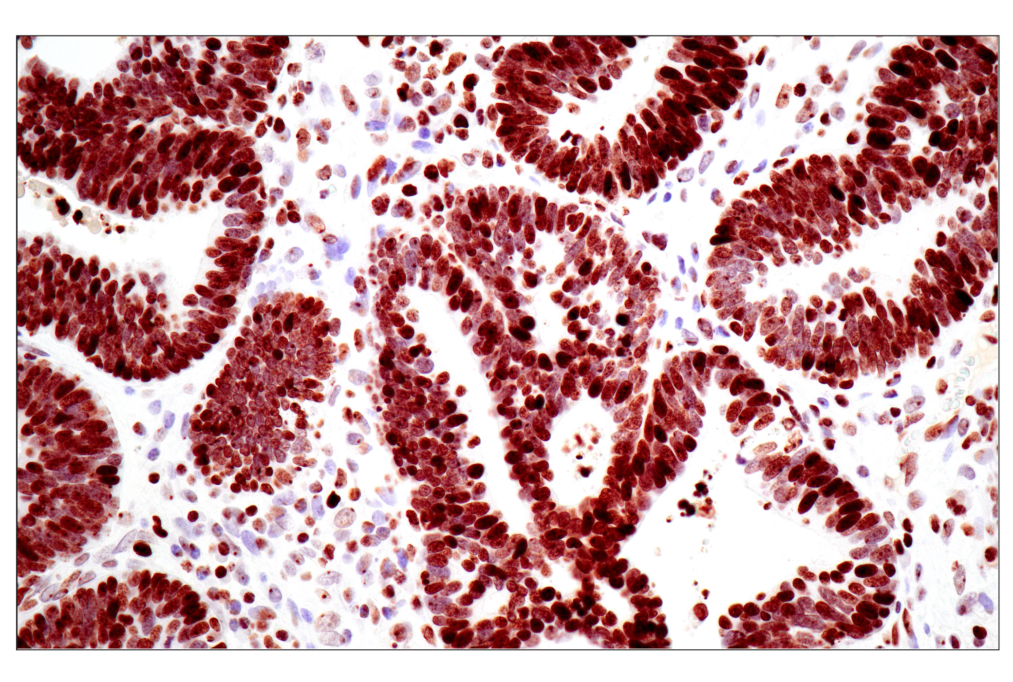
    Immunohistochemical analysis of paraffin-embedded human colon adenocarcinoma using Tri-Methyl-Histone H3 (Lys27) (C36B11) Rabbit mAb performed on the Leica® BOND Rx.
    Methyl-Histone H3 (Lys27) Antibody Sampler Kit: Image 13 Expand Image
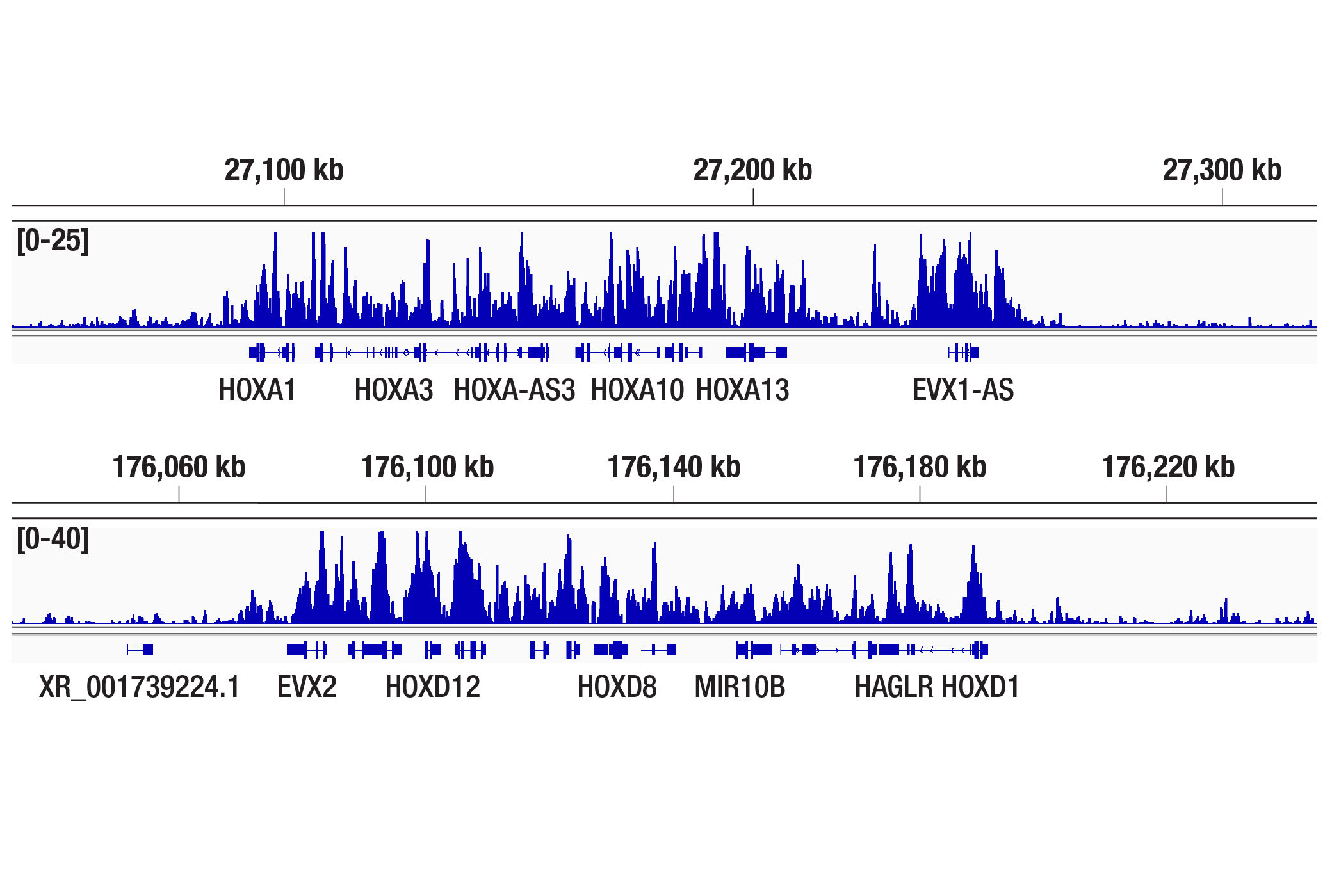
    CUT&Tag was performed with NCCIT cells and Tri-Methyl-Histone H3 (Lys27) (C36B11) Rabbit mAb, using CUT&Tag Assay Kit #77552. DNA library was prepared using CUT&Tag Dual Index Primers and PCR Master Mix for Illumina Systems #47415. The figures show binding across the HOXA gene cluster (upper), and HOXD gene cluster (lower).
    Methyl-Histone H3 (Lys27) Antibody Sampler Kit: Image 14 Expand Image
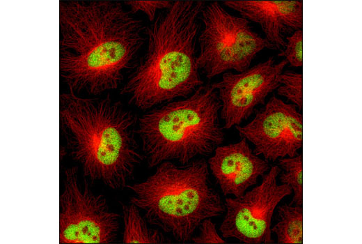
    Confocal immunofluorescent analysis of HeLa cells using Histone H3 (D1H2) XP® Rabbit mAb (green) and β-Tubulin (9F3) Rabbit mAb (Alexa Fluor® 555 Conjugate) #2116 (red).
    Methyl-Histone H3 (Lys27) Antibody Sampler Kit: Image 15 Expand Image
    Immunohistochemical analysis of paraffin-embedded LL/2 syngeneic tumor using Histone H3 (D1H2) XP® Rabbit mAb.
    Methyl-Histone H3 (Lys27) Antibody Sampler Kit: Image 16 Expand Image
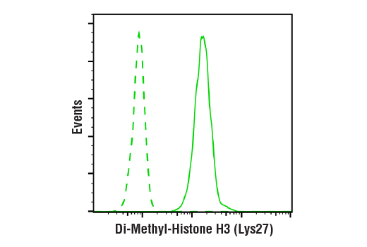
    Flow cytometric analysis of human peripheral blood mononuclear cells using Di-Methyl-Histone H3 (Lys27) (D18C8) XP® Rabbit mAb (solid line) compared to concentration-matched Rabbit (DA1E) mAb IgG XP® Isotype Control #3900 (dashed line). Anti-rabbit IgG (H+L), F(ab')2 Fragment (Alexa Fluor® 488 Conjugate) #4412 was used as a secondary antibody.
    Methyl-Histone H3 (Lys27) Antibody Sampler Kit: Image 17 Expand Image
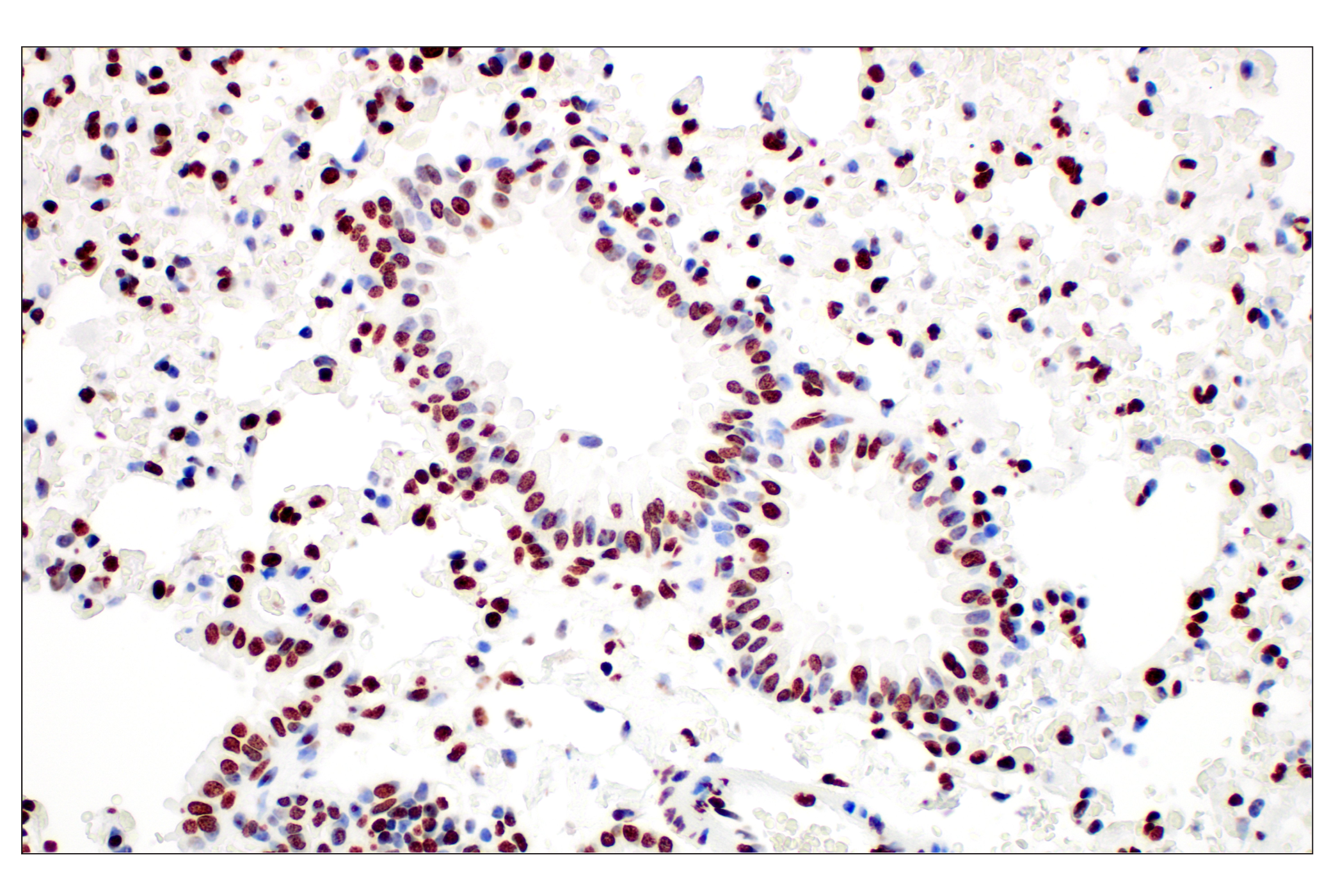
    Immunohistochemical analysis of paraffin-embedded mouse lung using Tri-Methyl-Histone H3 (Lys27) (C36B11) Rabbit mAb.
    Methyl-Histone H3 (Lys27) Antibody Sampler Kit: Image 18 Expand Image
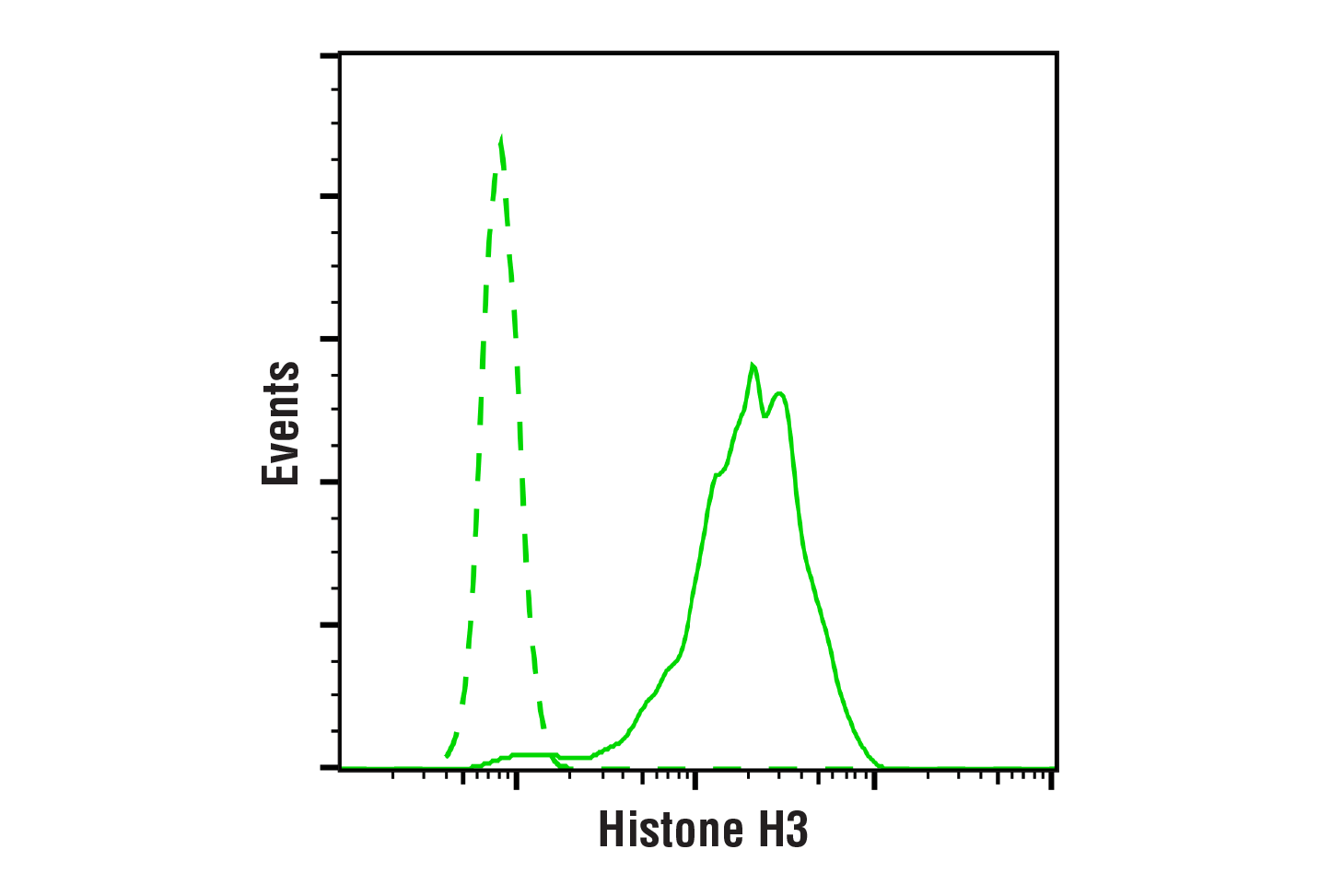
    Flow cytometric analysis of HeLa cells using Histone H3 (D1H2) XP® Rabbit mAb (solid line) or a concentration-matched Rabbit (DA1E) mAb IgG XP® Isotype Control #3900 (dashed line). Anti-rabbit IgG (H+L), F(ab')2 Fragment (Alexa Fluor® 488 Conjugate) #4412 was used as a secondary antibody.
    Methyl-Histone H3 (Lys27) Antibody Sampler Kit: Image 19 Expand Image
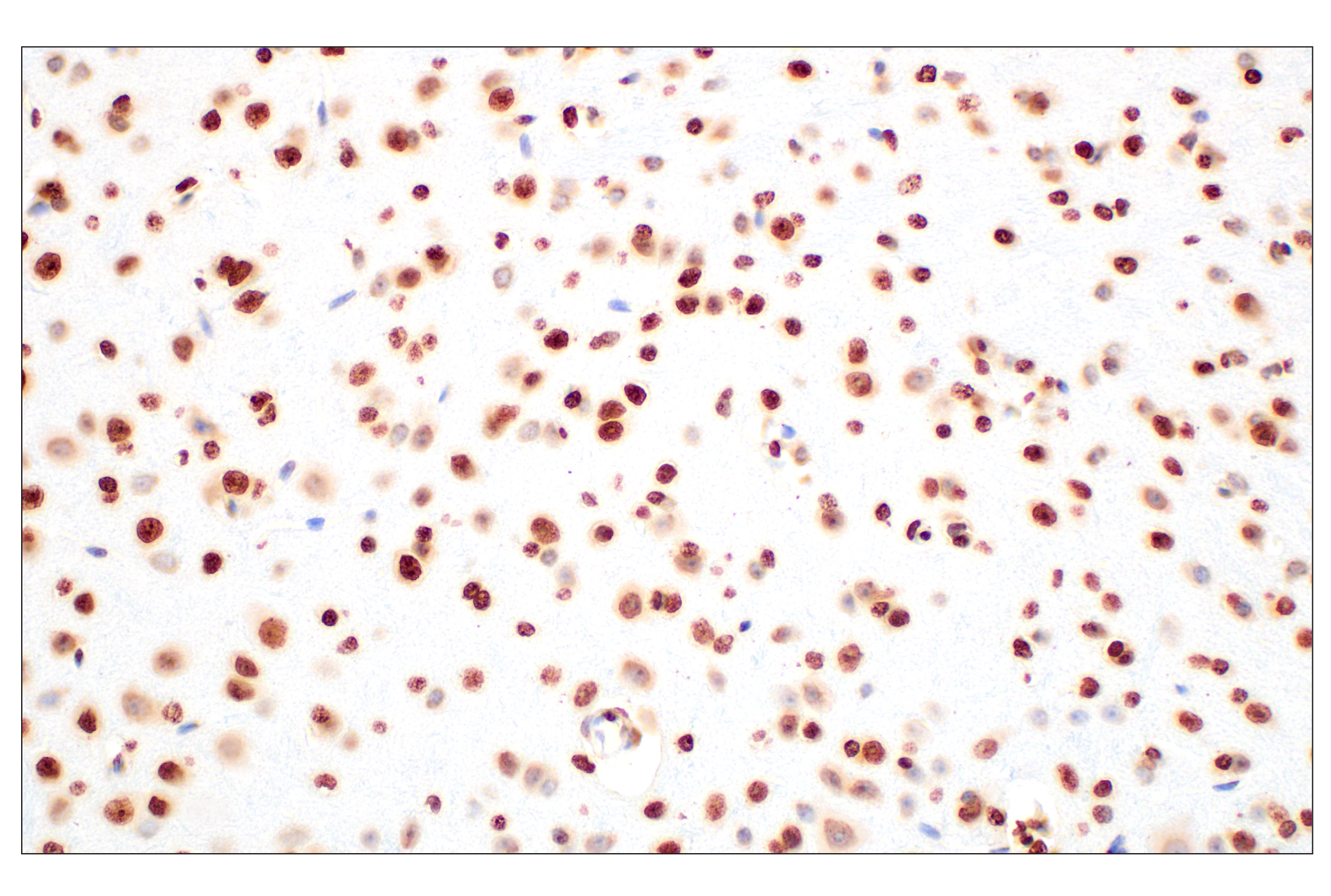
    Immunohistochemical analysis of paraffin-embedded mouse brain using Histone H3 (D1H2) XP® Rabbit mAb.
    Methyl-Histone H3 (Lys27) Antibody Sampler Kit: Image 20 Expand Image
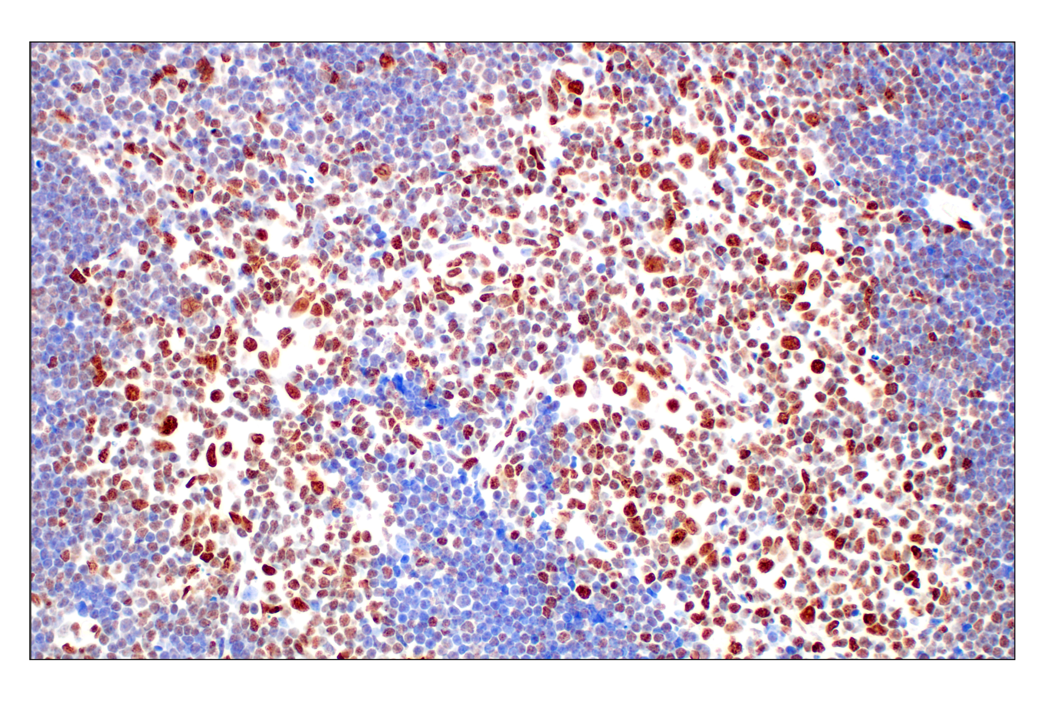
    Immunohistochemical analysis of paraffin-embedded mouse thymus using Tri-Methyl-Histone H3 (Lys27) (C36B11) Rabbit mAb.
    Methyl-Histone H3 (Lys27) Antibody Sampler Kit: Image 21 Expand Image
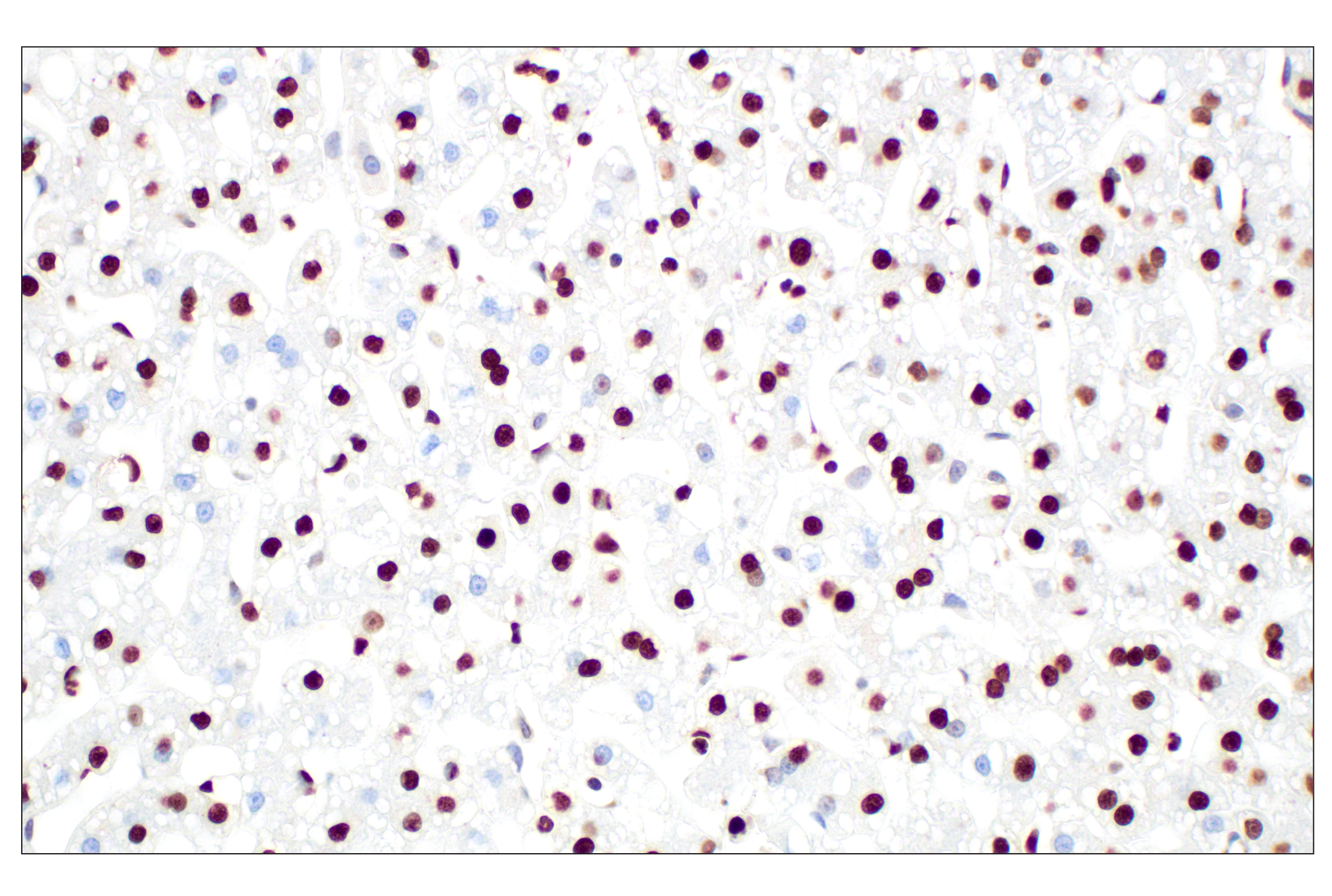
    Immunohistochemical analysis of paraffin-embedded rhesus monkey liver using Histone H3 (D1H2) XP® Rabbit mAb.
    Methyl-Histone H3 (Lys27) Antibody Sampler Kit: Image 22 Expand Image
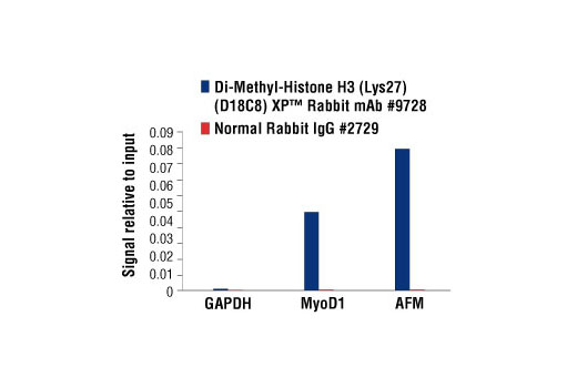
    Chromatin immunoprecipitations were performed with cross-linked chromatin from HeLa cells and either Di-Methyl-Histone H3 (Lys27) (D18C8) XP® Rabbit mAb or Normal Rabbit IgG #2729 using SimpleChIP® Enzymatic Chromatin IP Kit (Magnetic Beads) #9003. The enriched DNA was quantified by real-time PCR, using SimpleChIP® Human GAPDH Exon 1 Primers #5516, SimpleChIP® Human MyoD1 Exon 1 Primers #4490, and SimpleChIP® Human AFM Intron 1 Primers #5098. The amount of immunoprecipitated DNA in each sample is represented as signal relative to the total amount of input chromatin, which is equivalent to one.
    Methyl-Histone H3 (Lys27) Antibody Sampler Kit: Image 23 Expand Image
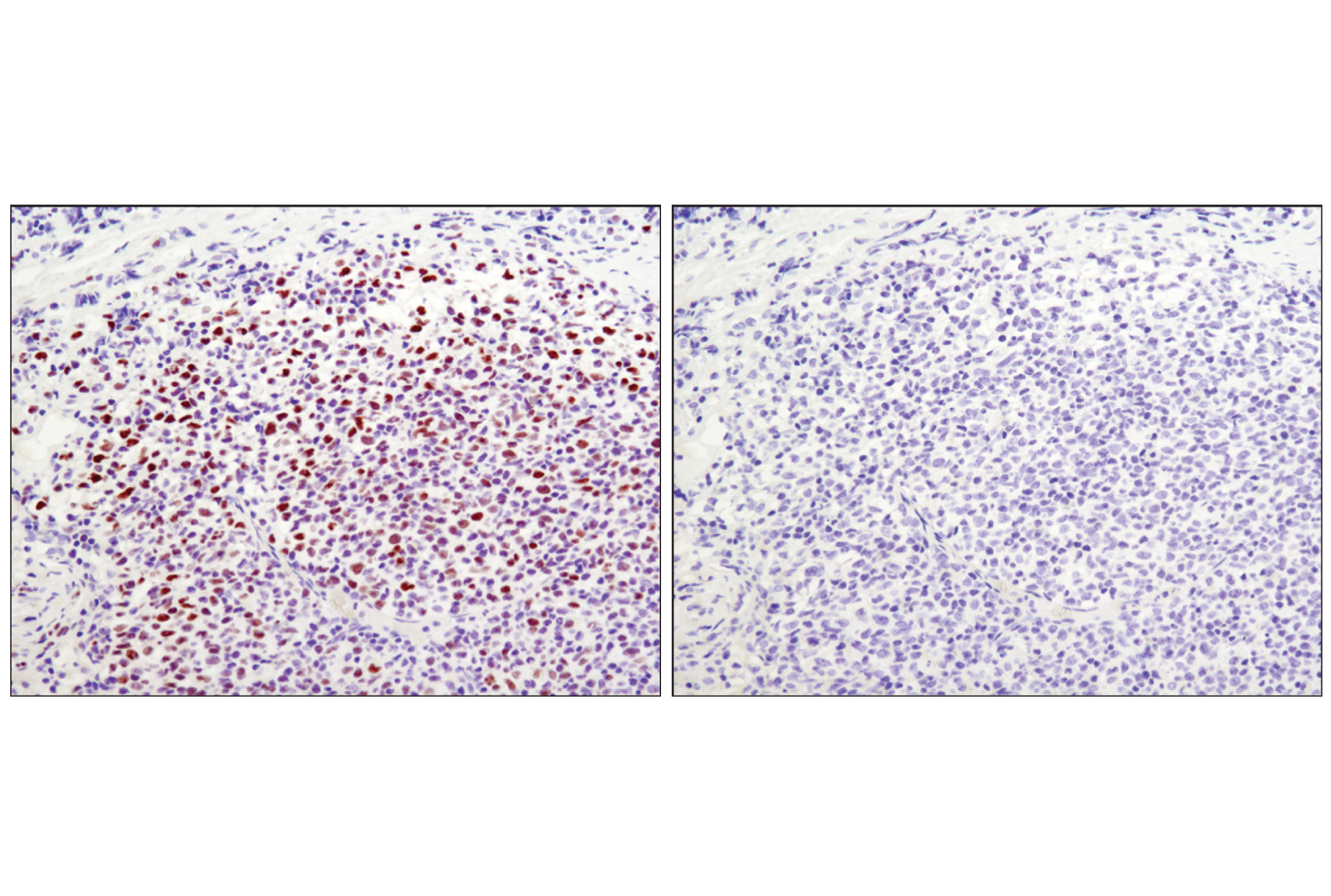
    Immunohistochemical analysis of paraffin-embedded human lymphoma using Tri-Methyl-Histone H3 (Lys27) (C36B11) Rabbit mAb in the presence of non-methyl peptide (left) or K27 tri-methyl peptide (right).
    Methyl-Histone H3 (Lys27) Antibody Sampler Kit: Image 24 Expand Image
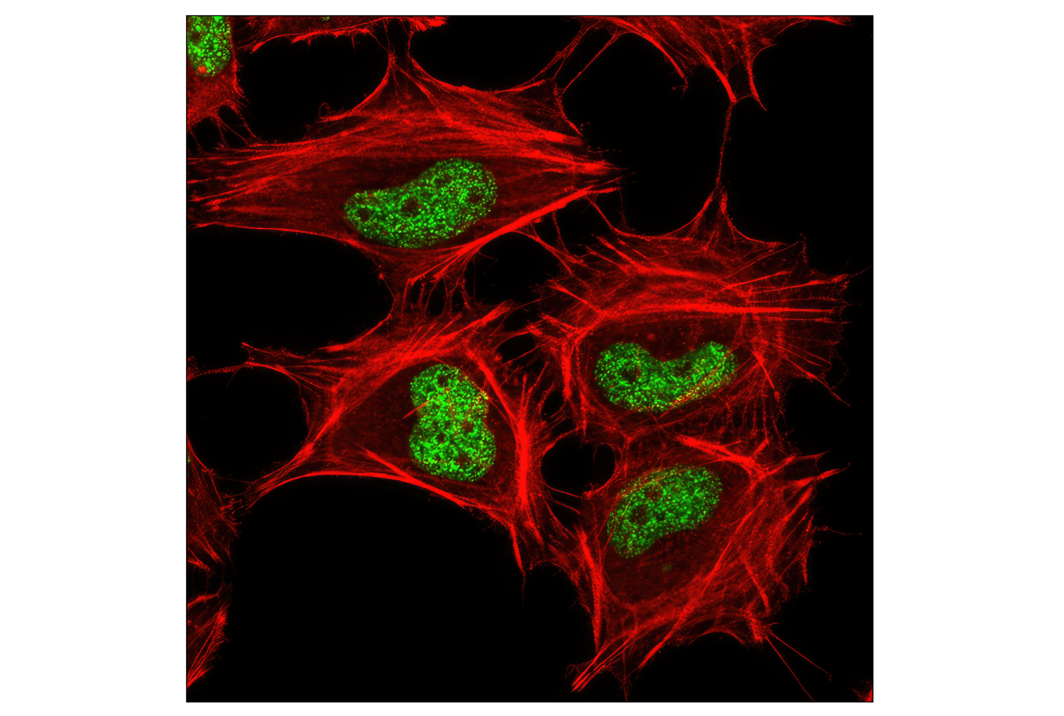
    Confocal immunofluorescent analysis of HeLa cells using Tri-Methyl-Histone H3 (Lys27) (C36B11) Rabbit mAb (green). Actin filaments have been labeled with DY-554 phalloidin (red).
    Methyl-Histone H3 (Lys27) Antibody Sampler Kit: Image 25 Expand Image
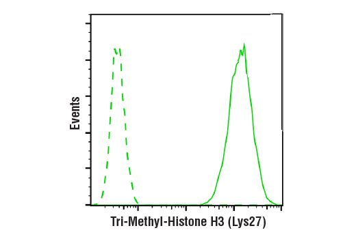
    Flow cytometric analysis of Jurkat cells using Tri-Methyl-Histone H3 (Lys27) (C36B11) Rabbit mAb (solid line) compared to concentration-matched Rabbit (DA1E) mAb IgG XP® Isotype Control #3900 (dashed line). Anti-rabbit IgG (H+L), F(ab')2 Fragment (Alexa Fluor® 488 Conjugate) #4412 was used as a secondary antibody.
    Methyl-Histone H3 (Lys27) Antibody Sampler Kit: Image 26 Expand Image
    Chromatin immunoprecipitations were performed with cross-linked chromatin from Hela cells and either Ezh2 (D2C9) XP® Rabbit mAb #5246 or Tri-Methyl-Histone H3 (Lys27) (C36B11) Rabbit mAb, using SimpleChIP® Enzymatic Chromatin IP Kit (Magnetic Beads) #9003. DNA Libraries were prepared using DNA Library Prep Kit for Illumina® (ChIP-seq, CUT&RUN) #56795. EZH2 and H3K27me3 are known to associate with each other on chromatin. The figure shows binding of both EZH2 and H3K27me3 across MYT1, a known target gene of H3K27me3 (see additional figure containing ChIP-qPCR data).
    Methyl-Histone H3 (Lys27) Antibody Sampler Kit: Image 27 Expand Image
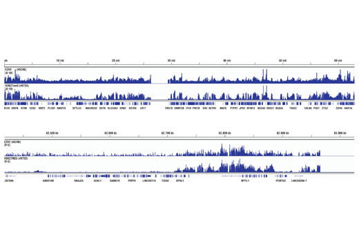
    Chromatin immunoprecipitations were performed with cross-linked chromatin from Hela cells and either Ezh2 (D2C9) XP® Rabbit mAb #5246 or Tri-Methyl-Histone H3 (Lys27) (C36B11) Rabbit mAb, using SimpleChIP® Enzymatic Chromatin IP Kit (Magnetic Beads) #9003. DNA Libraries were prepared using DNA Library Prep Kit for Illumina® (ChIP-seq, CUT&RUN) #56795. EZH2 and H3K27me3 are known to associate with each other on chromatin. The figure shows binding of both EZH2 and H3K27me3 across chromosome 20 (upper), including MYT1 (lower), a known target gene of H3K27me3 (see additional figure containing ChIP-qPCR data).
    Methyl-Histone H3 (Lys27) Antibody Sampler Kit: Image 28 Expand Image
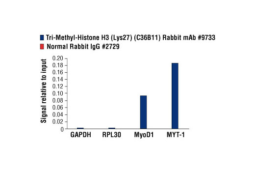
    Chromatin immunoprecipitations were performed with cross-linked chromatin from HeLa cells and either Tri-Methyl-Histone H3 (Lys27) (C36B11) Rabbit mAb, or Normal Rabbit IgG #2729, using SimpleChIP® Enzymatic Chromatin IP Kit (Magnetic Beads) #9003. The enriched DNA was quantified by real-time PCR using SimpleChIP® Human GAPDH Exon 1 Primers #5516, SimpleChIP® Human RPL30 Exon 3 Primers #7014, SimpleChIP® Human MyoD1 Exon 1 Primers #4490, and SimpleChIP® Human MYT-1 Exon 1 Primers #4493. The amount of immunoprecipitated DNA in each sample is represented as signal relative to the total amount of input chromatin, which is equivalent to one.
    Methyl-Histone H3 (Lys27) Antibody Sampler Kit: Image 29 Expand Image
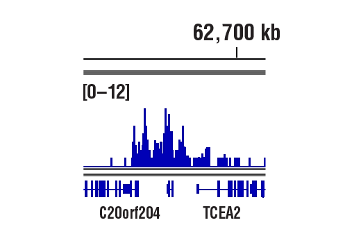
    CUT&RUN was performed with HeLa cells and Tri-Methyl-Histone H3 (Lys27) (C36B11) Rabbit mAb, using CUT&RUN Assay Kit #86652. DNA Libraries were prepared using DNA Library Prep Kit for Illumina® (ChIP-seq, CUT&RUN) #56795. The figure shows binding across TCEA2 gene.
    Methyl-Histone H3 (Lys27) Antibody Sampler Kit: Image 30 Expand Image
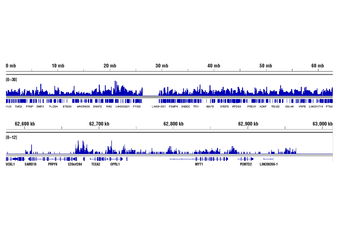
    CUT&RUN was performed with HeLa cells and Tri-Methyl-Histone H3 (Lys27) (C36B11) Rabbit mAb, using CUT&RUN Assay Kit #86652. DNA Libraries were prepared using DNA Library Prep Kit for Illumina® (ChIP-seq, CUT&RUN) #56795. The figures show binding across chromosome 20 (upper), including TCEA2 gene (lower).
    Methyl-Histone H3 (Lys27) Antibody Sampler Kit: Image 31 Expand Image
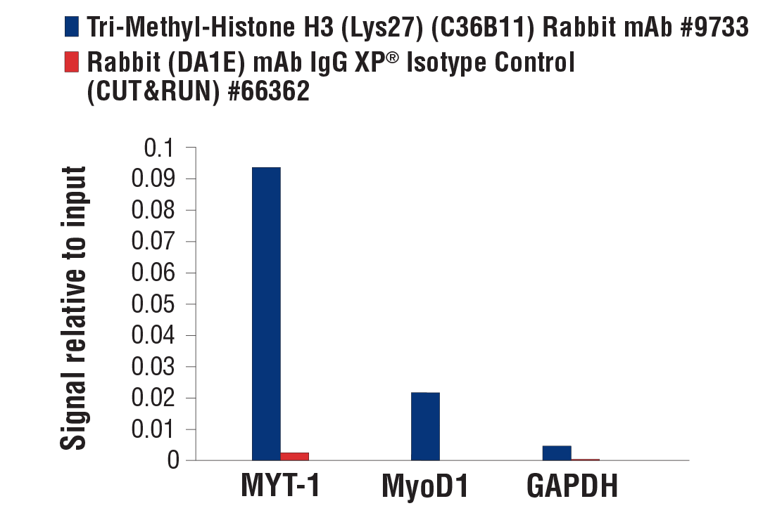
    CUT&RUN was performed with HeLa cells and either Tri-Methyl-Histone H3 (Lys27) (C36B11) Rabbit mAb or Rabbit (DA1E) mAb IgG XP® Isotype Control (CUT&RUN) #66362, using CUT&RUN Assay Kit #86652. The enriched DNA was quantified by real-time PCR using SimpleChIP® Human MYT-1 Exon 1 Primers, SimpleChIP® Human MyoD1 Exon 1 Primers, and SimpleChIP® Human GAPDH Exon 1 Primers. The amount of immunoprecipitated DNA in each sample is represented as signal relative to the total amount of input chromatin, which is equivalent to one.