| Product Includes | Product # | Quantity | Mol. Wt | Isotype/Source |
|---|---|---|---|---|
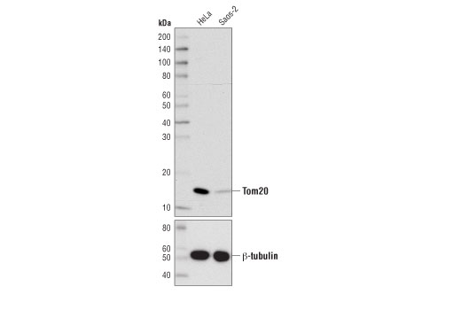
| Tom20 (D8T4N) Rabbit mAb | 42406 | 20 µl | 16 kDa | Rabbit IgG |
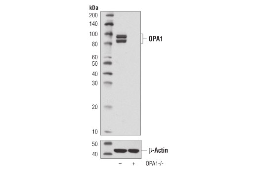
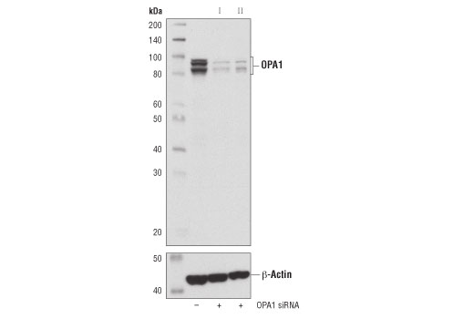
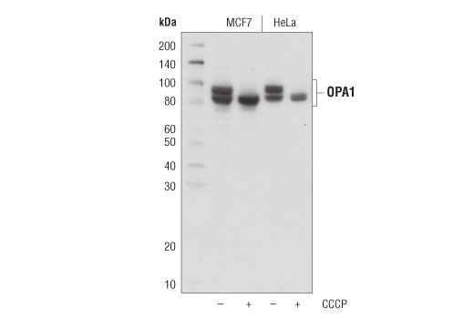
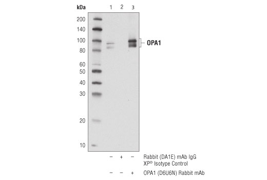
| OPA1 (D6U6N) Rabbit mAb | 80471 | 20 µl | 80-100 kDa | Rabbit IgG |
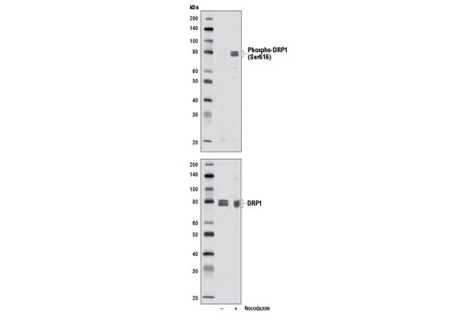
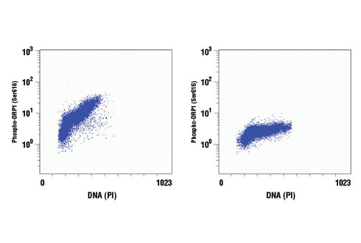
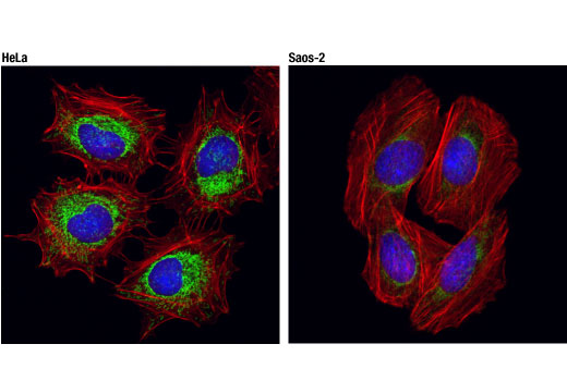
| Phospho-DRP1 (Ser616) (D9A1) Rabbit mAb | 4494 | 20 µl | 78-82 kDa | Rabbit IgG |
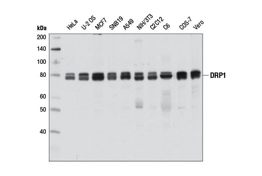
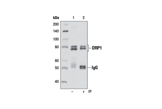
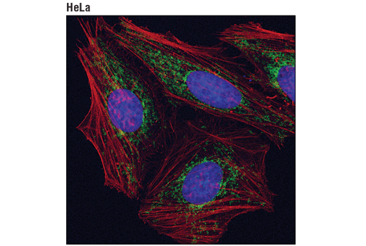
| DRP1 (D8H5) Rabbit mAb | 5391 | 20 µl | 78-82 kDa | Rabbit IgG |
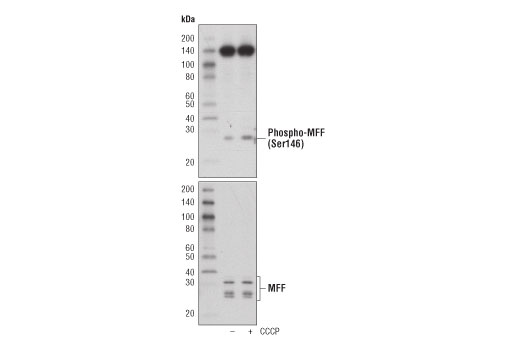
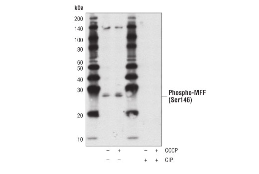
| Phospho-MFF (Ser146) Antibody | 49281 | 20 µl | 25, 27 kDa | Rabbit |
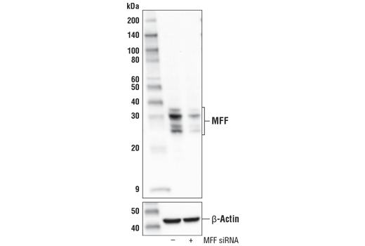
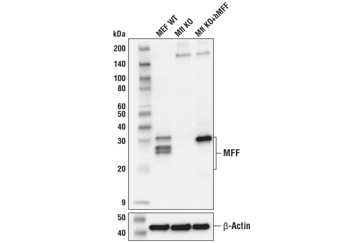
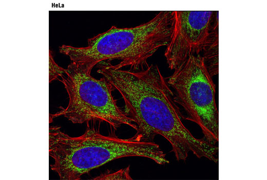
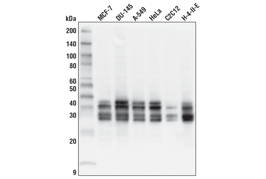
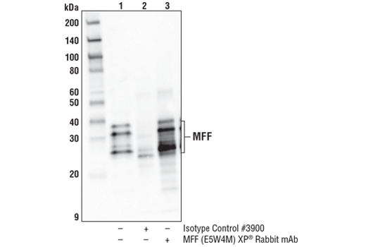
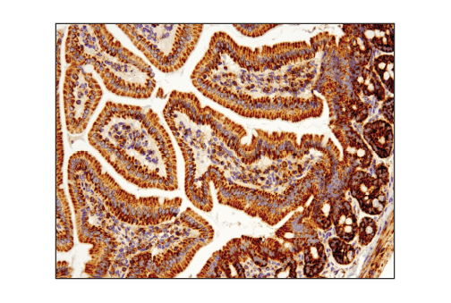
| MFF (E5W4M) XP® Rabbit mAb | 84580 | 20 µl | 25, 27, 30, 35 kDa | Rabbit IgG |
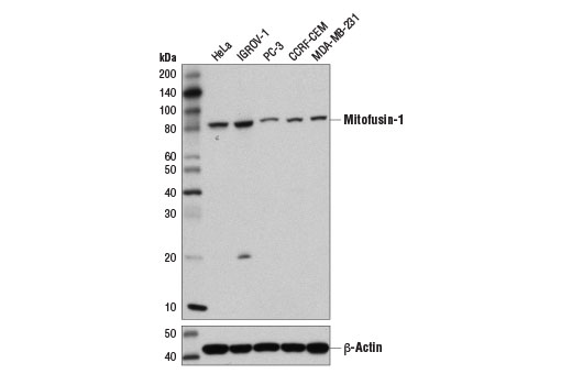
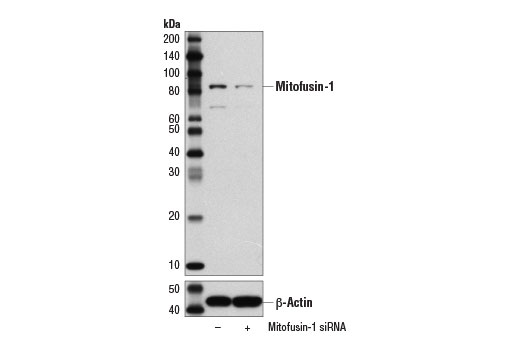
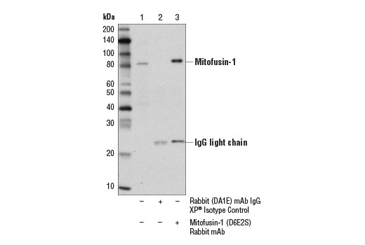
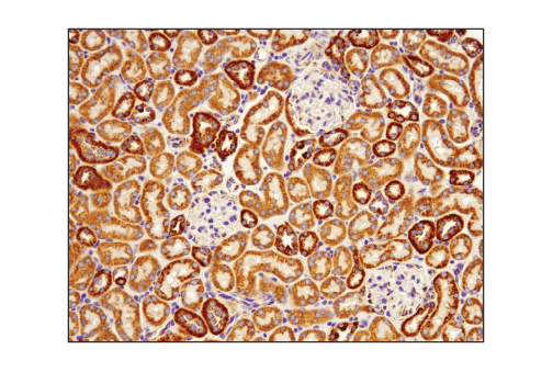
| Mitofusin-1 (D6E2S) Rabbit mAb | 14739 | 20 µl | 82 kDa | Rabbit IgG |
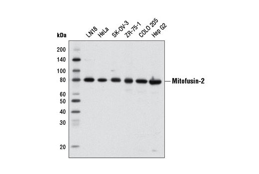
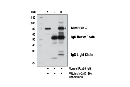
| Mitofusin-2 (D1E9) Rabbit mAb | 11925 | 20 µl | 80 kDa | Rabbit IgG |
| Anti-rabbit IgG, HRP-linked Antibody | 7074 | 100 µl | Goat |
Please visit cellsignal.com for individual component applications, species cross-reactivity, dilutions, protocols, and additional product information.
Description
The Mitochondrial Dynamics Antibody Sampler Kit provides an economical means to examine signaling involved in mitochondrial dynamics. The kit contains enough primary antibody to perform two western blot experiments.
Storage
Background
Import of proteins into the mitochondria is regulated by the translocase of the outer mitochondrial membrane (TOM) complex, which facilitates transport through the outer mitochondrial membrane, and a complementary translocase of the inner membrane (TIM) complex, responsible for protein transport to the mitochondrial matrix. The TOM complex consists of the receptors Tom20, Tom22, and Tom70, and the channel-forming protein Tom40 (1). Tom20 is localized in the outer mitochondrial membrane and initially recognizes precursors with a presequence to facilitate protein import across the outer mitochondrial membrane (2).
Changes in mitochondrial dynamics regulated by environmental cues affect mitochondrial size and shape and have been shown to dramatically impact mitochondrial metabolism, apoptosis, and autophagy (3). These processes are largely controlled by mitochondrial dynamin-related GTPases, including mitofusin-1, mitofusin-2, OPA1, and DRP1. DRP1 regulates mitochondrial fission, while the mitofusins and OPA1 control fusion at the outer and inner mitochondrial membrane, respectively. These proteins are tightly regulated. OPA1 activity is regulated through alternative splicing and post-translational modifications, including complex proteolytic processing by multiple proteases (4-9). In addition, OPA1 expression can be induced under conditions of metabolic demand through a pathway involving Parkin induced NF-κB activation (10). DRP1 is regulated in part through multiple phosphorylation sites (11). Phosphorylation of DRP1 at Ser616 by MAPK or during mitosis by CDKs stimulates mitochondrial fission (12-14). In contrast, PKA dependent phosphorylation of DRP1 at Ser637 inhibits its GTPase activity and mitochondrial fission (15,16). Mitochondrial fission factor (MFF) is a tail-anchored protein that resides within the outer mitochondrial membrane and is part of the mitochondrial fission complex. MFF participates in mitochondrial fission by serving as one of multiple receptors for the GTPase dynamin-related protein 1 (Drp1) (17-20). AMPK directly phosphorylates MFF at two sites to allow for enhanced recruitment of Drp1 to the mitochondria (21).
- Chacinska, A. et al. (2009) Cell 138, 628-44.
- Saitoh, T. et al. (2007) EMBO J 26, 4777-87.
- Kasahara, A. and Scorrano, L. (2014) Trends Cell Biol 24, 761-70.
- Delettre, C. et al. (2001) Hum Genet 109, 584-91.
- Olichon, A. et al. (2007) Cell Death Differ 14, 682-92.
- Ishihara, N. et al. (2006) EMBO J 25, 2966-77.
- Cipolat, S. et al. (2006) Cell 126, 163-75.
- Griparic, L. et al. (2007) J Cell Biol 178, 757-64.
- Merkwirth, C. et al. (2008) Genes Dev 22, 476-88.
- Müller-Rischart, A.K. et al. (2013) Mol Cell 49, 908-21.
- Knott, A.B. et al. (2008) Nat Rev Neurosci 9, 505-18.
- Kashatus, J.A. et al. (2015) Mol Cell 57, 537-51.
- Kashatus, D.F. et al. (2011) Nat Cell Biol 13, 1108-15.
- Taguchi, N. et al. (2007) J Biol Chem 282, 11521-9.
- Chang, C.R. and Blackstone, C. (2007) J Biol Chem 282, 21583-7.
- Cribbs, J.T. and Strack, S. (2007) EMBO Rep 8, 939-44.
- Liu, R. and Chan, D.C. (2015) Mol Biol Cell 26, 4466-77.
- Shen, Q. et al. (2014) Mol Biol Cell 25, 145-59.
- Losón, O.C. et al. (2013) Mol Biol Cell 24, 659-67.
- Otera, H. et al. (2010) J Cell Biol 191, 1141-58.
- Toyama, E.Q. et al. (2016) Science 351, 275-281.
Background References
Trademarks and Patents
使用に関する制限
法的な権限を与えられたCSTの担当者が署名した書面によって別途明示的に合意された場合を除き、 CST、その関連会社または代理店が提供する製品には以下の条件が適用されます。お客様が定める条件でここに定められた条件に含まれるものを超えるもの、 または、ここに定められた条件と異なるものは、法的な権限を与えられたCSTの担当者が別途書面にて受諾した場合を除き、拒絶され、 いかなる効力も効果も有しません。
研究専用 (For Research Use Only) またはこれに類似する表示がされた製品は、 いかなる目的についても FDA または外国もしくは国内のその他の規制機関により承認、認可または許可を受けていません。 お客様は製品を診断もしくは治療目的で使用してはならず、また、製品に表示された内容に違反する方法で使用してはなりません。 CST が販売または使用許諾する製品は、エンドユーザーであるお客様に対し、使途を研究および開発のみに限定して提供されるものです。 診断、予防もしくは治療目的で製品を使用することまたは製品を再販売 (単独であるか他の製品等の一部であるかを問いません) もしくはその他の商業的利用の目的で購入することについては、CST から別途許諾を得る必要があります。 お客様は以下の事項を遵守しなければなりません。(a) CST の製品 (単独であるか他の資材と一緒であるかを問いません) を販売、使用許諾、貸与、寄付もしくはその他の態様で第三者に譲渡したり使用させたりしてはなりません。また、商用の製品を製造するために CST の製品を使用してはなりません。(b) 複製、改変、リバースエンジニアリング、逆コンパイル、 分解または他の方法により製品の構造または技術を解明しようとしてはなりません。また、 CST の製品またはサービスと競合する製品またはサービスを開発する目的で CST の製品を使用してはなりません。(c) CST の製品の商標、商号、ロゴ、特許または著作権に関する通知または表示を除去したり改変したりしてはなりません。(d) CST の製品をCST 製品販売条件(CST’s Product Terms of Sale) および該当する書面のみに従って使用しなければなりません。(e) CST の製品に関連してお客様が使用する第三者の製品またはサービスに関する使用許諾条件、 サービス提供条件またはこれに類する合意事項を遵守しなければなりません。