WB, IP, IF-IC
H M R Mk
Endogenous
180
Rabbit IgG
#Q03164
4297
Product Information
Product Usage Information
| Application | Dilution |
|---|---|
| Western Blotting | 1:1000 |
| Immunoprecipitation | 1:50 |
| Immunofluorescence (Immunocytochemistry) | 1:200 |
Storage
Specificity / Sensitivity
Species Reactivity:
Human, Mouse, Rat, Monkey
Source / Purification
Monoclonal antibody is produced by immunizing animals with recombinant protein specific to the carboxy terminus of human MLL1 protein.
Background
The Set1 histone methyltransferase protein was first identified in yeast as part of the Set1/COMPASS histone methyltransferase complex, which methylates histone H3 at Lys4 and functions as a transcriptional co-activator (1). While yeast contain only one known Set1 protein, mammals contain six Set1-related proteins: SET1A, SET1B, MLL1, MLL2, MLL3, and MLL4, all of which assemble into COMPASS-like complexes and methylate histone H3 at Lys4 (2,3). These Set1-related proteins are each found in distinct protein complexes, all of which share the common subunits WDR5, RBBP5, ASH2L, CXXC1 and DPY30, which are required for proper complex assembly and modulation of histone methyltransferase activity (2-6). MLL1 and MLL2 complexes contain the additional protein subunit, menin (6).
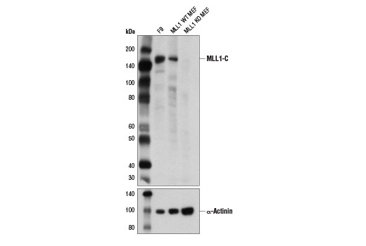
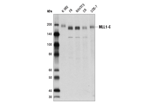
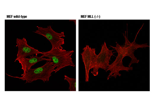
MLL1 functions as a master regulator of both embryogenesis and hematopoiesis, and is required for proper expression of Hox genes (7,8). MLL1 is a large, approximately 4000 amino acid, protein that is cleaved by the taspase 1 threonine endopeptidase to form N-terminal (MLL1-N) and C-terminal MLL1 (MLL1-C) fragments, both of which are subunits of the functional MLL1/COMPASS complex (9,10). MLL1-N, MLL1-C, WDR5, RBBP5 and ASH2L define the core catalytic component of the MLL1/COMPASS complex, which is recruited to target genes and methylates histone H3 lysine 4 to regulate transcriptional initiation (11). At least 60 different MLL1 translocation partners have been molecularly characterized and associated with various hematological malignancies. The most common translocation partners include AF4, AF9, ENL, AF10, ELL and AF6 (8,12,13). With the exception of AF6, all of these partners are nuclear proteins that function to positively regulate transcriptional elongation. AF4, AF9 and ENL are all components of the super elongation complex (SEC), while AF4, AF9, AF10 and ENL all interact with the histone H3 lysine 79 methyltransferase DOT1L. Many MLL1 target genes are normally regulated by promoter-proximal pausing, with the release of RNA polymerase and transcriptional elongation occurring in response to proper stimuli (14). The association of MLL1 translocation partners with SEC and DOT1L suggest that MLL1-fusion proteins may function to sustain specific gene expression programs by constitutively activating transcriptional elongation.
- Miller, T. et al. (2001) Proc Natl Acad Sci U S A 98, 12902-7.
- Shilatifard, A. (2008) Curr Opin Cell Biol 20, 341-8.
- Tenney, K. and Shilatifard, A. (2005) J Cell Biochem 95, 429-36.
- Lee, J.H. and Skalnik, D.G. (2005) J Biol Chem 280, 41725-31.
- Lee, J.H. et al. (2007) J Biol Chem 282, 13419-28.
- Hughes, C.M. et al. (2004) Mol Cell 13, 587-97.
- Eissenberg, J.C. and Shilatifard, A. (2010) Dev Biol 339, 240-9.
- Smith, E. et al. (2011) Genes Dev 25, 661-72.
- Takeda, S. et al. (2006) Genes Dev 20, 2397-409.
- Yokoyama, A. et al. (2002) Blood 100, 3710-8.
- Dou, Y. et al. (2006) Nat Struct Mol Biol 13, 713-9.
- Yip, B.H. and So, C.W. (2013) Exp Biol Med (Maywood) 238, 315-23.
- Neff, T. and Armstrong, S.A. (2013) Blood 121, 4847-53.
- Wang, P. et al. (2009) Mol Cell Biol 29, 6074-85.
Species Reactivity
Species reactivity is determined by testing in at least one approved application (e.g., western blot).
Western Blot Buffer
IMPORTANT: For western blots, incubate membrane with diluted primary antibody in 5% w/v BSA, 1X TBS, 0.1% Tween® 20 at 4°C with gentle shaking, overnight.
Applications Key
WB: Western Blotting IP: Immunoprecipitation IF-IC: Immunofluorescence (Immunocytochemistry)
Cross-Reactivity Key
H: human M: mouse R: rat Hm: hamster Mk: monkey Vir: virus Mi: mink C: chicken Dm: D. melanogaster X: Xenopus Z: zebrafish B: bovine Dg: dog Pg: pig Sc: S. cerevisiae Ce: C. elegans Hr: horse GP: Guinea Pig Rab: rabbit All: all species expected
Trademarks and Patents
使用に関する制限
法的な権限を与えられたCSTの担当者が署名した書面によって別途明示的に合意された場合を除き、 CST、その関連会社または代理店が提供する製品には以下の条件が適用されます。お客様が定める条件でここに定められた条件に含まれるものを超えるもの、 または、ここに定められた条件と異なるものは、法的な権限を与えられたCSTの担当者が別途書面にて受諾した場合を除き、拒絶され、 いかなる効力も効果も有しません。
研究専用 (For Research Use Only) またはこれに類似する表示がされた製品は、 いかなる目的についても FDA または外国もしくは国内のその他の規制機関により承認、認可または許可を受けていません。 お客様は製品を診断もしくは治療目的で使用してはならず、また、製品に表示された内容に違反する方法で使用してはなりません。 CST が販売または使用許諾する製品は、エンドユーザーであるお客様に対し、使途を研究および開発のみに限定して提供されるものです。 診断、予防もしくは治療目的で製品を使用することまたは製品を再販売 (単独であるか他の製品等の一部であるかを問いません) もしくはその他の商業的利用の目的で購入することについては、CST から別途許諾を得る必要があります。 お客様は以下の事項を遵守しなければなりません。(a) CST の製品 (単独であるか他の資材と一緒であるかを問いません) を販売、使用許諾、貸与、寄付もしくはその他の態様で第三者に譲渡したり使用させたりしてはなりません。また、商用の製品を製造するために CST の製品を使用してはなりません。(b) 複製、改変、リバースエンジニアリング、逆コンパイル、 分解または他の方法により製品の構造または技術を解明しようとしてはなりません。また、 CST の製品またはサービスと競合する製品またはサービスを開発する目的で CST の製品を使用してはなりません。(c) CST の製品の商標、商号、ロゴ、特許または著作権に関する通知または表示を除去したり改変したりしてはなりません。(d) CST の製品をCST 製品販売条件(CST’s Product Terms of Sale) および該当する書面のみに従って使用しなければなりません。(e) CST の製品に関連してお客様が使用する第三者の製品またはサービスに関する使用許諾条件、 サービス提供条件またはこれに類する合意事項を遵守しなければなりません。