Revision 3

#37495Store at -20C

1 Kit

(8 x 20 microliters)

Cell Signaling Technology

Orders: 877-616-CELL (2355) [email protected]

Support: 877-678-TECH (8324)

Web: [email protected] cellsignal.com

3 Trask LaneDanversMassachusetts01923USA
For Research Use Only. Not for Use in Diagnostic Procedures.
Product Includes Product # Quantity Mol. Wt Isotype/Source
CD4 (D7D2Z) Rabbit mAb 25229 20 µl 55 kDa Rabbit IgG
CD8α (D4W2Z) XP® Rabbit mAb 98941 20 µl 35-42 kDa Rabbit IgG
FoxP3 (D6O8R) Rabbit mAb 12653 20 µl Rabbit IgG
F4/80 (D2S9R) XP® Rabbit mAb 70076 20 µl 65-250 kDa Rabbit IgG
CD19 (Intracellular Domain) (D4V4B) XP® Rabbit mAb 90176 20 µl 95 kDa Rabbit IgG
CD11c (D1V9Y) Rabbit mAb 97585 20 µl 145 kDa Rabbit IgG
Granzyme B (E5V2L) Rabbit mAb 44153 20 µl 30 kDa Rabbit IgG
CD3ε (E4T1B) XP® Rabbit mAb 78588 20 µl 23 kDa Rabbit IgG

Please visit cellsignal.com for individual component applications, species cross-reactivity, dilutions, protocols, and additional product information.

Description

The Mouse Immune Cell Phenotyping IHC Antibody Sampler Kit provides an economical means of detecting the accumulation of immune cell types in formalin-fixed, paraffin-embedded tissue samples.

Storage

Supplied in 10 mM sodium HEPES (pH 7.5), 150 mM NaCl, 100 µg/ml BSA, 50% glycerol and less than 0.02% sodium azide. Store at –20°C. Do not aliquot the antibody.

Background

Cluster of Differentiation 3 (CD3) is a multiunit protein complex expressed on the surface of T-cells that directly associates with the T-cell receptor (TCR). CD3 is composed of four polypeptides: ζ, γ, ε and δ. Engagement of TCR complex with antigens presented in Major Histocompatibility Complexes (MHC) induces tyrosine phosphorylation in the immunoreceptor tyrosine-based activation motif (ITAM) of CD3 proteins. CD3 phosphorylation is required for downstream signaling through ZAP-70 and p85 subunit of PI-3 kinase, leading to T cell activation, proliferation, and effector functions (1). Cluster of Differentiation 8 (CD8) is a transmembrane glycoprotein expressed primarily on cytotoxic T cells, but has also been described on a subset of dendritic cells in mice (2,3). On T cells, CD8 is a co-receptor for the TCR, and these two distinct structures are required to recognize antigen bound to MHC Class I (2). Cluster of Differentiation 4 (CD4) is expressed on the surface of T helper cells, regulatory T cells, monocytes, macrophages, and dendritic cells, and plays an important role in the development and activation of T cells. On T cells, CD4 is the co-receptor for the TCR, and these two distinct structures recognize antigen bound to MHC Class II. CD8 and CD4 co-receptors ensure specificity of the TCR–antigen interaction, prolong the contact between the T cell and the antigen presenting cell, and recruit the tyrosine kinase Lck, which is essential for T cell activation (2). Granzyme B is a serine protease expressed by CD8+ cytotoxic T lymphocytes and natural killer (NK) cells and is a key component of the immune response to pathogens and transformed cancer cells (4). Forkhead box P3 (FoxP3) is crucial for the development of T cells with immunosuppressive regulatory properties and is a well-established marker for T regulatory cells (Tregs) (5). CD19 is a co-receptor expressed on B cells that amplifies the signaling cascade initiated by the B cell receptor (BCR) to induce activation. It is a biomarker of B lymphocyte development, lymphoma diagnosis, and can be utilized as a target for leukemia immunotherapies (6,7). F4/80 (EMR1) is a heavily glycosylated G-protein-coupled receptor and is a well-established marker for mouse macrophages (8). CD11c (integrin αX, ITGAX) is a transmembrane glycoprotein highly expressed by dendritic cells, and has also been observed on activated NK cells, subsets of B and T cells, monocytes, granulocytes, and some B cell malignancies including hairy cell leukemia (9,10).

  1. Kuhns, M.S. et al. (2006) Immunity 24, 133-9.
  2. Zamoyska, R. (1994) Immunity 1, 243-6.
  3. Shortman, K. and Heath, W.R. (2010) Immunol Rev 234, 18-31.
  4. Trapani, J.A. (2001) Genome Biol 2, REVIEWS3014.
  5. Ochs, H.D. et al. (2007) Immunol Res 38, 112-21.
  6. Tedder, T.F. et al. (1997) Immunity 6, 107-18.
  7. Scheuermann, R.H. and Racila, E. (1995) Leuk Lymphoma 18, 385-97.
  8. McKnight, A.J. et al. (1996) J Biol Chem 271, 486-9.
  9. Kohrgruber, N. et al. (1999) J Immunol 163, 3250-9.
  10. Qualai, J. et al. (2016) PLoS One 11, e0154253.

Background References

    Trademarks and Patents

    Cell Signaling Technology is a trademark of Cell Signaling Technology, Inc.
    SignalStain is a registered trademark of Cell Signaling Technology, Inc.
    XP is a registered trademark of Cell Signaling Technology, Inc.
    All other trademarks are the property of their respective owners. Visit cellsignal.com/trademarks for more information.

    使用に関する制限

    法的な権限を与えられたCSTの担当者が署名した書面によって別途明示的に合意された場合を除き、 CST、その関連会社または代理店が提供する製品には以下の条件が適用されます。お客様が定める条件でここに定められた条件に含まれるものを超えるもの、 または、ここに定められた条件と異なるものは、法的な権限を与えられたCSTの担当者が別途書面にて受諾した場合を除き、拒絶され、 いかなる効力も効果も有しません。

    研究専用 (For Research Use Only) またはこれに類似する表示がされた製品は、 いかなる目的についても FDA または外国もしくは国内のその他の規制機関により承認、認可または許可を受けていません。 お客様は製品を診断もしくは治療目的で使用してはならず、また、製品に表示された内容に違反する方法で使用してはなりません。 CST が販売または使用許諾する製品は、エンドユーザーであるお客様に対し、使途を研究および開発のみに限定して提供されるものです。 診断、予防もしくは治療目的で製品を使用することまたは製品を再販売 (単独であるか他の製品等の一部であるかを問いません) もしくはその他の商業的利用の目的で購入することについては、CST から別途許諾を得る必要があります。 お客様は以下の事項を遵守しなければなりません。(a) CST の製品 (単独であるか他の資材と一緒であるかを問いません) を販売、使用許諾、貸与、寄付もしくはその他の態様で第三者に譲渡したり使用させたりしてはなりません。また、商用の製品を製造するために CST の製品を使用してはなりません。(b) 複製、改変、リバースエンジニアリング、逆コンパイル、 分解または他の方法により製品の構造または技術を解明しようとしてはなりません。また、 CST の製品またはサービスと競合する製品またはサービスを開発する目的で CST の製品を使用してはなりません。(c) CST の製品の商標、商号、ロゴ、特許または著作権に関する通知または表示を除去したり改変したりしてはなりません。(d) CST の製品をCST 製品販売条件(CST’s Product Terms of Sale) および該当する書面のみに従って使用しなければなりません。(e) CST の製品に関連してお客様が使用する第三者の製品またはサービスに関する使用許諾条件、 サービス提供条件またはこれに類する合意事項を遵守しなければなりません。
    Revision 3
    #37495

    Mouse Immune Cell Phenotyping IHC Antibody Sampler Kit

    Mouse Immune Cell Phenotyping IHC Antibody Sampler Kit: Image 1 Expand Image
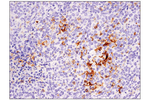
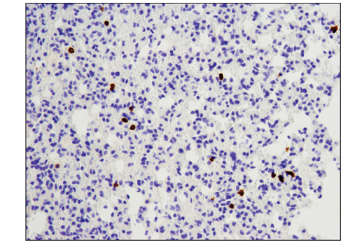
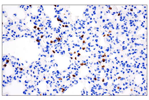
    Immunohistochemical analysis of paraffin-embedded 4T1 metastatic tumor in mouse lung using FoxP3 (D6O8R) Rabbit mAb performed on the Leica® BOND Rx.
    Mouse Immune Cell Phenotyping IHC Antibody Sampler Kit: Image 2 Expand Image
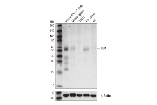
    Western blot analysis of extracts from various cell lines and tissues using CD4 (D7D2Z) Rabbit mAb (upper) and β-Actin (D6A8) Rabbit mAb #8457 (lower).
    Mouse Immune Cell Phenotyping IHC Antibody Sampler Kit: Image 3 Expand Image
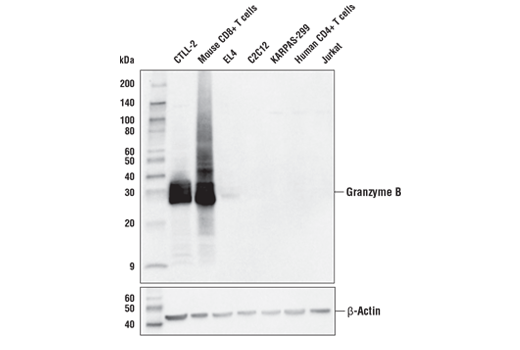
    Western blot analysis of extracts from various mouse and human cells using Granzyme B (E5V2L) Rabbit mAb (upper) or β-Actin (D6A8) Rabbit mAb #8457 (lower). As expected, Granzyme B (E5V2L) Rabbit mAb only detects Granzyme B in mouse primary CD8+ T cells and mouse T cell lines. CD8+ T cells were purified from mouse spleens or human blood and stimulated for 7 days using beads coated with CD3 and CD28 antibodies in the presence of Mouse Interleukin-2 (mIL-2) #5201 (20 ng/ml) or Human Interleukin-2 (hIL-2) #8907 (20 ng/ml). KARPAS cell line source: Dr. Abraham Karpas at the University of Cambridge.
    Mouse Immune Cell Phenotyping IHC Antibody Sampler Kit: Image 4 Expand Image
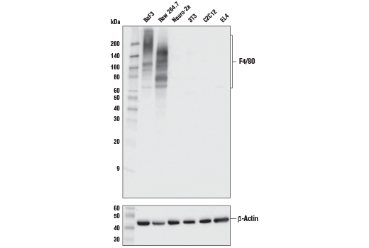
    Western blot analysis of extracts from various cell lines using F4/80 (D2S9R) XP® Rabbit mAb (upper) or β-Actin (D6A8) Rabbit mAb #8457 (lower).
    Mouse Immune Cell Phenotyping IHC Antibody Sampler Kit: Image 5 Expand Image
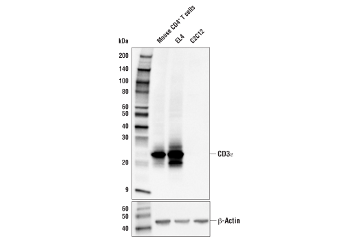
    Western blot analysis of extracts from mouse CD4+ T cells, EL4, and C2C12 cells using CD3ε (E4T1B) XP® Rabbit mAb (upper) and β-Actin (D6A8) Rabbit mAb #8457 (lower). CD4+ T cells were purified from mouse spleens and stimulated for 9 days using beads coated with mouse CD3 and CD28 antibodies in the presence of Mouse Interleukin-2 (mIL-2) #5201 (20 ng/ml).
    Mouse Immune Cell Phenotyping IHC Antibody Sampler Kit: Image 6 Expand Image
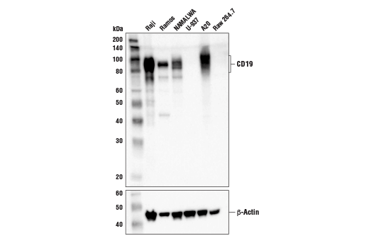
    Western blot analysis of extracts from various cell lines using CD19 (Intracellular Domain) (D4V4B) XP® Rabbit mAb (upper) and β-Actin (D6A8) Rabbit mAb #8457 (lower). As expected, CD19 protein is not detected in either U-937 cells or Raw 264.7 cells.
    Mouse Immune Cell Phenotyping IHC Antibody Sampler Kit: Image 7 Expand Image
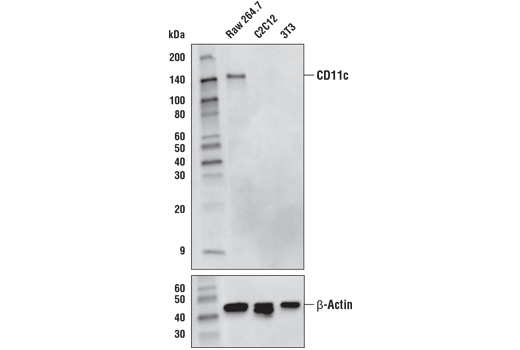
    Western blot analysis of extracts from Raw 264.7, C2C12, and 3T3 cells using CD11c (D1V9Y) Rabbit mAb (upper) or β-Actin (D6A8) Rabbit mAb #8457 (lower).
    Mouse Immune Cell Phenotyping IHC Antibody Sampler Kit: Image 8 Expand Image
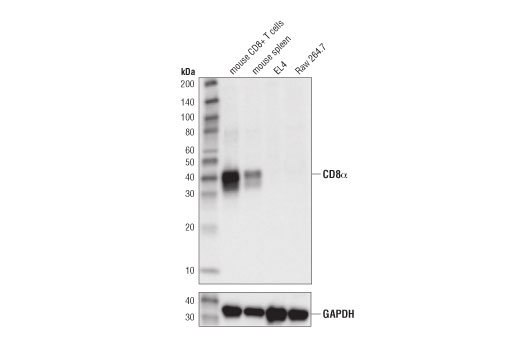
    Western blot analysis of extracts from mouse CD8+ T cells, mouse spleen, EL4 cells, and Raw264.7 cells using CD8α (D4W2Z) XP® Rabbit mAb (upper), and β-Actin (D6A8) Rabbit mAb #8457 (lower).
    Mouse Immune Cell Phenotyping IHC Antibody Sampler Kit: Image 9 Expand Image
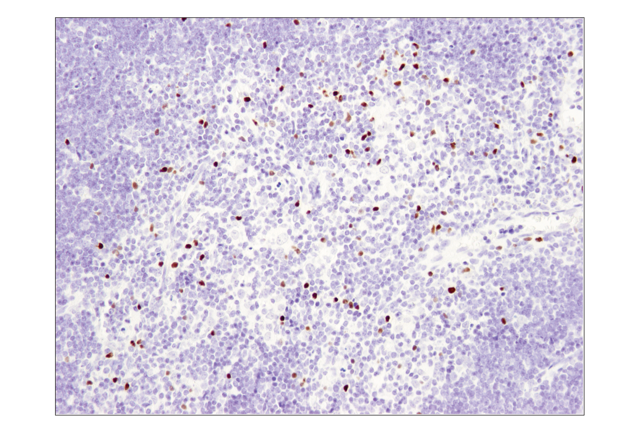
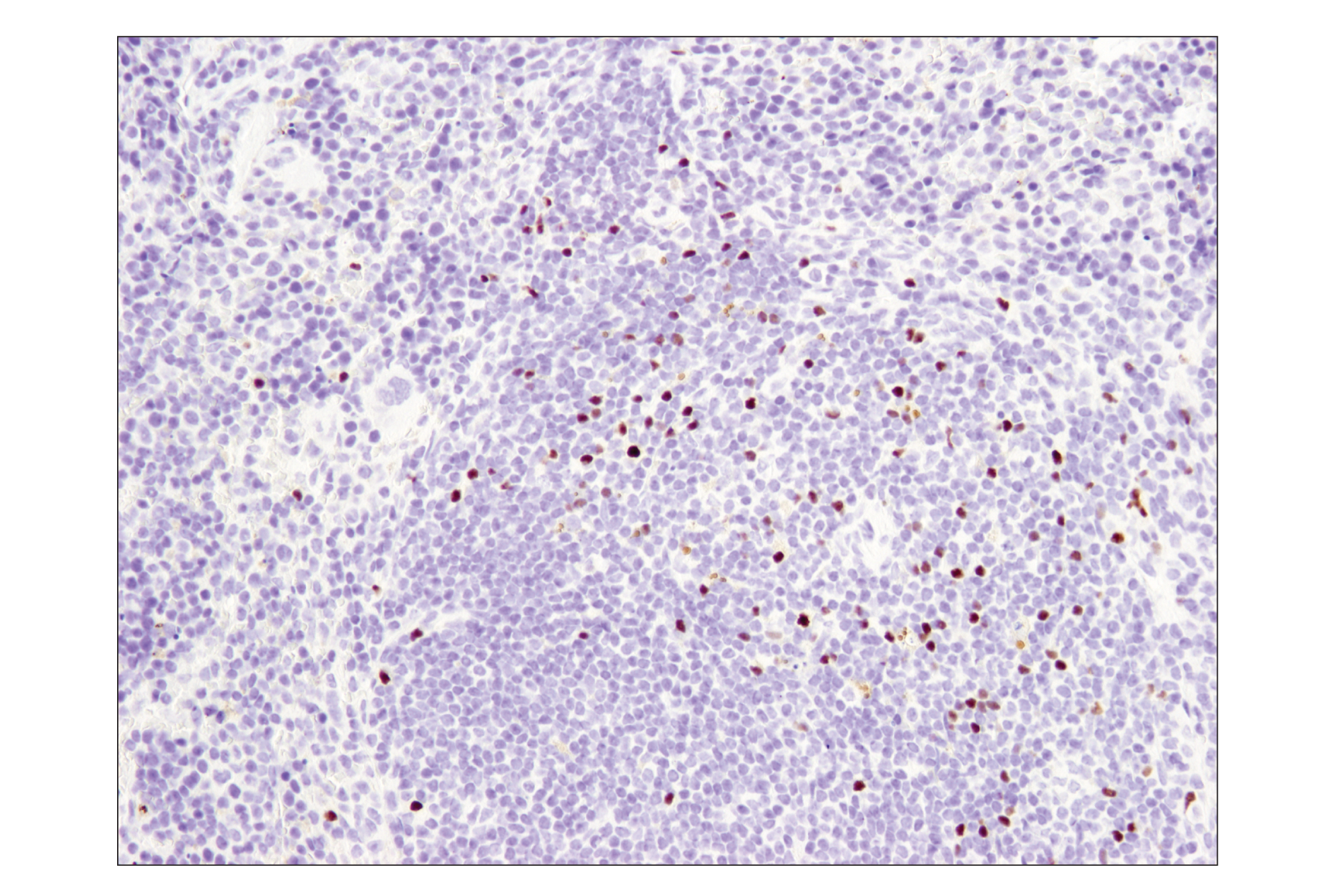
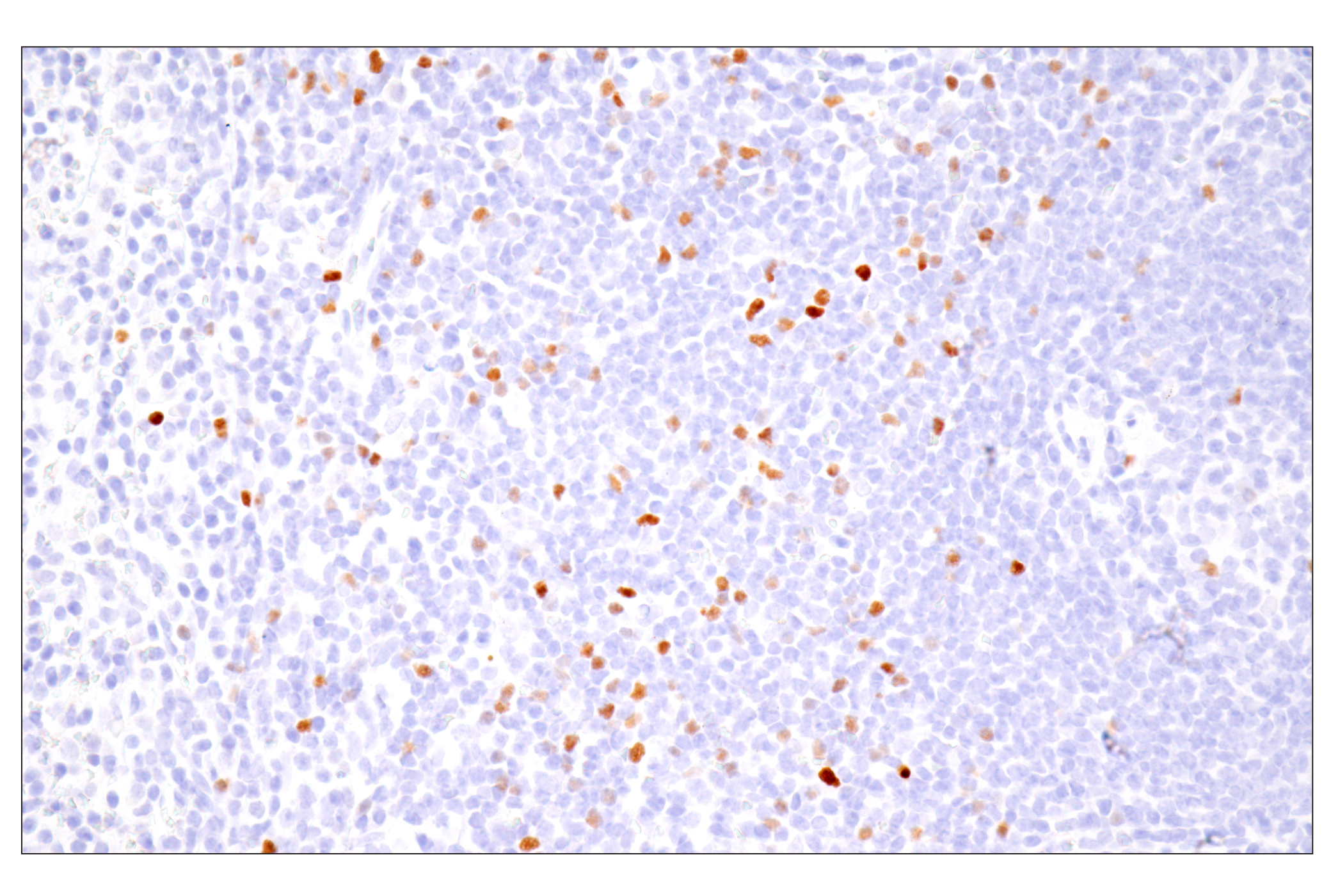
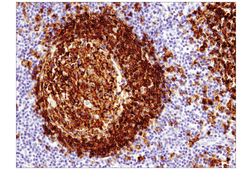
    Immunohistochemical analysis of paraffin-embedded mouse thymus using FoxP3 (D6O8R) Rabbit mAb.
    Mouse Immune Cell Phenotyping IHC Antibody Sampler Kit: Image 10 Expand Image
    Immunohistochemical analysis of paraffin-embedded 4T1 metastatic tumor in mouse lung using CD4 (D7D2Z) Rabbit mAb performed on the Leica® BOND Rx.
    Mouse Immune Cell Phenotyping IHC Antibody Sampler Kit: Image 11 Expand Image
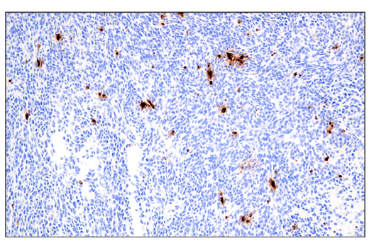
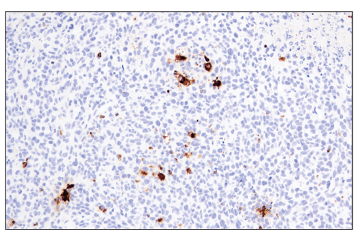
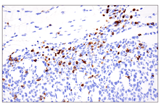
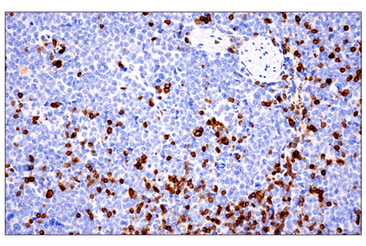
    Immunohistochemical analysis of paraffin-embedded CT26.WT syngeneic tumor using Granzyme B (E5V2L) Rabbit mAb performed on the Leica® BOND Rx.
    Mouse Immune Cell Phenotyping IHC Antibody Sampler Kit: Image 12 Expand Image
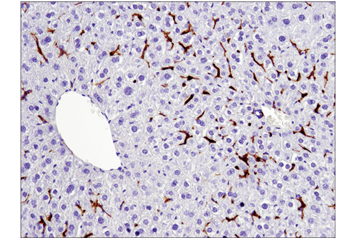
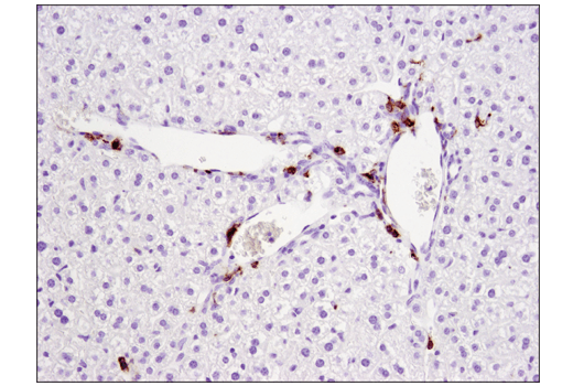
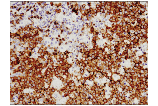
    Immunohistochemical analysis of paraffin-embedded mouse liver using F4/80 (D2S9R) XP® Rabbit mAb.
    Mouse Immune Cell Phenotyping IHC Antibody Sampler Kit: Image 13 Expand Image
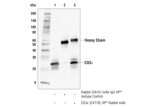
    Immunoprecipitation of CD3ε protein from EL4 cell extracts. Lane 1 is 10% input, lane 2 is Rabbit (DA1E) mAb IgG XP® Isotype Control #3900, and lane 3 is CD3ε (E4T1B) XP® Rabbit mAb. Western blot analysis was performed using CD3ε (E4T1B) XP® Rabbit mAb.
    Mouse Immune Cell Phenotyping IHC Antibody Sampler Kit: Image 14 Expand Image
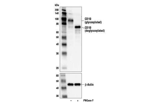
    Western blot analysis of extracts from Daudi cells, untreated (-) or treated with PNGase F (+), using CD19 (Intracellular Domain) (D4V4B) XP® Rabbit mAb (upper) and β-Actin (D6A8) Rabbit mAb #8457 (lower).
    Mouse Immune Cell Phenotyping IHC Antibody Sampler Kit: Image 15 Expand Image
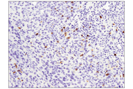
    Immunohistochemical analysis of paraffin-embedded 4T1 mammary tumor using CD11c (D1V9Y) Rabbit mAb performed on the Leica® Bond Rx.
    Mouse Immune Cell Phenotyping IHC Antibody Sampler Kit: Image 16 Expand Image
    Immunohistochemical analysis of paraffin-embedded mouse lung using CD8α (D4W2Z) XP® Rabbit mAb performed on the Leica® BOND Rx.
    Mouse Immune Cell Phenotyping IHC Antibody Sampler Kit: Image 17 Expand Image
    Immunohistochemical analysis of paraffin-embedded mouse spleen using FoxP3 (D6O8R) Rabbit mAb.
    Mouse Immune Cell Phenotyping IHC Antibody Sampler Kit: Image 18 Expand Image
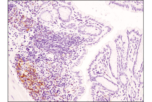
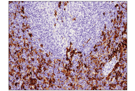
    Immunohistochemical analysis of paraffin-embedded mouse colon using CD4 (D7D2Z) Rabbit mAb.
    Mouse Immune Cell Phenotyping IHC Antibody Sampler Kit: Image 19 Expand Image
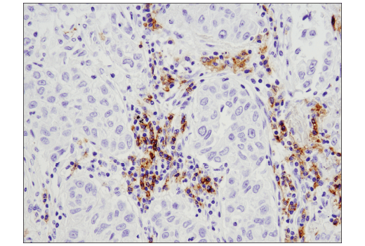
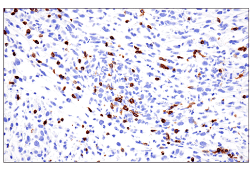
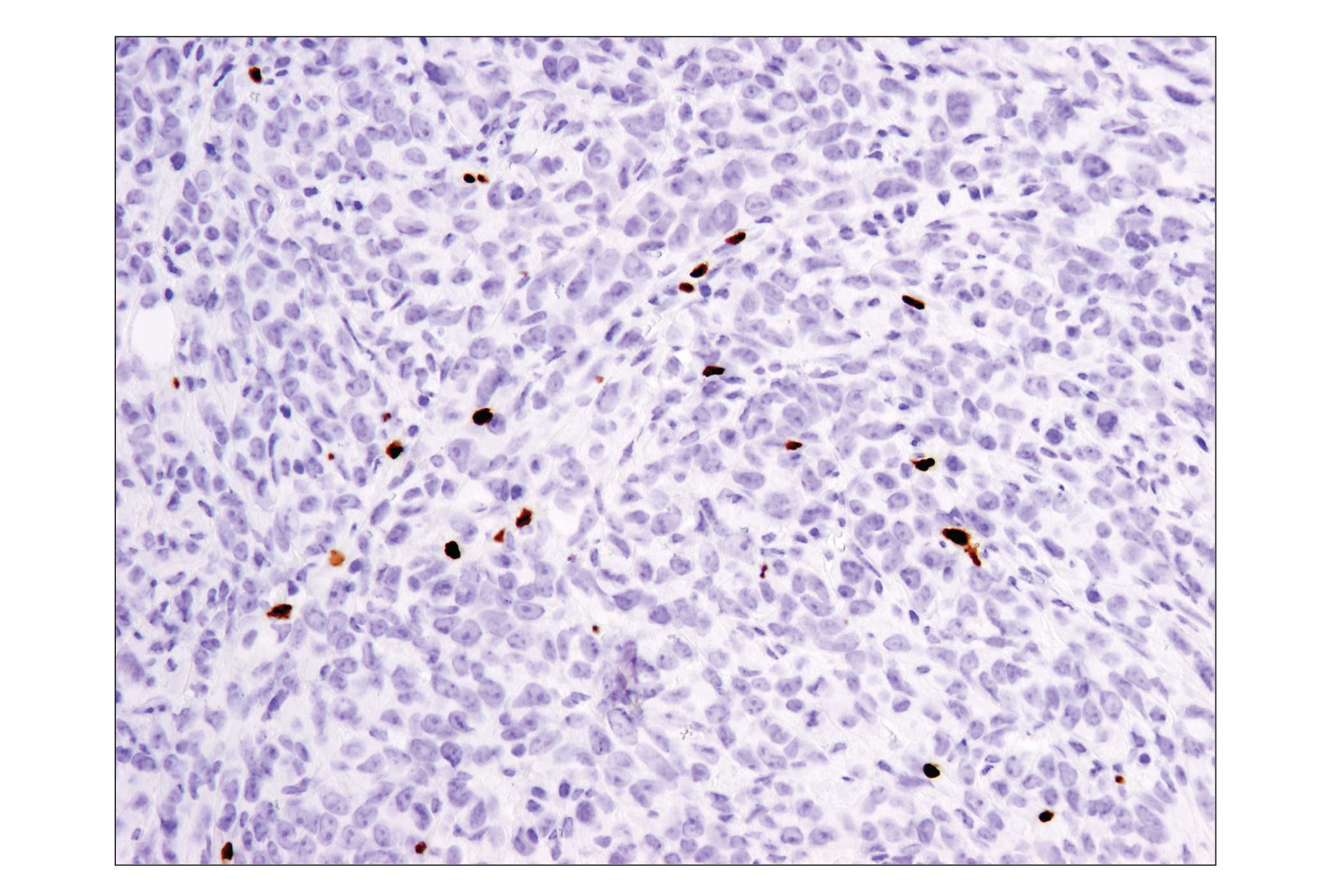
    Immunohistochemical analysis of paraffin-embedded 4T1 syngeneic mammary tumor using Granzyme B (E5V2L) Rabbit mAb.
    Mouse Immune Cell Phenotyping IHC Antibody Sampler Kit: Image 20 Expand Image
    Immunohistochemical analysis of paraffin-embedded mouse spleen using F4/80 (D2S9R) XP® Rabbit mAb.
    Mouse Immune Cell Phenotyping IHC Antibody Sampler Kit: Image 21 Expand Image
    Immunohistochemical analysis of paraffin-embedded mouse lung using CD3ε (E4T1B) XP® Rabbit mAb performed on the Leica® BOND Rx.
    Mouse Immune Cell Phenotyping IHC Antibody Sampler Kit: Image 22 Expand Image
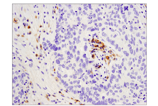
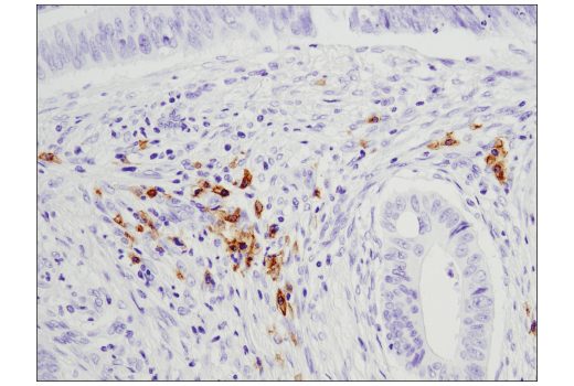
    Immunohistochemical analysis of paraffin-embedded human endometrioid carcinoma using CD19 (Intracellular Domain) (D4V4B) XP® Rabbit mAb performed on the Leica® Bond Rx.
    Mouse Immune Cell Phenotyping IHC Antibody Sampler Kit: Image 23 Expand Image
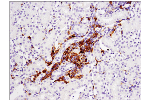
    Immunohistochemical analysis of paraffin-embedded mouse kidney using CD11c (D1V9Y) Rabbit mAb.
    Mouse Immune Cell Phenotyping IHC Antibody Sampler Kit: Image 24 Expand Image
    Immunohistochemical analysis of paraffin-embedded mouse spleen using CD8α (D4W2Z) XP® Rabbit mAb.
    Mouse Immune Cell Phenotyping IHC Antibody Sampler Kit: Image 25 Expand Image
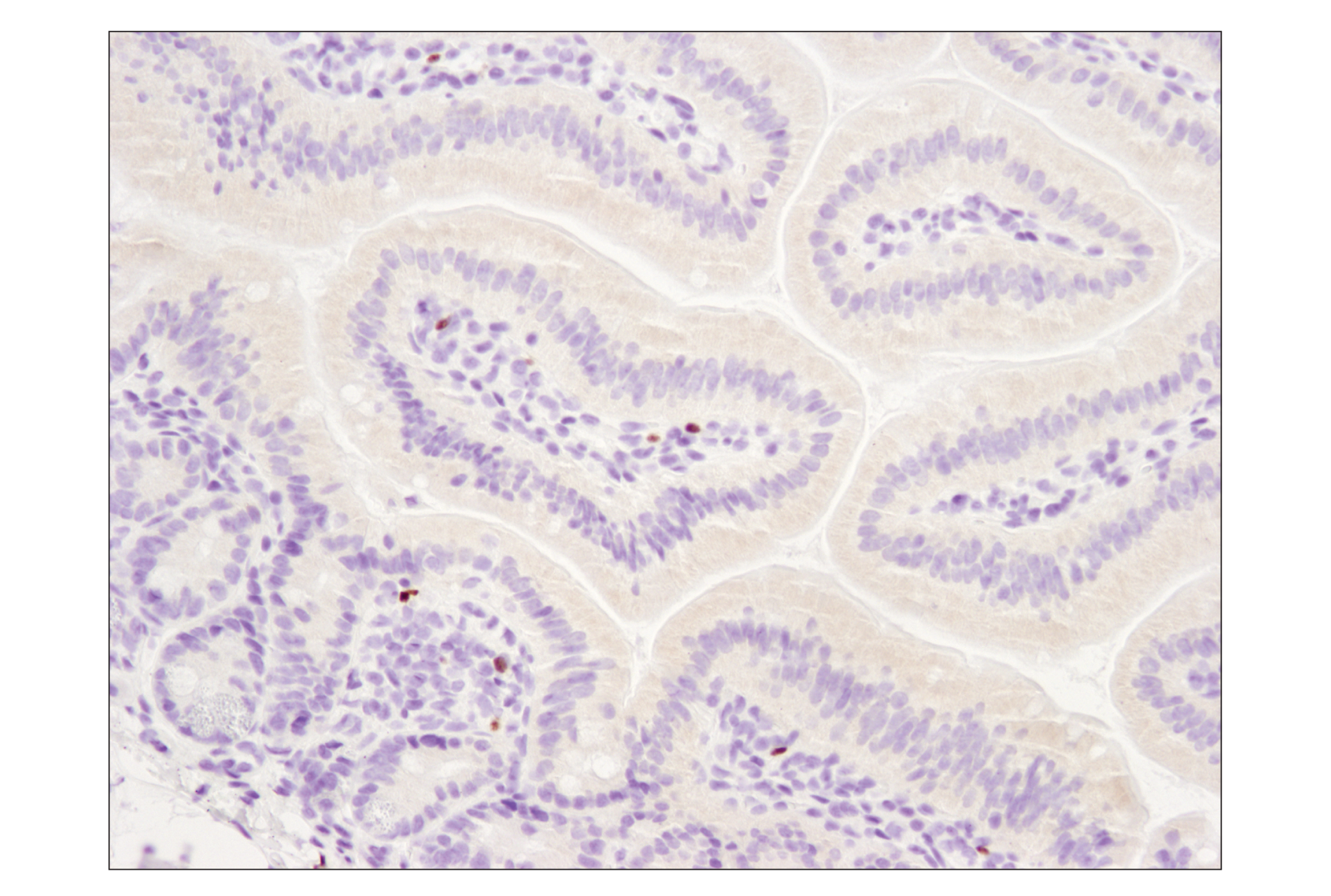
    Immunohistochemical analysis of paraffin-embedded mouse small intestine using FoxP3 (D6O8R) Rabbit mAb.
    Mouse Immune Cell Phenotyping IHC Antibody Sampler Kit: Image 26 Expand Image
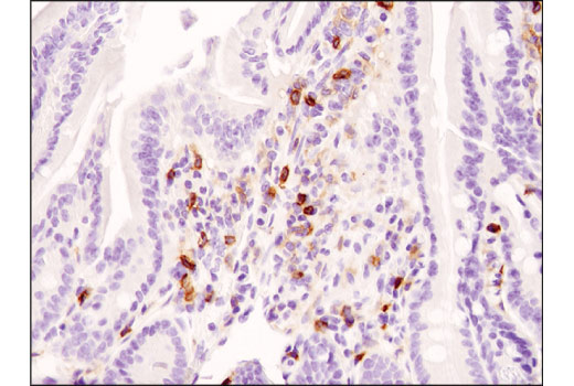
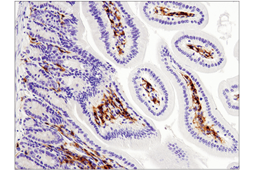
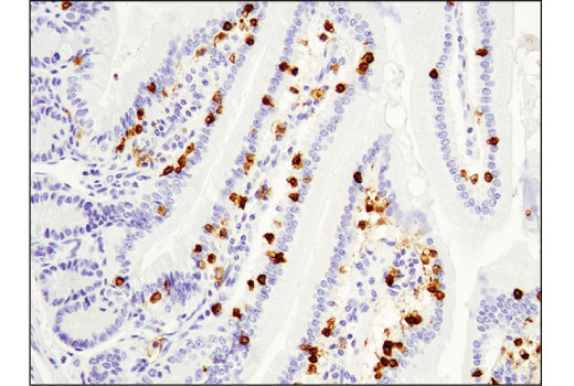
    Immunohistochemical analysis of paraffin-embedded mouse small intestine using CD4 (D7D2Z) Rabbit mAb.
    Mouse Immune Cell Phenotyping IHC Antibody Sampler Kit: Image 27 Expand Image
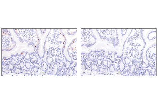
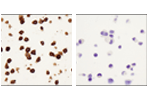
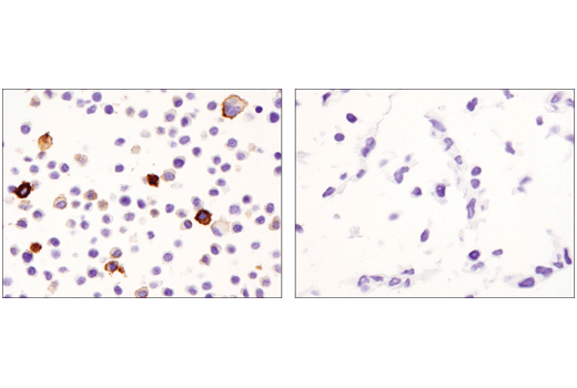
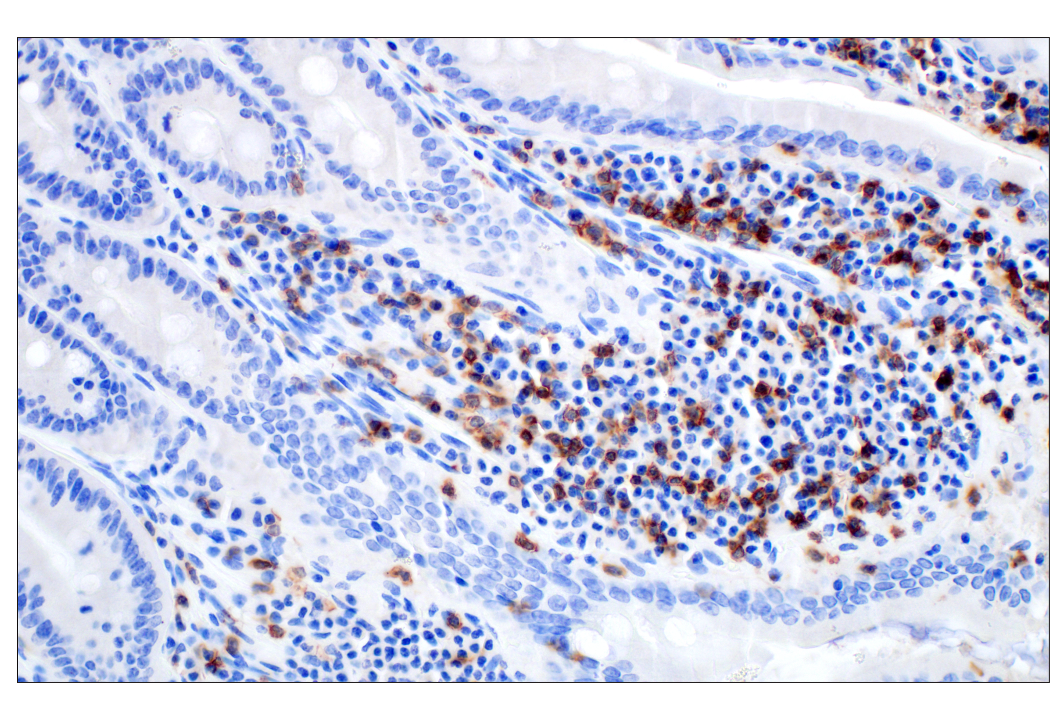
    Immunohistochemical analysis of paraffin-embedded mouse small intestine using Granzyme B (E5V2L) Rabbit mAb (left) compared to concentration matched Rabbit (DA1E) mAb IgG XP® Isotype Control #3900 (right).
    Mouse Immune Cell Phenotyping IHC Antibody Sampler Kit: Image 28 Expand Image
    Immunohistochemical analysis of paraffin-embedded mouse small intestine using F4/80 (D2S9R) XP® Rabbit mAb.
    Mouse Immune Cell Phenotyping IHC Antibody Sampler Kit: Image 29 Expand Image
    Immunohistochemical analysis of paraffin-embedded 4T1 syngeneic mammary tumor using CD3ε (E4T1B) XP® Rabbit mAb.
    Mouse Immune Cell Phenotyping IHC Antibody Sampler Kit: Image 30 Expand Image
    Immunohistochemical analysis of paraffin-embedded human colon carcinoma using CD19 (Intracellular Domain) (D4V4B) XP® Rabbit mAb.
    Mouse Immune Cell Phenotyping IHC Antibody Sampler Kit: Image 31 Expand Image
    Immunohistochemical analysis of paraffin-embedded mouse liver using CD11c (D1V9Y) Rabbit mAb.
    Mouse Immune Cell Phenotyping IHC Antibody Sampler Kit: Image 32 Expand Image
    Immunohistochemical analysis of paraffin-embedded mouse small intestine using CD8α (D4W2Z) XP® Rabbit mAb.
    Mouse Immune Cell Phenotyping IHC Antibody Sampler Kit: Image 33 Expand Image
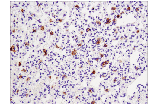
    Immunohistochemical analysis of paraffin-embedded mouse lung using FoxP3 (D6O8R) Rabbit mAb.
    Mouse Immune Cell Phenotyping IHC Antibody Sampler Kit: Image 34 Expand Image
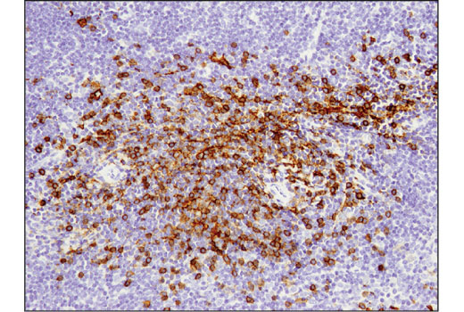
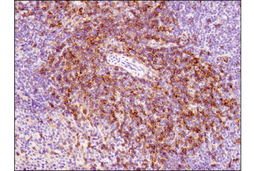
    Immunohistochemical analysis of paraffin-embedded mouse spleen using CD4 (D7D2Z) Rabbit mAb.
    Mouse Immune Cell Phenotyping IHC Antibody Sampler Kit: Image 35 Expand Image
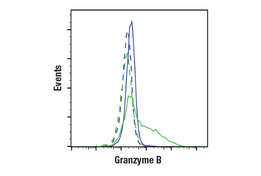
    Flow cytometric analysis of C2C12 cells (blue, negative) and CTLL-2 cells (green, positive) using Granzyme B (E5V2L) Rabbit mAb (solid lines) or a concentration-matched Rabbit (DA1E) mAb IgG XP® Isotype Control #3900 (dashed lines). Anti-rabbit IgG (H+L), F(ab')2 Fragment (Alexa Fluor® 488 Conjugate) #4412 was used as a secondary antibody.
    Mouse Immune Cell Phenotyping IHC Antibody Sampler Kit: Image 36 Expand Image
    Immunohistochemical analysis of paraffin-embedded Raw 264.7 cell pellet (left, positive) or Neuro-2A cell pellet (right, negative) using F4/80 (D2S9R) XP® Rabbit mAb.
    Mouse Immune Cell Phenotyping IHC Antibody Sampler Kit: Image 37 Expand Image
    Immunohistochemical analysis of paraffin-embedded A20 syngeneic tumor using CD3ε (E4T1B) XP® Rabbit mAb.
    Mouse Immune Cell Phenotyping IHC Antibody Sampler Kit: Image 38 Expand Image
    Immunohistochemical analysis of paraffin-embedded human lung carcinoma using CD19 (Intracellular Domain) (D4V4B) XP® Rabbit mAb.
    Mouse Immune Cell Phenotyping IHC Antibody Sampler Kit: Image 39 Expand Image
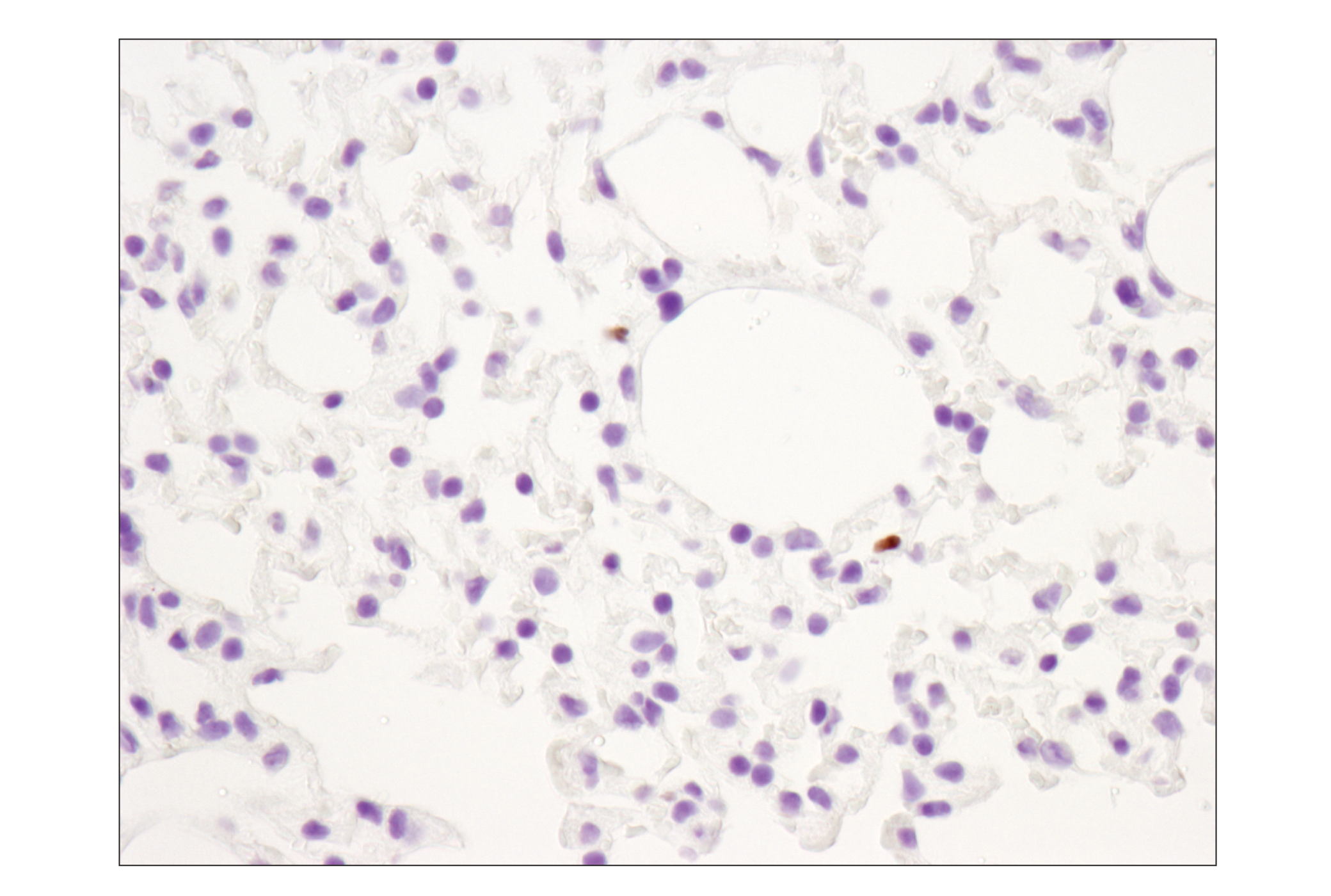
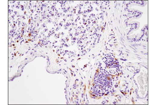
    Immunohistochemical analysis of paraffin-embedded mouse lung using CD11c (D1V9Y) Rabbit mAb.
    Mouse Immune Cell Phenotyping IHC Antibody Sampler Kit: Image 40 Expand Image
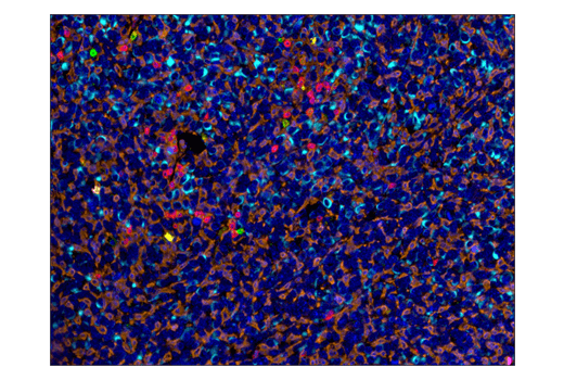
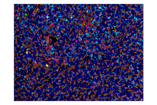
    Multiplex immunohistochemical analysis of paraffin-embedded mouse LL2 syngeneic tumor tissue using CD8α (D4W2Z) XP® rabbit mAb (magenta), F4/80 (D2S9R) XP® rabbit mAb #70076 (orange), PD-1 (D7D5W) XP® rabbit mAb #84651 (green), PD-L1 (D5V3B) rabbit mAb #64988 (yellow), CD3ε (D4V8L) rabbit mAb #99940 (red) and pan keratin (C11) mouse mAb #4279 (cyan).
    Mouse Immune Cell Phenotyping IHC Antibody Sampler Kit: Image 41 Expand Image
    Immunohistochemical analysis of paraffin-embedded normal rhesus monkey spleen using FoxP3 (D6O8R) Rabbit mAb.
    Mouse Immune Cell Phenotyping IHC Antibody Sampler Kit: Image 42 Expand Image
    Immunohistochemical analysis of paraffin-embedded rat lung using CD4 (D7D2Z) Rabbit mAb.
    Mouse Immune Cell Phenotyping IHC Antibody Sampler Kit: Image 43 Expand Image
    Multiplex immunohistochemical analysis of paraffin-embedded mouse LL2 syngeneic tumor tissue using F4/80 (D2S9R) XP® rabbit mAb (orange), PD-1 (D7D5W) XP® rabbit mAb #84651 (green), PD-L1 (D5V3B) rabbit mAb #64988 (yellow), CD3ε (D4V8L) rabbit mAb #99940 (red), CD8α (D4W2Z) XP® rabbit mAb #98941 (magenta) and pan keratin (C11) mouse mAb #4279 (cyan).
    Mouse Immune Cell Phenotyping IHC Antibody Sampler Kit: Image 44 Expand Image
    Immunohistochemical analysis of paraffin-embedded MC38 syngeneic tumor using CD3ε (E4T1B) XP® Rabbit mAb.
    Mouse Immune Cell Phenotyping IHC Antibody Sampler Kit: Image 45 Expand Image
    Immunohistochemical analysis of paraffin-embedded human lymphoma using CD19 (Intraceullular Domain) (D4V4B) XP® Rabbit mAb.
    Mouse Immune Cell Phenotyping IHC Antibody Sampler Kit: Image 46 Expand Image
    Immunohistochemical analysis of paraffin-embedded Raw 264.7 cell pellet (left, positive) or C2C12 cell pellet (right, negative) using CD11c (D1V9Y) Rabbit mAb.
    Mouse Immune Cell Phenotyping IHC Antibody Sampler Kit: Image 47 Expand Image
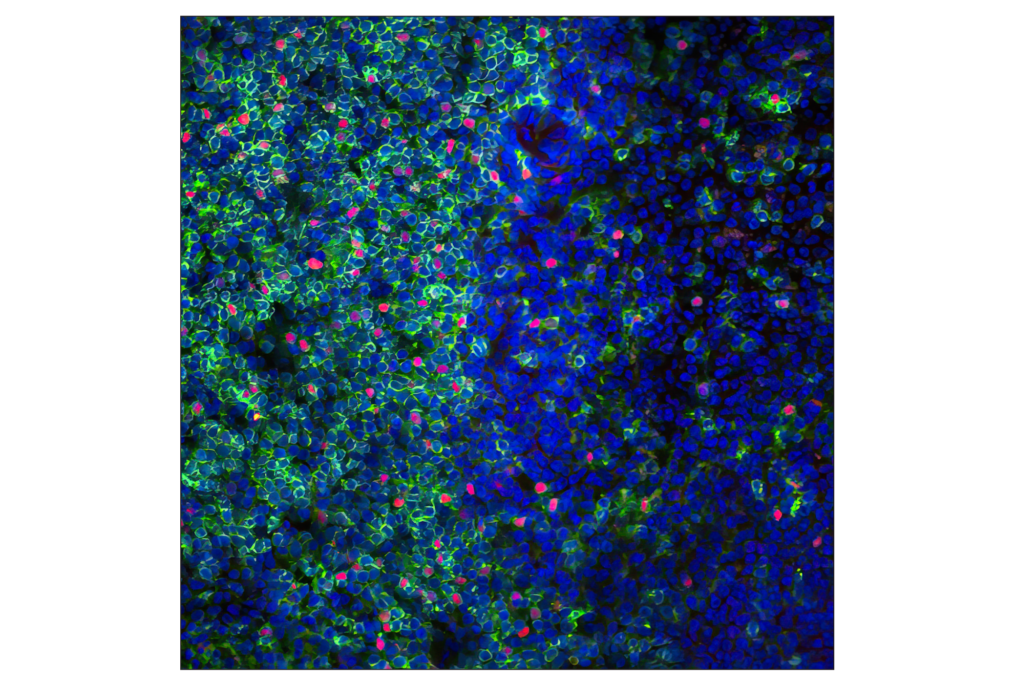
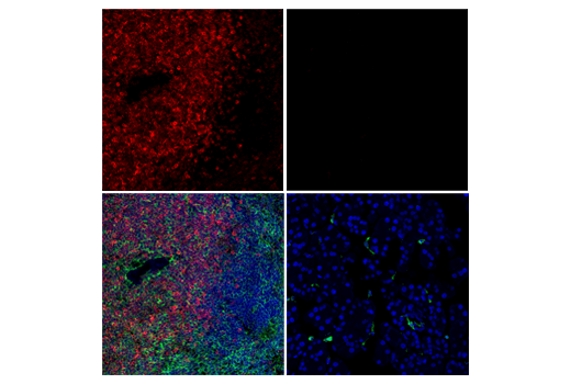
    Immunofluorescent analysis of mouse spleen using FoxP3 (D6O8R) Rabbit mAb (red) and a CD4 antibody (green). Blue = DRAQ5® #4084 (fluorescent DNA dye).Note: FoxP3 (D6O8R) Rabbit mAb has been validated on freshly-prepared frozen tissue sections that were fixed for 15 minutes in 4% formaldehyde. Over-fixation diminishes the performance of this antibody.
    Mouse Immune Cell Phenotyping IHC Antibody Sampler Kit: Image 48 Expand Image
    Immunohistochemical analysis of paraffin-embedded Syrian hamster small intestine using CD4 (D7D2Z) Rabbit mAb.
    Mouse Immune Cell Phenotyping IHC Antibody Sampler Kit: Image 49 Expand Image
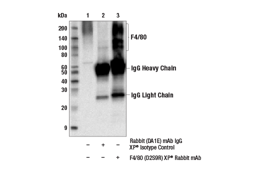
    Immunoprecipitation of F4/80 from BaF3 cell extracts. Lane 1 is 10% input, lane 2 is precipitated with Rabbit (DA1E) mAb IgG XP® Isotype Control #3900, and lane 3 is F4/80 (D2S9R) XP® Rabbit mAb, #70076. Western blot was performed using F4/80 (D2S9R) XP® Rabbit mAb.
    Mouse Immune Cell Phenotyping IHC Antibody Sampler Kit: Image 50 Expand Image
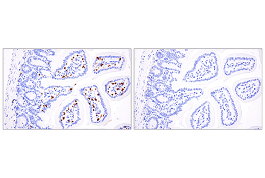
    Immunohistochemical analysis of paraffin-embedded mouse small intestine using CD3ε (E4T1B) XP® Rabbit mAb (left) compared to concentration-matched Rabbit (DA1E) mAb IgG XP® Isotype Control #3900 (right).
    Mouse Immune Cell Phenotyping IHC Antibody Sampler Kit: Image 51 Expand Image
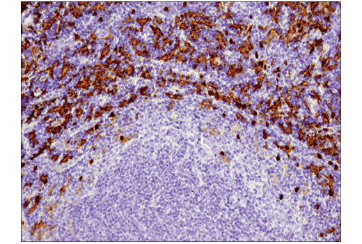
    Immunohistochemical analysis of paraffin-embedded human tonsil using CD19 (Intracellular Domain) (D4V4B) XP® Rabbit mAb.
    Mouse Immune Cell Phenotyping IHC Antibody Sampler Kit: Image 52 Expand Image
    Immunohistochemical analysis of paraffin-embedded mouse spleen using CD11c (D1V9Y) Rabbit mAb.
    Mouse Immune Cell Phenotyping IHC Antibody Sampler Kit: Image 53 Expand Image
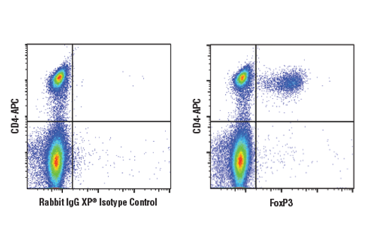
    Flow cytometric analysis of mouse splenocytes using FoxP3 (D6O8R) Rabbit mAb (right) and co-stained with CD4 (RM4-5) Rat mAb (APC Conjugate) #82116, compared to a concentration-matched Rabbit (DA1E) mAb IgG XP® Isotype Control #3900 (left). Anti-rabbit IgG (H+L), F(ab')2 Fragment (Alexa Fluor® 488 Conjugate) was used as a secondary antibody.
    Mouse Immune Cell Phenotyping IHC Antibody Sampler Kit: Image 54 Expand Image
    Confocal immunofluorescent analysis of mouse spleen (left, positive) or pancreas (right, negative) using CD3ε (E4T1B) XP® Rabbit mAb (red). After blocking free secondary antibody binding sites with Rabbit (DA1E) mAb IgG XP® Isotype Control #3900, the tissue was then labeled using HS1 (D5A9) XP® Rabbit mAb (Rodent Specific) (Alexa Fluor® 488 Conjugate) #68206 (green). Sections were mounted in ProLong® Gold Antifade Reagent with DAPI #8961 (blue).
    Mouse Immune Cell Phenotyping IHC Antibody Sampler Kit: Image 55 Expand Image
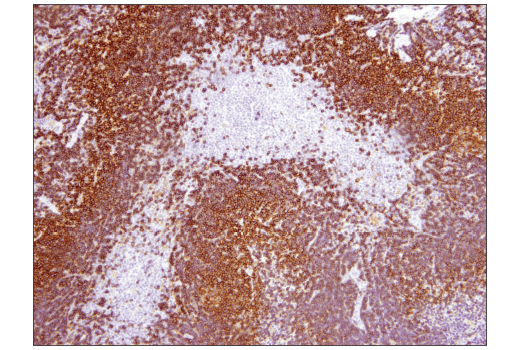
    Immunohistochemical analysis of paraffin-embedded mouse spleen using CD19 (Intracellular Domain) (D4V4B) XP® Rabbit mAb.
    Mouse Immune Cell Phenotyping IHC Antibody Sampler Kit: Image 56 Expand Image
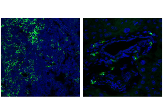
    Confocal immunofluorescent analysis of mouse spleen (left) and mouse kidney (right) using CD11c (D1V9Y) Rabbit mAb. Nuclei were labeled with ProLong® Gold Antifade reagent with DAPI #8961 (blue).
    Mouse Immune Cell Phenotyping IHC Antibody Sampler Kit: Image 57 Expand Image
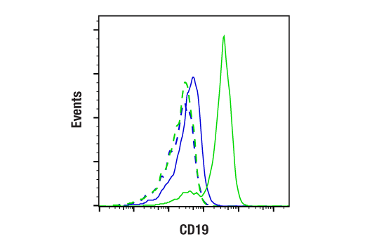
    Flow cytometric analysis of U-937 (negative, blue) and Raji (positive, green) cells using CD19 (D4V4B) Rabbit mAb (solid line) or a concentration-matched Rabbit (DA1E) mAb IgG XP® Isotype Control #3900 (dashed lines). Anti-rabbit IgG (H+L), F(ab')2 Fragment (Alexa Fluor® 647 Conjugate) #4414 was used as a secondary antibody.
    Mouse Immune Cell Phenotyping IHC Antibody Sampler Kit: Image 58 Expand Image
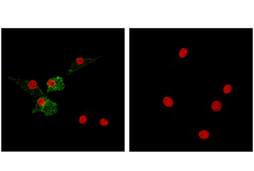
    Confocal immunofluorescent analysis of Raw 264.7 cells (left, positive) and 3T3 cells (right, negative) using CD11c (D1V9Y) Rabbit mAb (green). Red = Propidium Iodide (PI)/RNase Staining Solution #4087.
    Mouse Immune Cell Phenotyping IHC Antibody Sampler Kit: Image 59 Expand Image
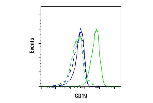
    Flow cytometric analysis of Raw264.7 (negative, blue) and A20 (positive, green) cells using CD19 (D4V4B) Rabbit mAb (solid line) or a concentration-matched Rabbit DA1E) mAb IgG XP® Isotype Control #3900 (dashed lines). Anti-rabbit IgG (H+L), F(ab')2 Fragment (Alexa Fluor® 647 Conjugate) #4414 was used as a secondary antibody.