| Product Includes | Product # | Quantity | Mol. Wt | Isotype/Source |
|---|---|---|---|---|
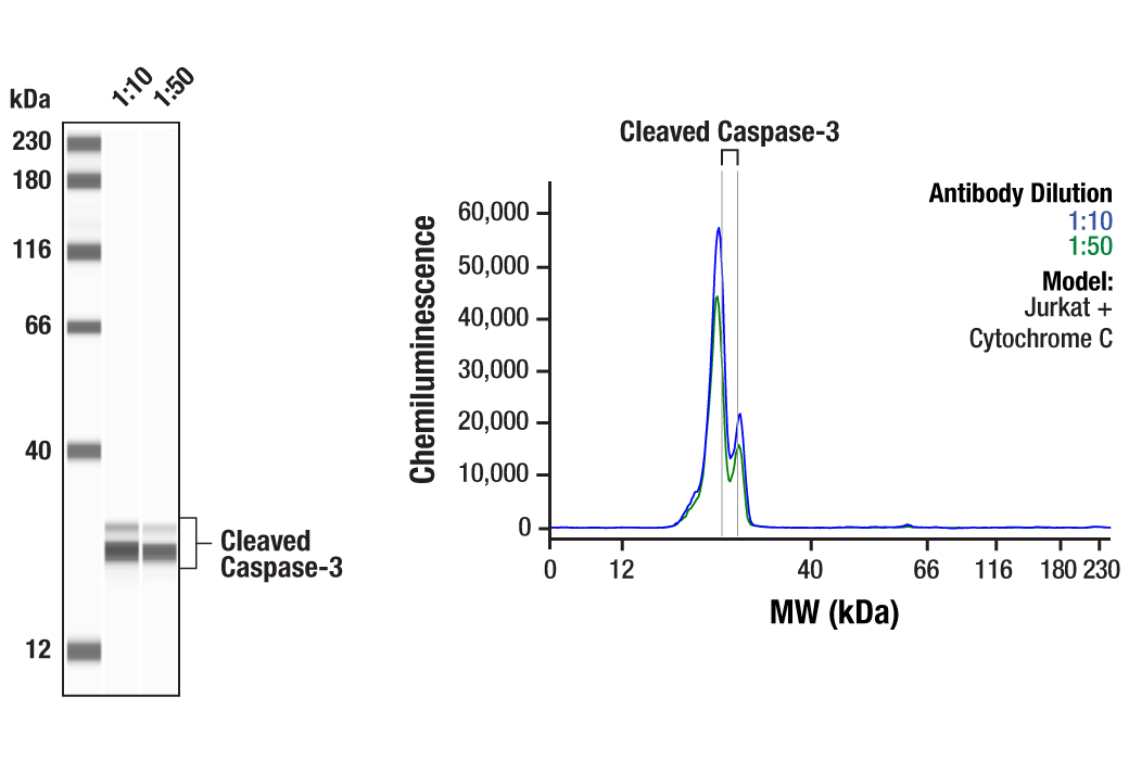
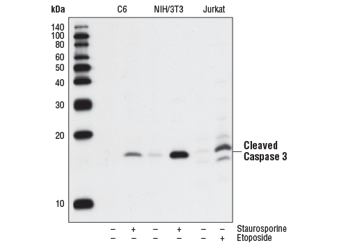
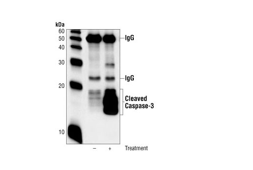
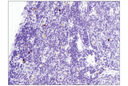
| Cleaved Caspase-3 (Asp175) (5A1E) Rabbit mAb | 9664 | 20 µl | 17, 19 kDa | Rabbit IgG |
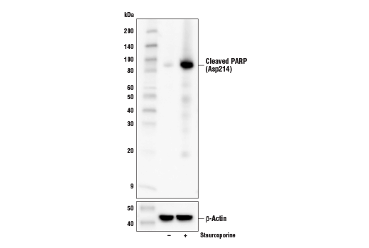
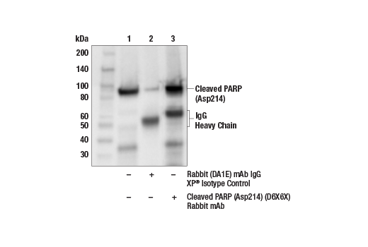
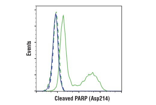
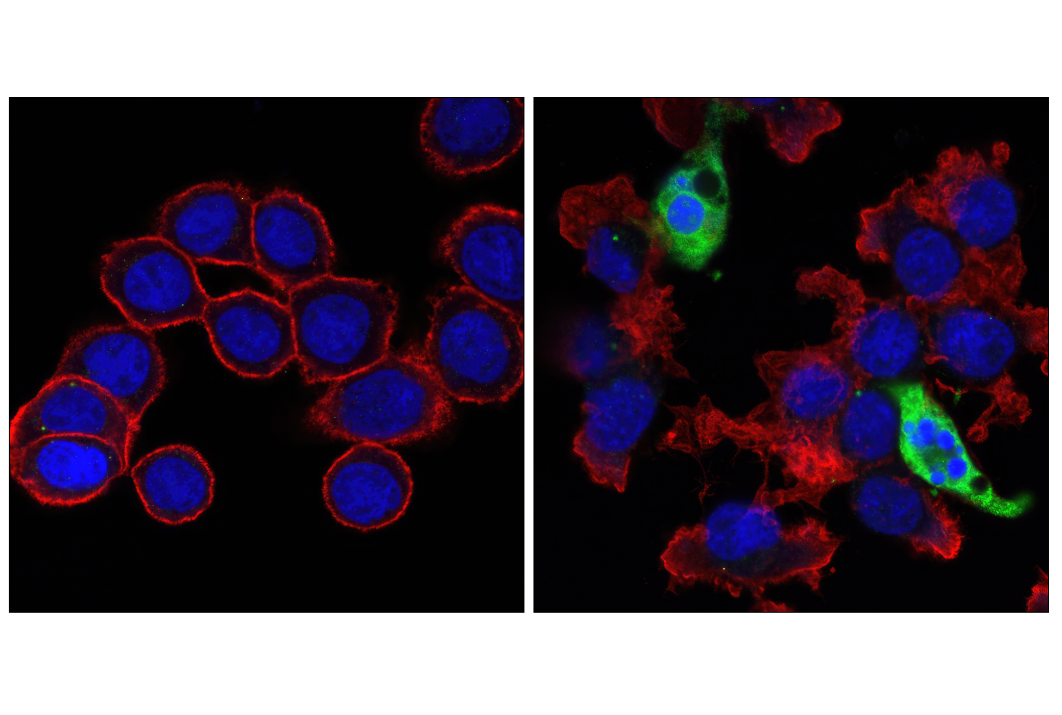
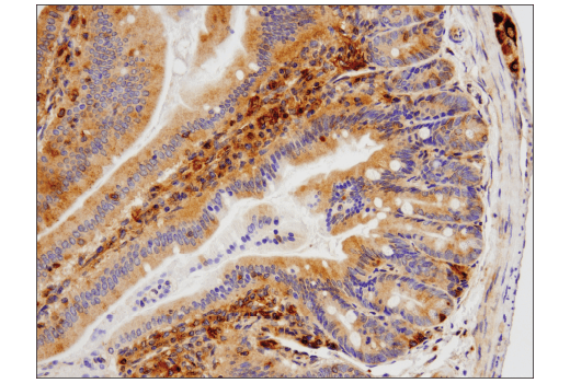
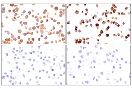
| Cleaved PARP (Asp214) (D6X6X) Rabbit mAb | 94885 | 20 µl | 89 kDa | Rabbit IgG |
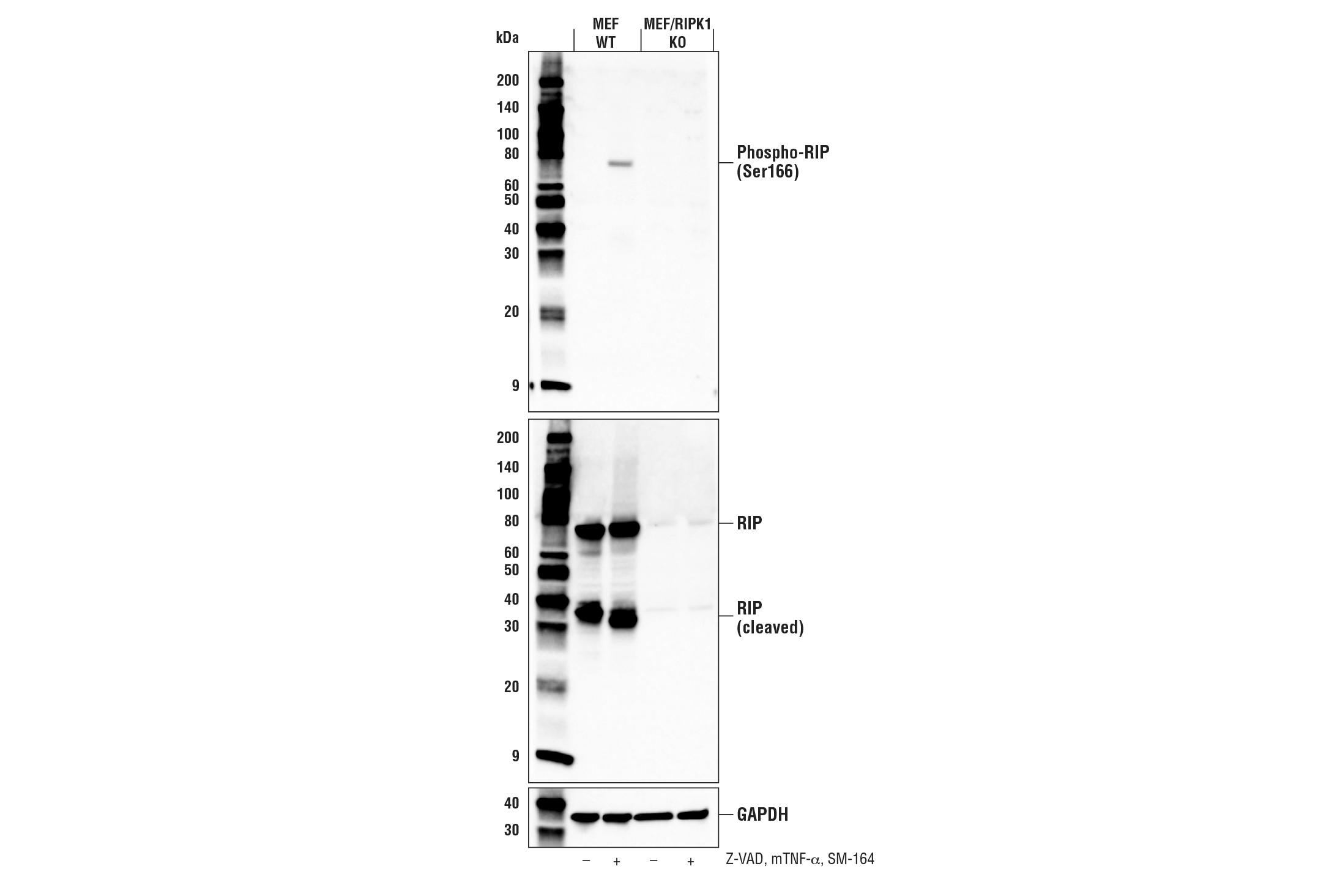
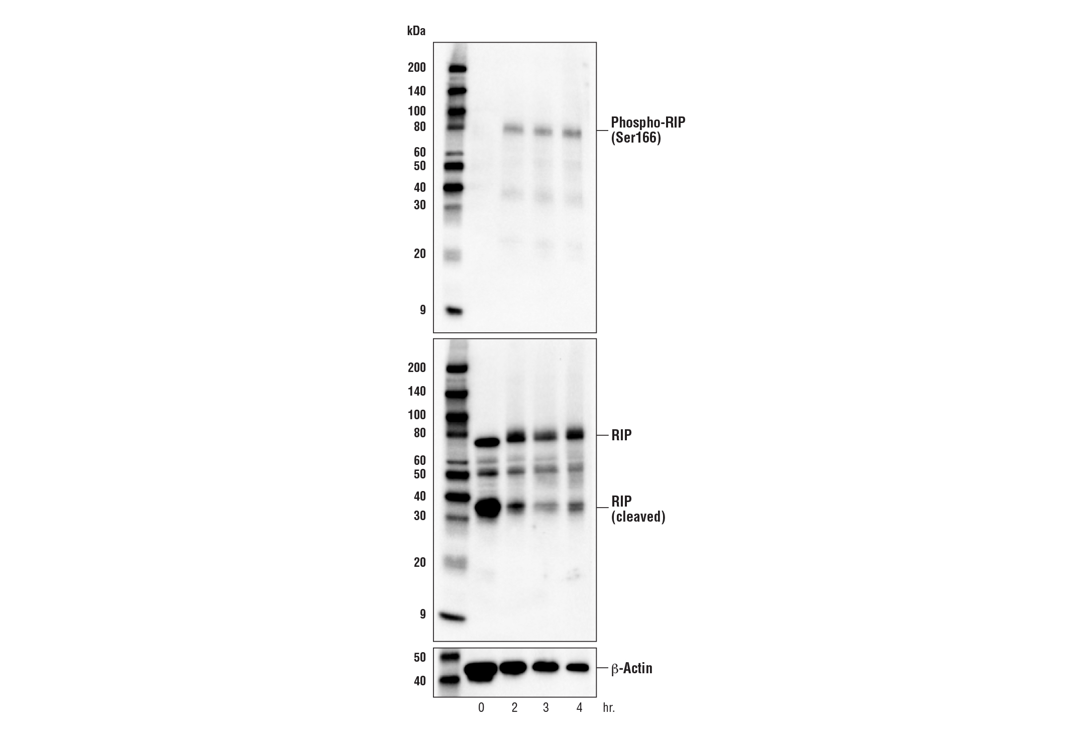
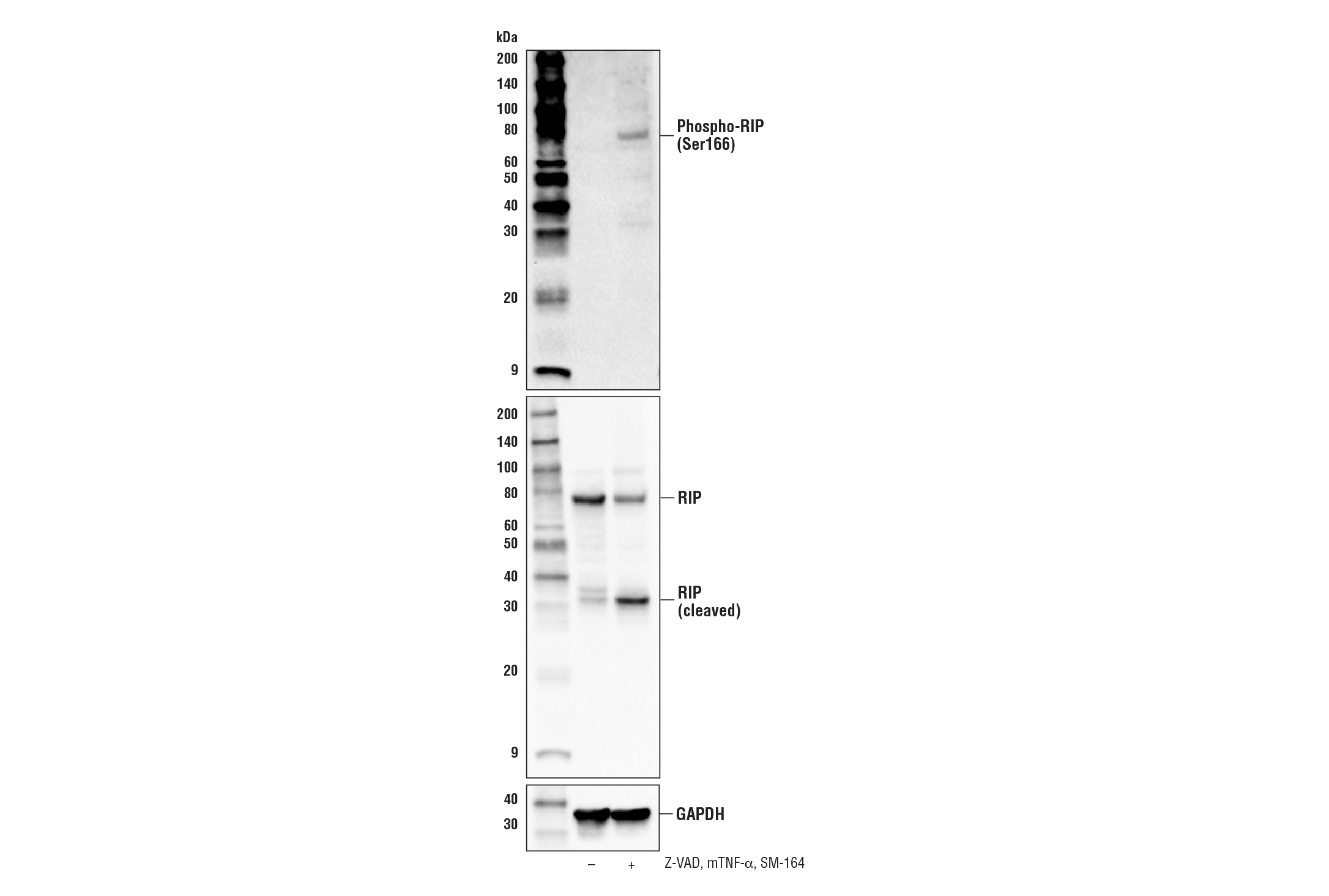
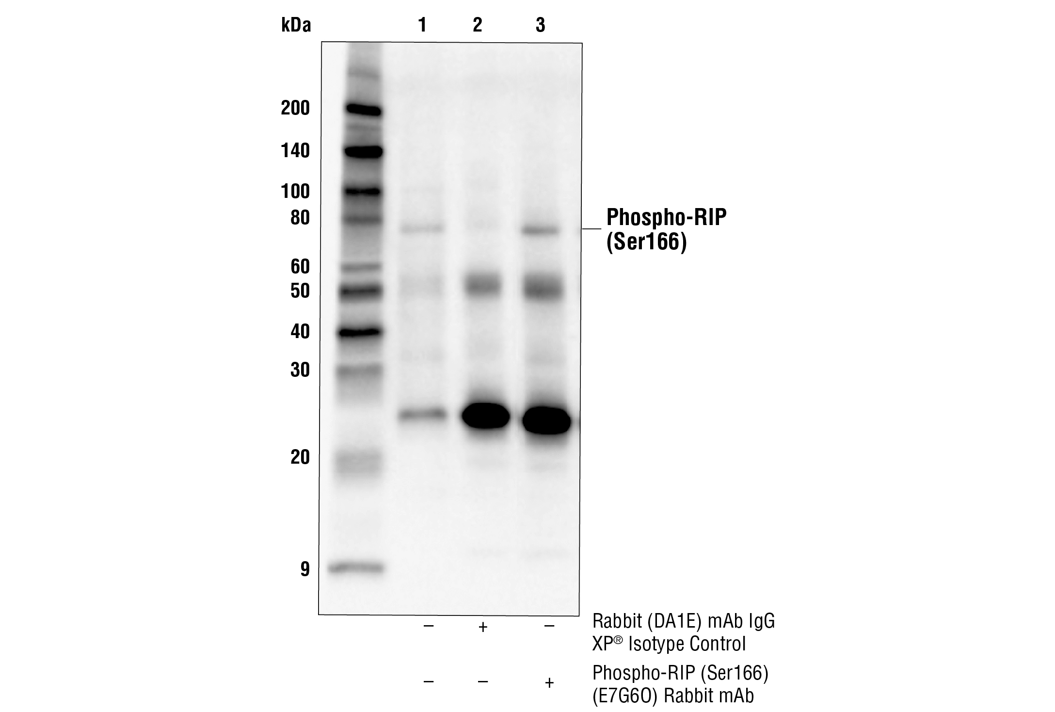
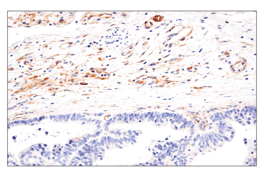
| Phospho-RIP (Ser166) (E7G6O) Rabbit mAb | 53286 | 20 µl | 78 kDa | Rabbit IgG |
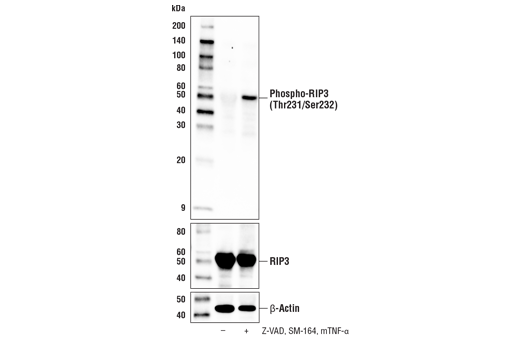
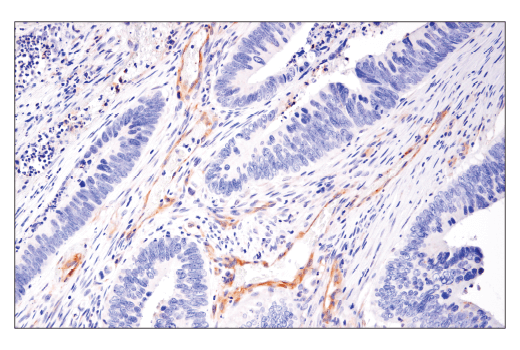
| Phospho-RIP3 (Thr231/Ser232) (E7S1R) Rabbit mAb | 91702 | 20 µl | 46-62 kDa | Rabbit IgG |
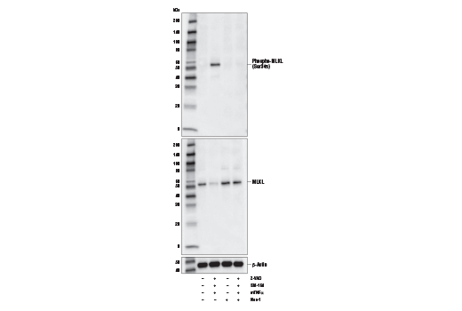
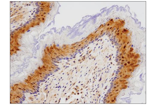
| Phospho-MLKL (Ser345) (D6E3G) Rabbit mAb | 37333 | 20 µl | 54 kDa | Rabbit IgG |
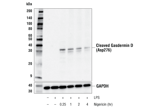
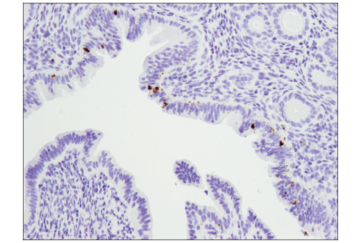
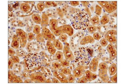
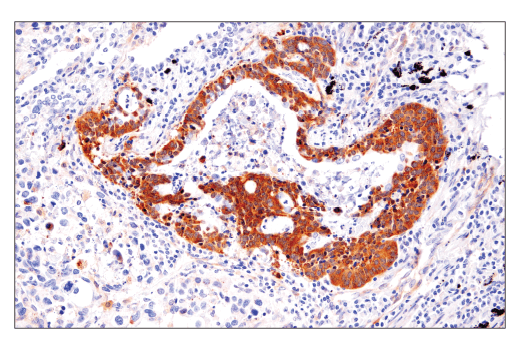
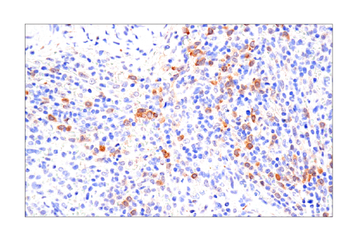
| Cleaved Gasdermin D (Asp276) (E3E3P) Rabbit mAb | 10137 | 20 µl | 31 kDa | Rabbit IgG |
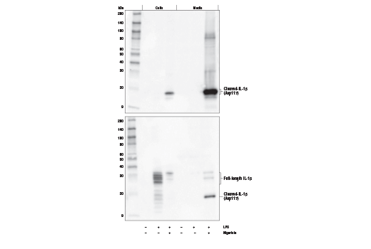
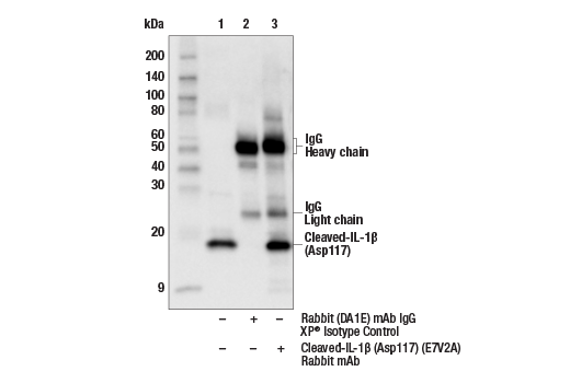
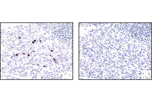
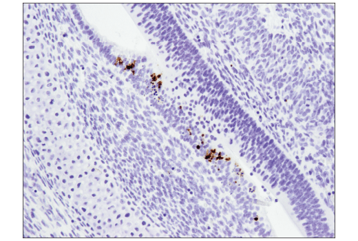
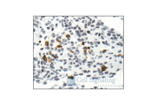
| Cleaved-IL-1β (Asp117) (E7V2A) Rabbit mAb | 63124 | 20 µl | 17 kDa | Rabbit IgG |
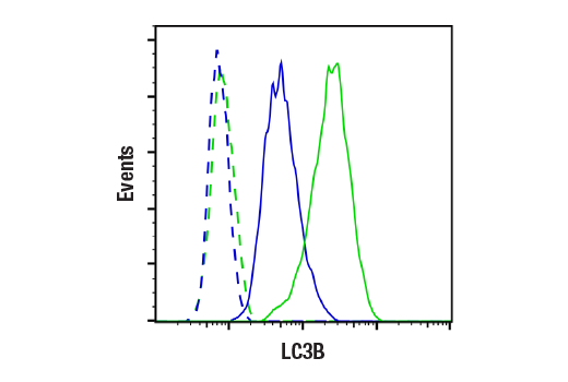
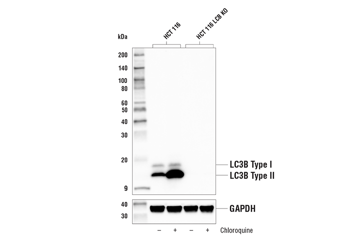
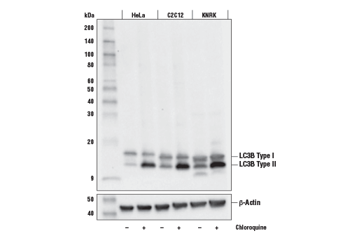
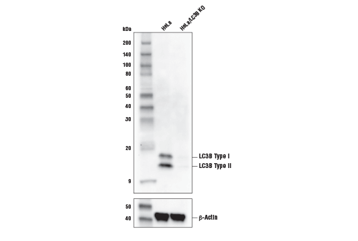
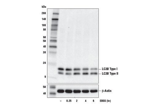
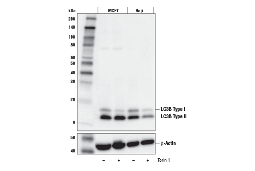
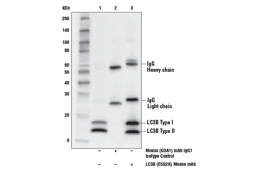
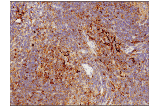
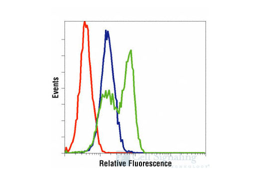
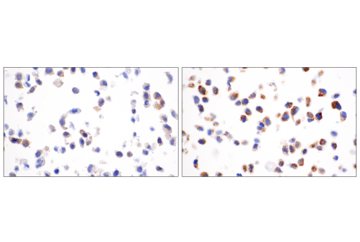
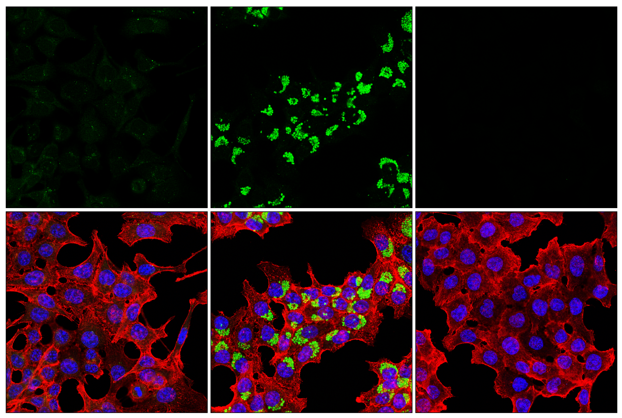
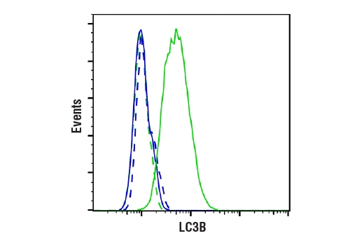
| LC3B (E5Q2K) Mouse mAb | 83506 | 20 µl | 14, 16 kDa | Mouse IgG2b |
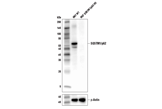
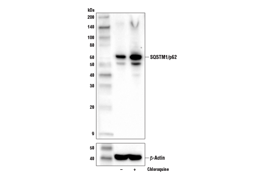
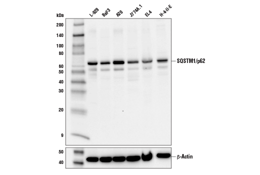
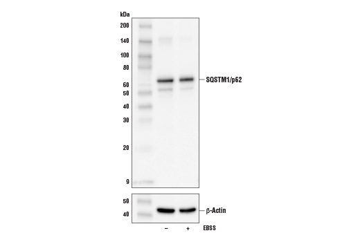
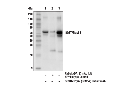
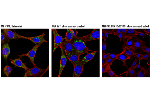
| SQSTM1/p62 (D6M5X) Rabbit mAb | 23214 | 20 µl | 62 kDa | Rabbit IgG |
| Anti-rabbit IgG, HRP-linked Antibody | 7074 | 100 µl | Goat |
Please visit cellsignal.com for individual component applications, species cross-reactivity, dilutions, protocols, and additional product information.
Description
The Mouse Reactive Cell Death and Autophagy Antibody Sampler Kit provides an economical means of detecting common readouts in apoptosis, necroptosis, pyroptosis, and autophagy. The kit includes enough antibodies to perform two western blot experiments with each primary antibody.
Storage
Background
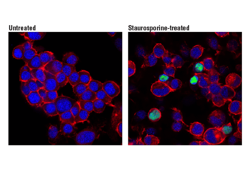
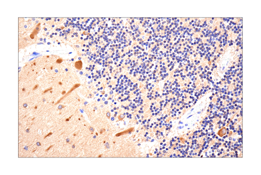
Regulated cell death has been classified based on distinct morphological and biochemical pathways (1). Type I cell death, or apoptosis, is characterized by cytoplasmic shrinkage, chromatin condensation, nuclear fragmentation, plasma membrane blebbing, and phagocytic update of dead cells. Apoptosis can occur through extrinsic pathways involving extracellular factors, including the activation of death receptors, or through intrinsic pathways involving intracellular perturbations, including mitochondrial outer membrane permeabilization (2). Both of these apoptotic pathways lead to activation of caspases, a family of cysteine acid proteases that are synthesized as inactive zymogens containing pro-domains, followed by large (p20) and small (p10) subunits which are proteolytically activated in a cascade-like fashion. Caspase-3 is a key downstream protease activated by both extrinsic and intrinsic apoptotic pathways and cleaves a large number of proteins involved in the disassembly of the cell, including poly(ADP-ribose) polymerase (PARP), a protein involved in the DNA damage response.
Type II cell death, or autophagy, manifests with extensive cytoplasmic vacuolization, and like apoptosis, can include phagocytic update. Autophagy is a catabolic process for the degradation of cellular components including protein aggregates, damaged organelles, and pathogens (3). The process involves the engulfment of these components into a double membrane structure, the autophagosome, which fuses to the lysosome for degradation. Autophagy requires, and can be monitored by, the conversion of LC3 family members, such as LC3B, from a type I form to a lipidated type II form that is incorporated into the autophagosome membrane and binds to a variety of cargo receptors. Cargo receptors such as SQSTM1/p62 bind LC3 along with ubiquitinated proteins that are targeted for degradation. SQSTM1/p62 is also degraded during this process, and thus its expression is frequently used to monitor this process.
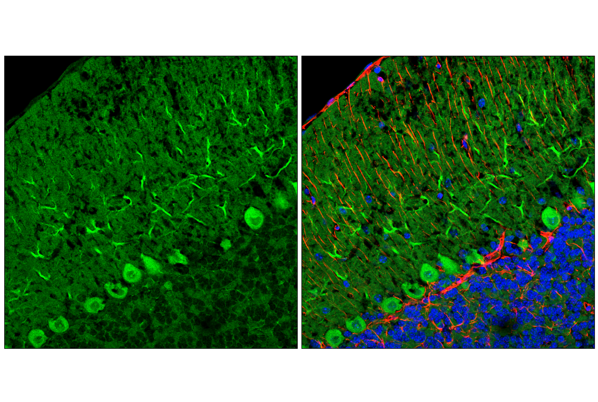
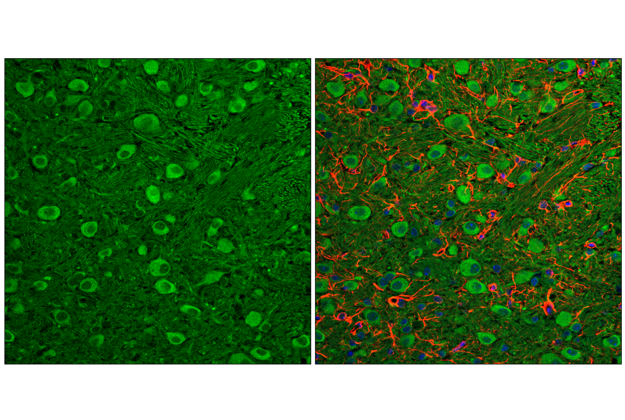
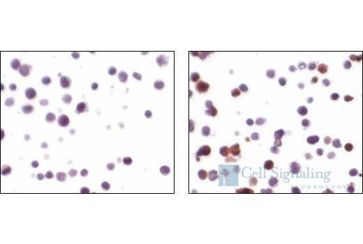
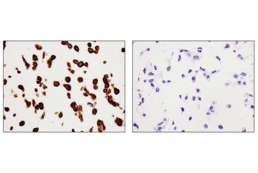
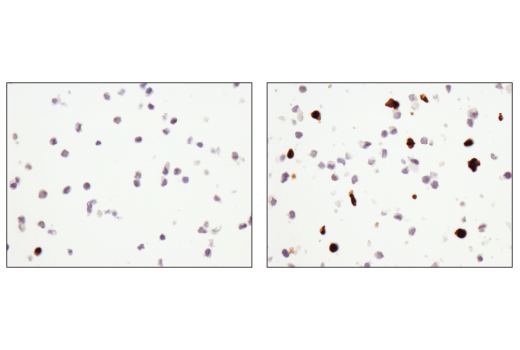
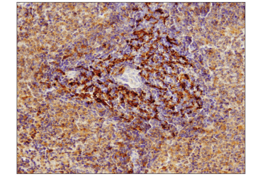
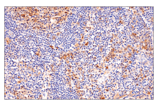
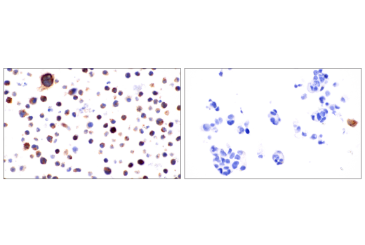
Type III cell death, or necrosis, manifests with plasma membrane permeability with cellular swelling and fragmentation, and lacks a clear phagocytic response which then leads to an inflammatory signaling with the release of damage-associated molecular patterns (DAMPs). Necrosis can be triggered by multiple regulated pathways including necroptosis and pyroptosis. Necroptosis is regulated by the kinase activities of RIP and RIP3 and the pore forming ability of MLKL (4). Necroptosis requires the activation of RIP3 which then phosphorylates MLKL at Ser358 (Ser345 in mouse). Phosphorylation of MLKL leads to generation of a pore complex involved in cell swelling and the secretion of DAMPs. RIP3 activation is triggered through several RIP homotypic interaction motif (RHIM) domain interactions including RIP, TRIF, and ZBP1 and results in the phosphorylation of RIP3 at Ser227 (Thr231/Ser232 in mouse). Canonical necroptosis signaling is mediated by RIP, and this can be inhibited by necrostatins, small molecules that directly inhibit RIP kinase activity. Activation of RIP can be monitored through autophosphorylation sites including Ser166. Pyroptosis is generally induced in cells of the innate immune system, and is characterized by cleavage of Gasdermin D (5). The amino-terminal fragment of Gasdermin D produced following cleavage by inflammatory caspases (Caspase-1, -4, -5), oligomerizes to form a pore. Canonical cleavage of Gasdermin D occurs through a two-step process. The first step involves transcriptional regulation of targets such as NLRP3 and the pro-forms of IL-1β and IL-18. In the second execution step, Caspase-1 is activated through formation of inflammasome complexes. Activated Caspase-1 cleaves Gasdermin D as well as IL-1β and IL-18 to their mature forms, and these active cytokines are secreted through pores formed by Gasdermin D.
Background References
Trademarks and Patents
使用に関する制限
法的な権限を与えられたCSTの担当者が署名した書面によって別途明示的に合意された場合を除き、 CST、その関連会社または代理店が提供する製品には以下の条件が適用されます。お客様が定める条件でここに定められた条件に含まれるものを超えるもの、 または、ここに定められた条件と異なるものは、法的な権限を与えられたCSTの担当者が別途書面にて受諾した場合を除き、拒絶され、 いかなる効力も効果も有しません。
研究専用 (For Research Use Only) またはこれに類似する表示がされた製品は、 いかなる目的についても FDA または外国もしくは国内のその他の規制機関により承認、認可または許可を受けていません。 お客様は製品を診断もしくは治療目的で使用してはならず、また、製品に表示された内容に違反する方法で使用してはなりません。 CST が販売または使用許諾する製品は、エンドユーザーであるお客様に対し、使途を研究および開発のみに限定して提供されるものです。 診断、予防もしくは治療目的で製品を使用することまたは製品を再販売 (単独であるか他の製品等の一部であるかを問いません) もしくはその他の商業的利用の目的で購入することについては、CST から別途許諾を得る必要があります。 お客様は以下の事項を遵守しなければなりません。(a) CST の製品 (単独であるか他の資材と一緒であるかを問いません) を販売、使用許諾、貸与、寄付もしくはその他の態様で第三者に譲渡したり使用させたりしてはなりません。また、商用の製品を製造するために CST の製品を使用してはなりません。(b) 複製、改変、リバースエンジニアリング、逆コンパイル、 分解または他の方法により製品の構造または技術を解明しようとしてはなりません。また、 CST の製品またはサービスと競合する製品またはサービスを開発する目的で CST の製品を使用してはなりません。(c) CST の製品の商標、商号、ロゴ、特許または著作権に関する通知または表示を除去したり改変したりしてはなりません。(d) CST の製品をCST 製品販売条件(CST’s Product Terms of Sale) および該当する書面のみに従って使用しなければなりません。(e) CST の製品に関連してお客様が使用する第三者の製品またはサービスに関する使用許諾条件、 サービス提供条件またはこれに類する合意事項を遵守しなければなりません。