| Product Includes | Product # | Quantity | Mol. Wt | Isotype/Source |
|---|---|---|---|---|
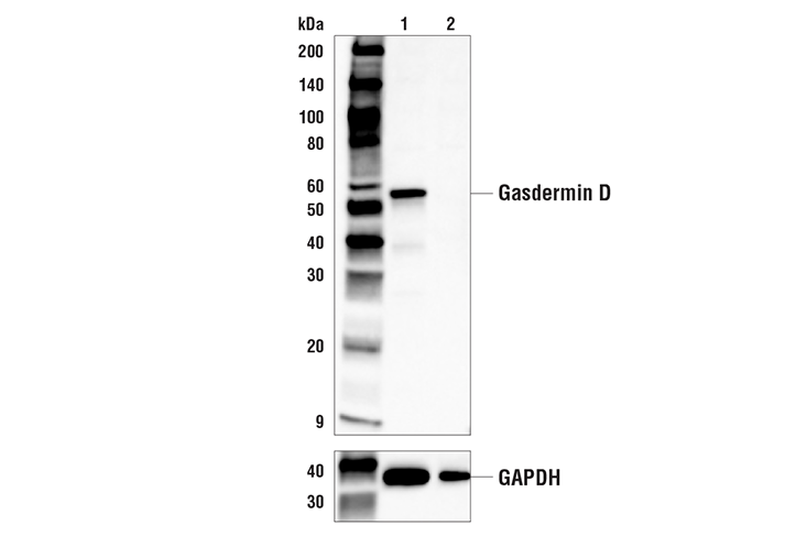
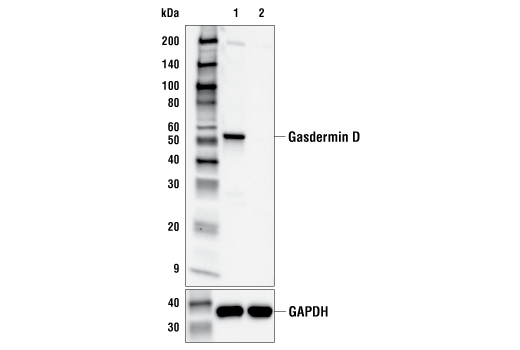
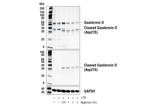
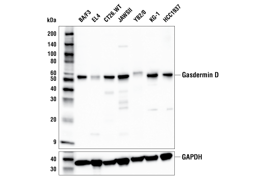
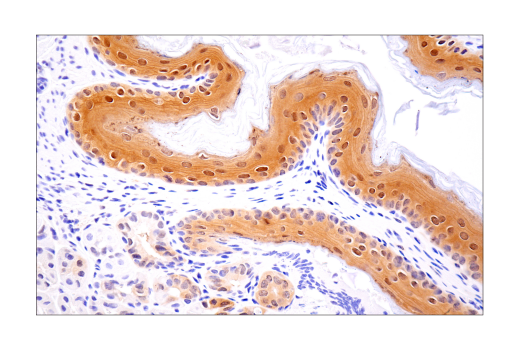
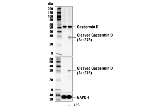
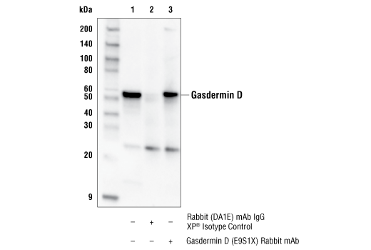
| Gasdermin D (E9S1X) Rabbit mAb | 39754 | 20 µl | 53, 30 kDa | Rabbit IgG |
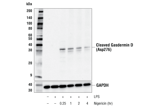
| Cleaved Gasdermin D (Asp276) (E3E3P) Rabbit mAb | 10137 | 20 µl | 31 kDa | Rabbit IgG |
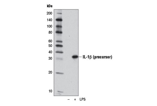
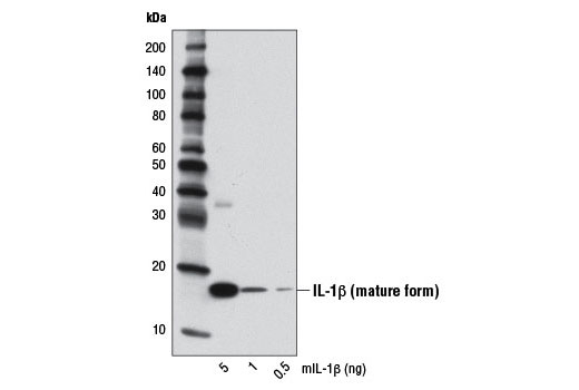
| IL-1β (D3H1Z) Rabbit mAb | 12507 | 20 µl | 17,31 kDa | Rabbit IgG |
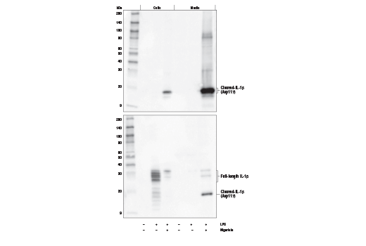
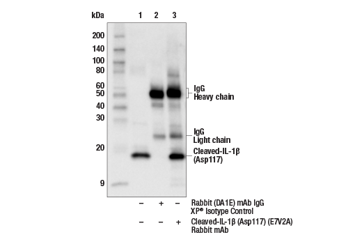
| Cleaved-IL-1β (Asp117) (E7V2A) Rabbit mAb | 63124 | 20 µl | 17 kDa | Rabbit IgG |
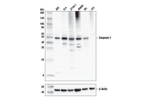
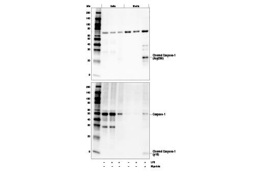
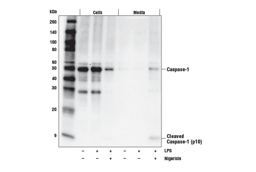
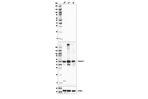
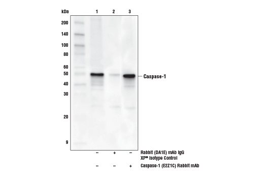
| Caspase-1 (E2Z1C) Rabbit mAb | 24232 | 20 µl | 48, 10 kDa | Rabbit IgG |
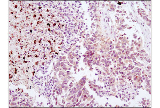
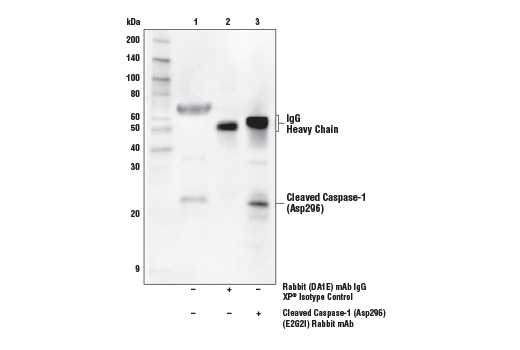
| Cleaved Caspase-1 (Asp296) (E2G2I) Rabbit mAb | 89332 | 20 µl | 22 kDa | Rabbit IgG |
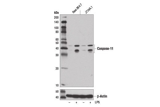
| Caspase-11 (17D9) Rat mAb | 14340 | 20 µl | 38, 43 kDa | Rat IgG2a |
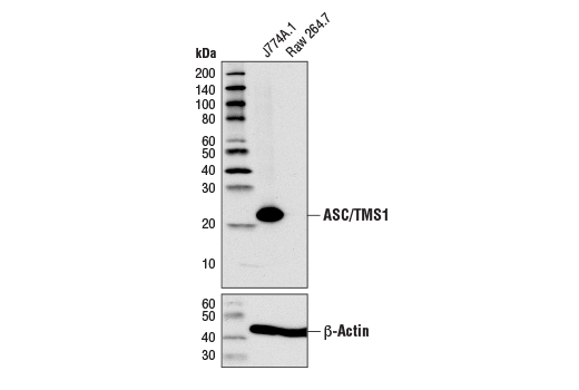
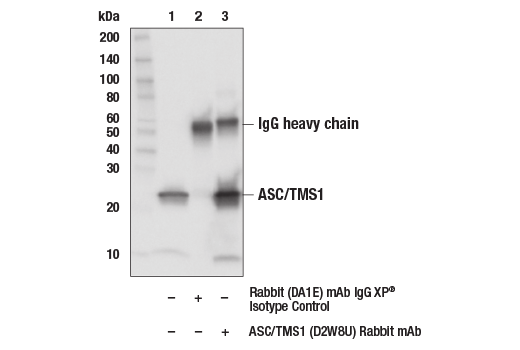
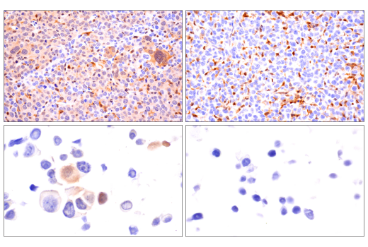
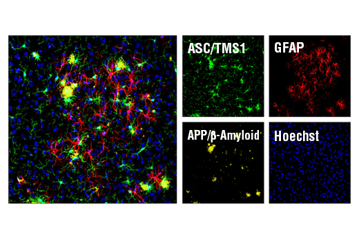
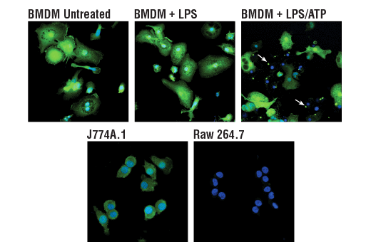
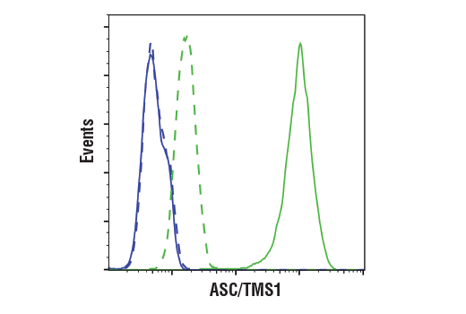
| ASC/TMS1 (D2W8U) Rabbit mAb | 67824 | 20 µl | 22 kDa | Rabbit IgG |
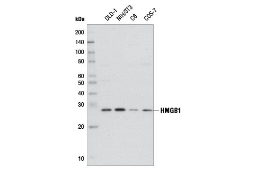
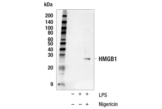
| HMGB1 (D3E5) Rabbit mAb | 6893 | 20 µl | 29 kDa | Rabbit IgG |
| Anti-rabbit IgG, HRP-linked Antibody | 7074 | 100 µl | Goat |
Please visit cellsignal.com for individual component applications, species cross-reactivity, dilutions, protocols, and additional product information.
Description
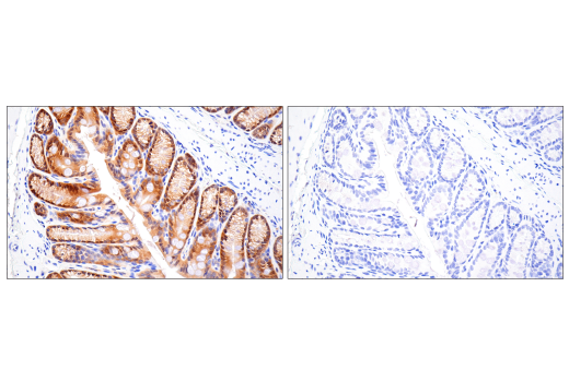
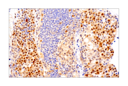
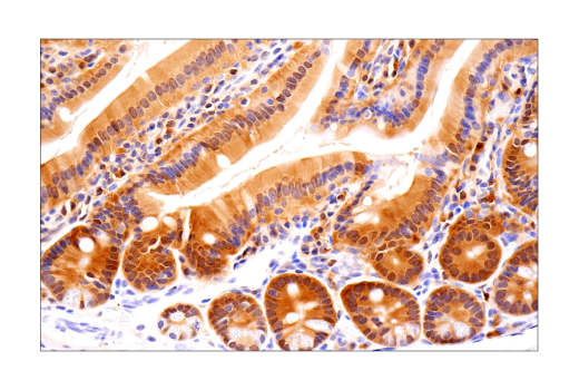
The Mouse Reactive Pyroptosis Antibody Sampler Kit provides an economical means of detecting proteins that are used as readouts for pyroptosis. The kit includes enough antibodies to perform two western blot experiments with each primary antibody.
Storage
Background
Pyroptosis is a regulated pathway of cell death with morphological features of necrosis, including cell swelling, plasma membrane pore formation, and engagement of an inflammatory response with the release of a number of damage-associated molecular patterns (DAMPs), such as HMGB1 and inflammatory cytokines like IL-1β and IL-18 (1,2). Pyroptosis is generally induced in cells of the innate immune system, such as monocytes, macrophages, and dendritic cells in the presence of pathogen-associated molecular patterns (PAMPs) expressed on microbial pathogens or by cell-derived DAMPs. It is induced through assembly of inflammasomes triggering proteolytic activation of caspase-1 which then cleaves inflammatory cytokines like IL-1β and IL-18 to their mature forms (3). A critical feature of pyroptosis is the cleavage of Gasdermin D by caspase-1 and mouse caspase-11 (or human caspase-4/5) (4-6). Upon cleavage, the N-terminal fragment of Gasdermin D oligomerizes to form a pore, allowing secretion of inflammatory DAMPs and cytokines. Canonical inflammasome assembly typically consists of a cytosolic-pattern recognition receptor (PPR; a nucleotide binding domain and leucine-rich repeat [NLR] or AIM2-like family members), an adaptor protein (ASC/TMS1), and pro-caspase-1. Distinct inflammasome complexes can recognize distinct PAMPs and DAMPs to trigger pyroptosis. The best characterized pathway triggered by the NLR, NLRP3, occurs through a two-step process. The first step is a priming signal, NF-κB is activated to induce the expression of a number of inflammasome components including NLRP3, pro-IL-1β, and pro-IL-18. In the second activation step, caspase-1 is activated and Gasdermin D and cytokines are proteolytically activated. In a non-canonical pathway, caspase-4 and caspase-5 can directly trigger Gasdermin D cleavage in monocytes following LPS stimulation (5,7).
- Frank, D. and Vince, J.E. (2019) Cell Death Differ 26, 99-114.
- Shi, J. et al. (2017) Trends Biochem Sci 42, 245-54.
- Malik, A. and Kanneganti, T.D. (2017) J Cell Sci 130, 3955-63.
- He, W.T. et al. (2015) Cell Res 25, 1285-98.
- Shi, J. et al. (2015) Nature 526, 660-5.
- Kayagaki, N. et al. (2015) Nature 526, 666-71.
- Viganò, E. et al. (2015) Nat Commun 6, 8761.
Background References
Trademarks and Patents
使用に関する制限
法的な権限を与えられたCSTの担当者が署名した書面によって別途明示的に合意された場合を除き、 CST、その関連会社または代理店が提供する製品には以下の条件が適用されます。お客様が定める条件でここに定められた条件に含まれるものを超えるもの、 または、ここに定められた条件と異なるものは、法的な権限を与えられたCSTの担当者が別途書面にて受諾した場合を除き、拒絶され、 いかなる効力も効果も有しません。
研究専用 (For Research Use Only) またはこれに類似する表示がされた製品は、 いかなる目的についても FDA または外国もしくは国内のその他の規制機関により承認、認可または許可を受けていません。 お客様は製品を診断もしくは治療目的で使用してはならず、また、製品に表示された内容に違反する方法で使用してはなりません。 CST が販売または使用許諾する製品は、エンドユーザーであるお客様に対し、使途を研究および開発のみに限定して提供されるものです。 診断、予防もしくは治療目的で製品を使用することまたは製品を再販売 (単独であるか他の製品等の一部であるかを問いません) もしくはその他の商業的利用の目的で購入することについては、CST から別途許諾を得る必要があります。 お客様は以下の事項を遵守しなければなりません。(a) CST の製品 (単独であるか他の資材と一緒であるかを問いません) を販売、使用許諾、貸与、寄付もしくはその他の態様で第三者に譲渡したり使用させたりしてはなりません。また、商用の製品を製造するために CST の製品を使用してはなりません。(b) 複製、改変、リバースエンジニアリング、逆コンパイル、 分解または他の方法により製品の構造または技術を解明しようとしてはなりません。また、 CST の製品またはサービスと競合する製品またはサービスを開発する目的で CST の製品を使用してはなりません。(c) CST の製品の商標、商号、ロゴ、特許または著作権に関する通知または表示を除去したり改変したりしてはなりません。(d) CST の製品をCST 製品販売条件(CST’s Product Terms of Sale) および該当する書面のみに従って使用しなければなりません。(e) CST の製品に関連してお客様が使用する第三者の製品またはサービスに関する使用許諾条件、 サービス提供条件またはこれに類する合意事項を遵守しなければなりません。