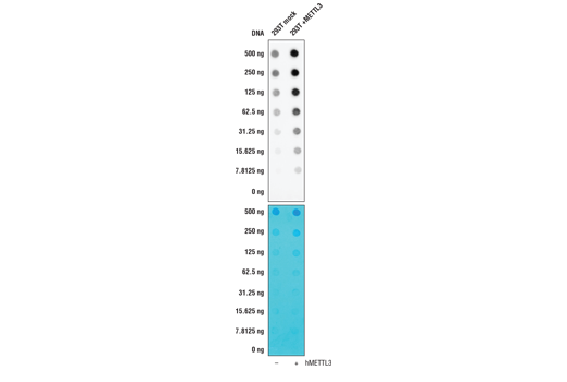
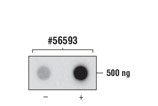
R Dot Blot
All
Endogenous
Rabbit IgG
Product Information
Product Usage Information
This antibody has been shown by an independent laboratory to work in RNA-IP-seq. Please use at an assay-dependent dilution.
Storage
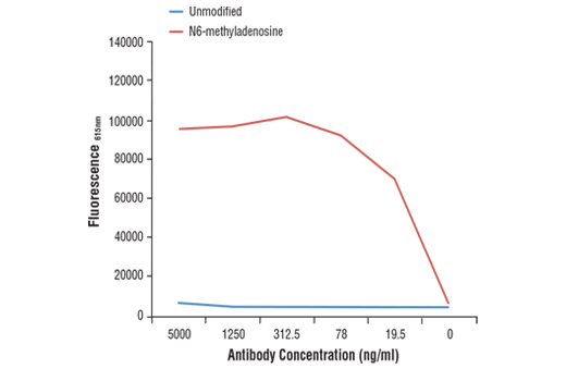
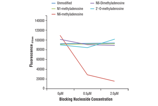
Specificity / Sensitivity
Species Reactivity:
All Species Expected
Source / Purification
Monoclonal antibody is produced by immunizing animals with N6-methyladenosine.
Background
N6-methyladenosine (m6A) is a post-transcriptional modification found in various RNA subtypes. While the presence of m6A in RNA was described decades ago, the lack of tools has made interrogating the epitranscriptomic landscape challenging (1,2). With the emergence of new technologies such as miCLIP and NG-RNA-seq, researchers have been able to show that m6A is a biologically relevant mark in mRNA that is enriched in 3’ UTRs and stop codons (3,4). The m6A writer complex consists of a core heterodimer of methyltransferase-like protein 3 (METTL3) and methytransferase-like protein 14 (METTL14), and the additional regulatory proteins Virlizer/VIRMA and Wilms tumor 1-associated protein (WTAP) (5). METTL3 is the catalytic methyltransferase subunit and METTL14 is the target recognition subunit that binds to RNA (6). The Virilzer/VIRMA protein directs m6A methylation to the 3’ UTRs and stop codons, and WTAP targets the complex to nuclear speckles, which are sites of RNA processing (7). Less is known about readers and erasers of m6A, and while the fat mass and obesity-associated protein FTO was the first discovered m6A demethylase, subsequent studies demonstrated that this enzyme may prefer the closely related m6Am mark in vivo (8,9). ALKBH5 was later shown to be a bona fide m6A demethylase enzyme, contributing to the idea that the m6A modification is dynamically regulated (10). Readers of the m6A mark include the YTH protein family, which can bind to m6A and influence mRNA stability and translation efficiency (3,11-13). The m6A mark and machinery have been shown to regulate a variety of cellular functions, including RNA splicing, translational control, pluripotency and cell fate determination, neuronal function, and disease (1, 14-17). The m6A writer complex has been linked to various cancer types including AML and endometrial cancers (18,19). Additionally, m6A has been implicated in resistance to chemotherapy (20).
- Meyer, K.D. and Jaffrey, S.R. (2017) Annu Rev Cell Dev Biol 33, 319-42.
- Desrosiers, R. et al. (1974) Proc Natl Acad Sci U S A 71, 3971-5.
- Dominissini, D. et al. (2012) Nature 485, 201-6.
- Meyer, K.D. et al. (2012) Cell 149, 1635-46.
- Liu, J. et al. (2014) Nat Chem Biol 10, 93-5.
- Wang, X. et al. (2016) Nature 534, 575-8.
- Ping, X.L. et al. (2014) Cell Res 24, 177-89.
- Jia, G. et al. (2011) Nat Chem Biol 7, 885-7.
- Mauer, J. et al. (2017) Nature 541, 371-75.
- Zheng, G. et al. (2013) Mol Cell 49, 18-29.
- Schwartz, S. et al. (2013) Cell 155, 1409-21.
- Wang, X. et al. (2014) Nature 505, 117-20.
- Wang, X. et al. (2015) Cell 161, 1388-99.
- Batista, P.J. et al. (2014) Cell Stem Cell 15, 707-19.
- Batista, P.J. (2017) Genomics Proteomics Bioinformatics 15, 154-63.
- Patil, D.P. et al. (2016) Nature 537, 369-73.
- Wang, C.X. et al. (2018) PLoS Biol 16, e2004880.
- Barbieri, I. et al. (2017) Nature 552, 126-31.
- Liu, J. et al. (2018) Nat Cell Biol 20, 1074-83.
- Dai, D. et al. (2018) Cell Death Dis 9, 124.
Species Reactivity
Species reactivity is determined by testing in at least one approved application (e.g., western blot).
Applications Key
R Dot Blot: RNA Dot Blot
Cross-Reactivity Key
H: human M: mouse R: rat Hm: hamster Mk: monkey Vir: virus Mi: mink C: chicken Dm: D. melanogaster X: Xenopus Z: zebrafish B: bovine Dg: dog Pg: pig Sc: S. cerevisiae Ce: C. elegans Hr: horse GP: Guinea Pig Rab: rabbit All: all species expected
Trademarks and Patents
使用に関する制限
法的な権限を与えられたCSTの担当者が署名した書面によって別途明示的に合意された場合を除き、 CST、その関連会社または代理店が提供する製品には以下の条件が適用されます。お客様が定める条件でここに定められた条件に含まれるものを超えるもの、 または、ここに定められた条件と異なるものは、法的な権限を与えられたCSTの担当者が別途書面にて受諾した場合を除き、拒絶され、 いかなる効力も効果も有しません。
研究専用 (For Research Use Only) またはこれに類似する表示がされた製品は、 いかなる目的についても FDA または外国もしくは国内のその他の規制機関により承認、認可または許可を受けていません。 お客様は製品を診断もしくは治療目的で使用してはならず、また、製品に表示された内容に違反する方法で使用してはなりません。 CST が販売または使用許諾する製品は、エンドユーザーであるお客様に対し、使途を研究および開発のみに限定して提供されるものです。 診断、予防もしくは治療目的で製品を使用することまたは製品を再販売 (単独であるか他の製品等の一部であるかを問いません) もしくはその他の商業的利用の目的で購入することについては、CST から別途許諾を得る必要があります。 お客様は以下の事項を遵守しなければなりません。(a) CST の製品 (単独であるか他の資材と一緒であるかを問いません) を販売、使用許諾、貸与、寄付もしくはその他の態様で第三者に譲渡したり使用させたりしてはなりません。また、商用の製品を製造するために CST の製品を使用してはなりません。(b) 複製、改変、リバースエンジニアリング、逆コンパイル、 分解または他の方法により製品の構造または技術を解明しようとしてはなりません。また、 CST の製品またはサービスと競合する製品またはサービスを開発する目的で CST の製品を使用してはなりません。(c) CST の製品の商標、商号、ロゴ、特許または著作権に関する通知または表示を除去したり改変したりしてはなりません。(d) CST の製品をCST 製品販売条件(CST’s Product Terms of Sale) および該当する書面のみに従って使用しなければなりません。(e) CST の製品に関連してお客様が使用する第三者の製品またはサービスに関する使用許諾条件、 サービス提供条件またはこれに類する合意事項を遵守しなければなりません。