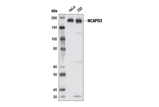
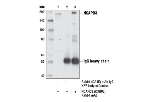
WB, IP
H
Endogenous
170
Rabbit IgG
#P42695
23310
Product Information
Product Usage Information
| Application | Dilution |
|---|---|
| Western Blotting | 1:1000 |
| Immunoprecipitation | 1:100 |
Storage
Specificity / Sensitivity
Species Reactivity:
Human
Source / Purification
Monoclonal antibody is produced by immunizing animals with a synthetic peptide corresponding to residues near the carboxy terminus of human NCAPD3 protein.
Background
The structural maintenance of chromosomes 2 (SMC2) and 4 (SMC4) proteins are condensin complex subunits that enable chromosome condensation and compaction during migration to opposite poles during anaphase (1,2). Condensin is a general regulator of chromosome architecture that may also regulate gene expression and DNA repair. Condensin complex subunits SMC2 and SMC4 form a functional ATPase essential for chromatin condensation, while three auxiliary subunits regulate ATPase activity. Both SMC2 and SMC4 are found within two distinct condensin complexes (condensin I and II) in higher eukaryotes. Condensin I contains auxiliary subunits NCAPD2, NCAPG, and NCAPH, while condensin II contains related auxiliary proteins NCAPD3, NCAPG2, and NCAPH2 (1,2).
Each condensin complex exhibits different localization patterns during the cell cycle and provides for distinct functions during mitosis (3-5). Condensin I is cytoplasmic during interphase and binds chromatin following the breakdown of the nuclear envelope at the end of prophase. Condensin I is required for complete dissociation of cohesin from chromosome arms, for chromosome shortening, and for normal timing of progression through pro-metaphase and metaphase. Mutations in corresponding condensin I genes result in cytokinesis defects due to the persistence of anaphase fibers. Condensin II is nuclear during interphase, but does not bind to chromatin until early prophase where it remains bound until the end of telophase. Condensin II is required for initial chromatin condensation during early prophase. Mutations in corresponding condensin II genes produce high numbers of anaphase bridges resulting from incomplete chromosome segregation. Condensin II complex subunit D3 (NCAPD3) plays a pivotal role in the loading of condensin II onto chromatin and the regulation of chromatin condensation (6,7). NCAPD3 protein contains HEAT repeat clusters that bind to mono-methyl histone H4 Lys20, a histone mark prevalent during mitosis and important for DNA repair and chromatin condensation (6). Increased mono-methyl histone H4 Lys20 levels caused by dissociation of the histone demethylase PHF8 from chromatin and increased expression of the methyltransferase SET8, leads to increased binding of NCAPD3 and condensin II to chromosomes early in mitosis (6). Phosphorylation of NCAPD3 at Thr1415 by CDK1 kinase (cdc2) leads to the recruitment of PLK1 kinase, which hyperphosphorylates condensin II and facilitates mitotic chromosome assembly (7).
- Losada, A. and Hirano, T. (2005) Genes Dev 19, 1269-87.
- Hudson, D.F. et al. (2009) Chromosome Res 17, 131-44.
- Hirota, T. et al. (2004) J Cell Sci 117, 6435-45.
- Ono, T. et al. (2004) Mol Biol Cell 15, 3296-308.
- Green, L.C. et al. (2012) J Cell Sci 125, 1591-604.
- Liu, W. et al. (2010) Nature 466, 508-12.
- Abe, S. et al. (2011) Genes Dev 25, 863-74.
Species Reactivity
Species reactivity is determined by testing in at least one approved application (e.g., western blot).
Western Blot Buffer
IMPORTANT: For western blots, incubate membrane with diluted primary antibody in 5% w/v BSA, 1X TBS, 0.1% Tween® 20 at 4°C with gentle shaking, overnight.
Applications Key
WB: Western Blotting IP: Immunoprecipitation
Cross-Reactivity Key
H: human M: mouse R: rat Hm: hamster Mk: monkey Vir: virus Mi: mink C: chicken Dm: D. melanogaster X: Xenopus Z: zebrafish B: bovine Dg: dog Pg: pig Sc: S. cerevisiae Ce: C. elegans Hr: horse GP: Guinea Pig Rab: rabbit All: all species expected
Trademarks and Patents
使用に関する制限
法的な権限を与えられたCSTの担当者が署名した書面によって別途明示的に合意された場合を除き、 CST、その関連会社または代理店が提供する製品には以下の条件が適用されます。お客様が定める条件でここに定められた条件に含まれるものを超えるもの、 または、ここに定められた条件と異なるものは、法的な権限を与えられたCSTの担当者が別途書面にて受諾した場合を除き、拒絶され、 いかなる効力も効果も有しません。
研究専用 (For Research Use Only) またはこれに類似する表示がされた製品は、 いかなる目的についても FDA または外国もしくは国内のその他の規制機関により承認、認可または許可を受けていません。 お客様は製品を診断もしくは治療目的で使用してはならず、また、製品に表示された内容に違反する方法で使用してはなりません。 CST が販売または使用許諾する製品は、エンドユーザーであるお客様に対し、使途を研究および開発のみに限定して提供されるものです。 診断、予防もしくは治療目的で製品を使用することまたは製品を再販売 (単独であるか他の製品等の一部であるかを問いません) もしくはその他の商業的利用の目的で購入することについては、CST から別途許諾を得る必要があります。 お客様は以下の事項を遵守しなければなりません。(a) CST の製品 (単独であるか他の資材と一緒であるかを問いません) を販売、使用許諾、貸与、寄付もしくはその他の態様で第三者に譲渡したり使用させたりしてはなりません。また、商用の製品を製造するために CST の製品を使用してはなりません。(b) 複製、改変、リバースエンジニアリング、逆コンパイル、 分解または他の方法により製品の構造または技術を解明しようとしてはなりません。また、 CST の製品またはサービスと競合する製品またはサービスを開発する目的で CST の製品を使用してはなりません。(c) CST の製品の商標、商号、ロゴ、特許または著作権に関する通知または表示を除去したり改変したりしてはなりません。(d) CST の製品をCST 製品販売条件(CST’s Product Terms of Sale) および該当する書面のみに従って使用しなければなりません。(e) CST の製品に関連してお客様が使用する第三者の製品またはサービスに関する使用許諾条件、 サービス提供条件またはこれに類する合意事項を遵守しなければなりません。