WB, IP, IHC-P, IF-IC, ChIP
H
Endogenous
270
Rabbit IgG
#O75376
9611
Product Information
Product Usage Information
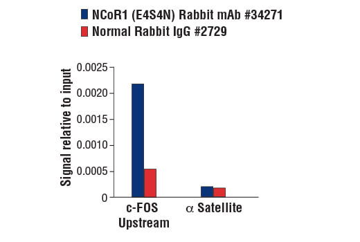
For optimal ChIP and ChIP-seq results, use 10 μl of antibody and 10 μg of chromatin (approximately 4 × 10^6 cells) per IP. This antibody has been validated using SimpleChIP® Enzymatic Chromatin IP Kits.
| Application | Dilution |
|---|---|
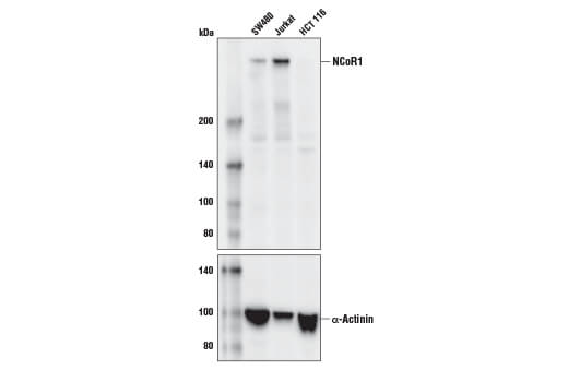
| Western Blotting | 1:1000 |
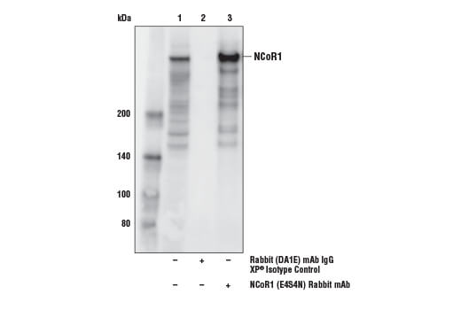
| Immunoprecipitation | 1:200 |
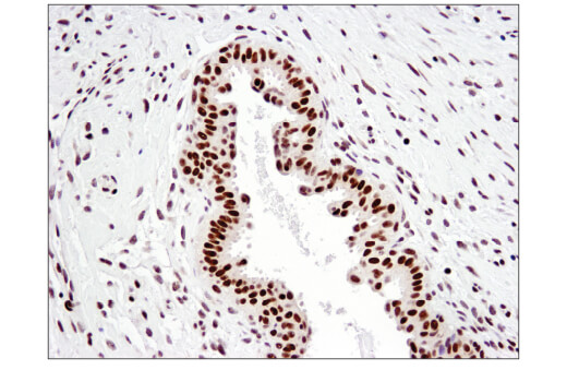
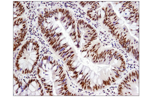
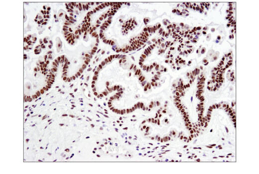
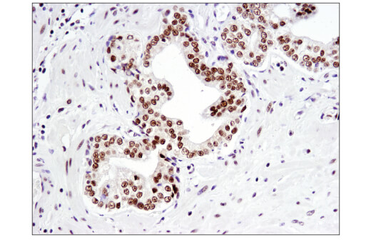
| Immunohistochemistry (Paraffin) | 1:100 |
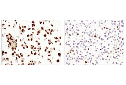
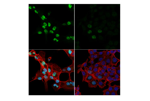
| Immunofluorescence (Immunocytochemistry) | 1:800 |
| Chromatin IP | 1:50 |
Storage
Specificity / Sensitivity
Species Reactivity:
Human
Source / Purification
Monoclonal antibody is produced by immunizing animals with recombinant protein specific to the carboxy terminus of human NCoR1 protein.
Background
The most well characterized nuclear receptor corepressors are SMRT (silencing mediator for retinoic acid and thyroid hormone receptors) and its close paralog NCoR1 (nuclear receptor corepressor) (1,2). NCoR1 functions to transcriptionally silence various unliganded, DNA bound non-steroidal nuclear receptors by serving as a large molecular scaffold that bridges the receptors with multiple chromatin remodeling factors that repress nuclear receptor-mediated gene transcription, in part, through deacetylation of core histones surrounding target promoters. Indeed, the N-terminal portion of NCoR1 possesses multiple distinct transcriptional repression domains (RDs) reponsible for the recruitment of additional components of the corepressor complex such as HDACs, mSin3, GPS2, and TBL1/TBLR1. In between the RDs lies a pair of potent repressor motifs known as SANT motifs (SWI3, ADA2, N-CoR, and TFIIIB), which recruit HDAC3 and histones to the repressor complex in order to enhance HDAC3 activity (3). The C-terminal portion of NCoR1 contains multiple nuclear receptor interaction domains (NDs), each of which contains a conserved CoRNR box (or L/I-X-X-I/V-I) motif that allow for binding to various unliganded nuclear hormone receptors such as thyroid hormone (THR) and retinoic acid (RAR) receptors (4,5).
Recent genetic studies in mice have not only corroborated the wealth of biochemical studies involving NCoR1 but have also provided significant insight regarding the function of NCoR1 in mammalian development and physiology. Although it has been observed that loss of Ncor1 does not affect early embyonic development, likely due to compensation by Smrt, embryonic lethality ultimately results during mid-gestation, largely due to defects in erythropoesis and thymopoesis (6). Another study demonstrated that the NDs of NCoR1 are critical for its ability to function in a physiological setting as a transcriptional repressor of hepatic THR and Liver X Receptor (LXR) (7).
- Chen, J.D. and Evans, R.M. (1995) Nature 377, 454-7.
- Hörlein, A.J. et al. (1995) Nature 377, 397-404.
- Jones, P.L. and Shi, Y.B. (2003) Curr Top Microbiol Immunol 274, 237-68.
- Downes, M. et al. (1996) Nucleic Acids Res 24, 4379-86.
- Wong, C.W. and Privalsky, M.L. (1998) Mol Cell Biol 18, 5724-33.
- Jepsen, K. et al. (2000) Cell 102, 753-63.
- Astapova, I. et al. (2008) Proc Natl Acad Sci U S A 105, 19544-9.
Species Reactivity
Species reactivity is determined by testing in at least one approved application (e.g., western blot).
Western Blot Buffer
IMPORTANT: For western blots, incubate membrane with diluted primary antibody in 5% w/v BSA, 1X TBS, 0.1% Tween® 20 at 4°C with gentle shaking, overnight.
Applications Key
WB: Western Blotting IP: Immunoprecipitation IHC-P: Immunohistochemistry (Paraffin) IF-IC: Immunofluorescence (Immunocytochemistry) ChIP: Chromatin IP
Cross-Reactivity Key
H: human M: mouse R: rat Hm: hamster Mk: monkey Vir: virus Mi: mink C: chicken Dm: D. melanogaster X: Xenopus Z: zebrafish B: bovine Dg: dog Pg: pig Sc: S. cerevisiae Ce: C. elegans Hr: horse GP: Guinea Pig Rab: rabbit All: all species expected
Trademarks and Patents
使用に関する制限
法的な権限を与えられたCSTの担当者が署名した書面によって別途明示的に合意された場合を除き、 CST、その関連会社または代理店が提供する製品には以下の条件が適用されます。お客様が定める条件でここに定められた条件に含まれるものを超えるもの、 または、ここに定められた条件と異なるものは、法的な権限を与えられたCSTの担当者が別途書面にて受諾した場合を除き、拒絶され、 いかなる効力も効果も有しません。
研究専用 (For Research Use Only) またはこれに類似する表示がされた製品は、 いかなる目的についても FDA または外国もしくは国内のその他の規制機関により承認、認可または許可を受けていません。 お客様は製品を診断もしくは治療目的で使用してはならず、また、製品に表示された内容に違反する方法で使用してはなりません。 CST が販売または使用許諾する製品は、エンドユーザーであるお客様に対し、使途を研究および開発のみに限定して提供されるものです。 診断、予防もしくは治療目的で製品を使用することまたは製品を再販売 (単独であるか他の製品等の一部であるかを問いません) もしくはその他の商業的利用の目的で購入することについては、CST から別途許諾を得る必要があります。 お客様は以下の事項を遵守しなければなりません。(a) CST の製品 (単独であるか他の資材と一緒であるかを問いません) を販売、使用許諾、貸与、寄付もしくはその他の態様で第三者に譲渡したり使用させたりしてはなりません。また、商用の製品を製造するために CST の製品を使用してはなりません。(b) 複製、改変、リバースエンジニアリング、逆コンパイル、 分解または他の方法により製品の構造または技術を解明しようとしてはなりません。また、 CST の製品またはサービスと競合する製品またはサービスを開発する目的で CST の製品を使用してはなりません。(c) CST の製品の商標、商号、ロゴ、特許または著作権に関する通知または表示を除去したり改変したりしてはなりません。(d) CST の製品をCST 製品販売条件(CST’s Product Terms of Sale) および該当する書面のみに従って使用しなければなりません。(e) CST の製品に関連してお客様が使用する第三者の製品またはサービスに関する使用許諾条件、 サービス提供条件またはこれに類する合意事項を遵守しなければなりません。