WB, IF-F
H M R
Endogenous
200
Rabbit IgG
#O94856
23114
Product Information
Product Usage Information
This formulation is ideal for use with technologies requiring specialized or custom antibody labeling, including fluorophores, metals, lanthanides, and oligonucleotides. It is not recommended for ChIP, ChIP-seq, CUT&RUN or CUT&Tag assays. If you require a carrier free formulation for chromatin profiling, please contact us. Optimal dilutions/concentrations should be determined by the end user.
Formulation
Storage
Specificity / Sensitivity
Species Reactivity:
Human, Mouse, Rat
Source / Purification
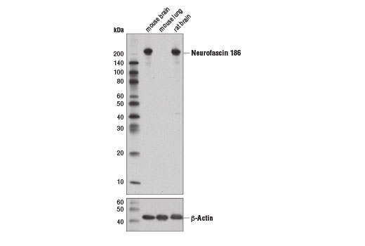
Monoclonal antibody is produced by immunizing animals with a synthetic peptide corresponding to residues surrounding Thr1108 of human neurofascin 186 protein.
Background
Myelinated axons contain un-myelinated gaps called nodes of Ranvier. These regularly spaced gaps are critical for the proper propagation and rapid conduction of nerve impulses in the central and peripheral nervous system (1). The structure and organization of the nodes of Ranvier is dictated by interaction between the axon and glial cells (2). Voltage-gated sodium channels concentrated at the nodes and potassium channels clustered at the paranodes are responsible for propagation of the action potentials (3,4). Other proteins that contribute to the architecture and function of the nodes of Ranvier include βIV spectrin (5), ankyrin-G (6), and the L1 cell adhesion molecules, neurofascin and NrCAM (7,8).
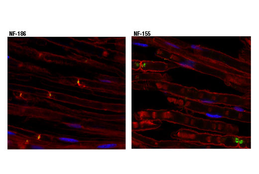
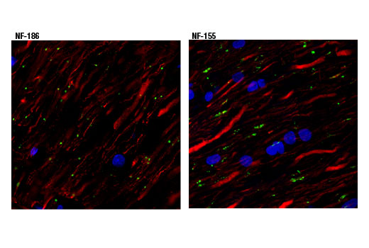
Alternative splicing produces several neurofascin isoforms that differ in temporal and spatial expression. Neurofascin 186 is expressed in axons where it is concentrated at the nodes. Research studies indicate that neurofascin 186 is responsible for nodal assembly and clustering of sodium channels (9). Neurofascin 155 is expressed in glial cells and is localized to myelin paranodes. Interactions between neurofascin 155 and the contactin-associated protein (Caspr) tether the myelin sheath to the axon (10). N-linked glycosylation results in two forms of neurofascin 155 (high and low) that are differentially expressed during development (11).
- Black, J.A. et al. (1990) Trends Neurosci 13, 48-54.
- Salzer, J.L. (1997) Neuron 18, 843-6.
- Waxman, S.G. et al. (1989) Proc Natl Acad Sci U S A 86, 1406-10.
- Ritchie, J.M. (1992) Trends Neurosci 15, 345-51.
- Berghs, S. et al. (2000) J Cell Biol 151, 985-1002.
- Zhou, D. et al. (1998) J Cell Biol 143, 1295-304.
- Davis, J.Q. et al. (1996) J Cell Biol 135, 1355-67.
- Ratcliffe, C.F. et al. (2001) J Cell Biol 154, 427-34.
- Thaxton, C. et al. (2011) Neuron 69, 244-57.
- Charles, P. et al. (2002) Curr Biol 12, 217-20.
- Pomicter, A.D. et al. (2010) Brain 133, 389-405.
Species Reactivity
Species reactivity is determined by testing in at least one approved application (e.g., western blot).
Applications Key
WB: Western Blotting IF-F: Immunofluorescence (Frozen)
Cross-Reactivity Key
H: human M: mouse R: rat Hm: hamster Mk: monkey Vir: virus Mi: mink C: chicken Dm: D. melanogaster X: Xenopus Z: zebrafish B: bovine Dg: dog Pg: pig Sc: S. cerevisiae Ce: C. elegans Hr: horse GP: Guinea Pig Rab: rabbit All: all species expected
Trademarks and Patents
使用に関する制限
法的な権限を与えられたCSTの担当者が署名した書面によって別途明示的に合意された場合を除き、 CST、その関連会社または代理店が提供する製品には以下の条件が適用されます。お客様が定める条件でここに定められた条件に含まれるものを超えるもの、 または、ここに定められた条件と異なるものは、法的な権限を与えられたCSTの担当者が別途書面にて受諾した場合を除き、拒絶され、 いかなる効力も効果も有しません。
研究専用 (For Research Use Only) またはこれに類似する表示がされた製品は、 いかなる目的についても FDA または外国もしくは国内のその他の規制機関により承認、認可または許可を受けていません。 お客様は製品を診断もしくは治療目的で使用してはならず、また、製品に表示された内容に違反する方法で使用してはなりません。 CST が販売または使用許諾する製品は、エンドユーザーであるお客様に対し、使途を研究および開発のみに限定して提供されるものです。 診断、予防もしくは治療目的で製品を使用することまたは製品を再販売 (単独であるか他の製品等の一部であるかを問いません) もしくはその他の商業的利用の目的で購入することについては、CST から別途許諾を得る必要があります。 お客様は以下の事項を遵守しなければなりません。(a) CST の製品 (単独であるか他の資材と一緒であるかを問いません) を販売、使用許諾、貸与、寄付もしくはその他の態様で第三者に譲渡したり使用させたりしてはなりません。また、商用の製品を製造するために CST の製品を使用してはなりません。(b) 複製、改変、リバースエンジニアリング、逆コンパイル、 分解または他の方法により製品の構造または技術を解明しようとしてはなりません。また、 CST の製品またはサービスと競合する製品またはサービスを開発する目的で CST の製品を使用してはなりません。(c) CST の製品の商標、商号、ロゴ、特許または著作権に関する通知または表示を除去したり改変したりしてはなりません。(d) CST の製品をCST 製品販売条件(CST’s Product Terms of Sale) および該当する書面のみに従って使用しなければなりません。(e) CST の製品に関連してお客様が使用する第三者の製品またはサービスに関する使用許諾条件、 サービス提供条件またはこれに類する合意事項を遵守しなければなりません。