| Product Includes | Product # | Quantity | Mol. Wt | Isotype/Source |
|---|---|---|---|---|
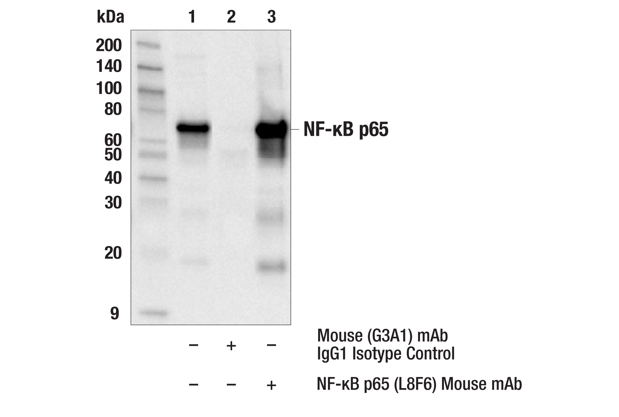
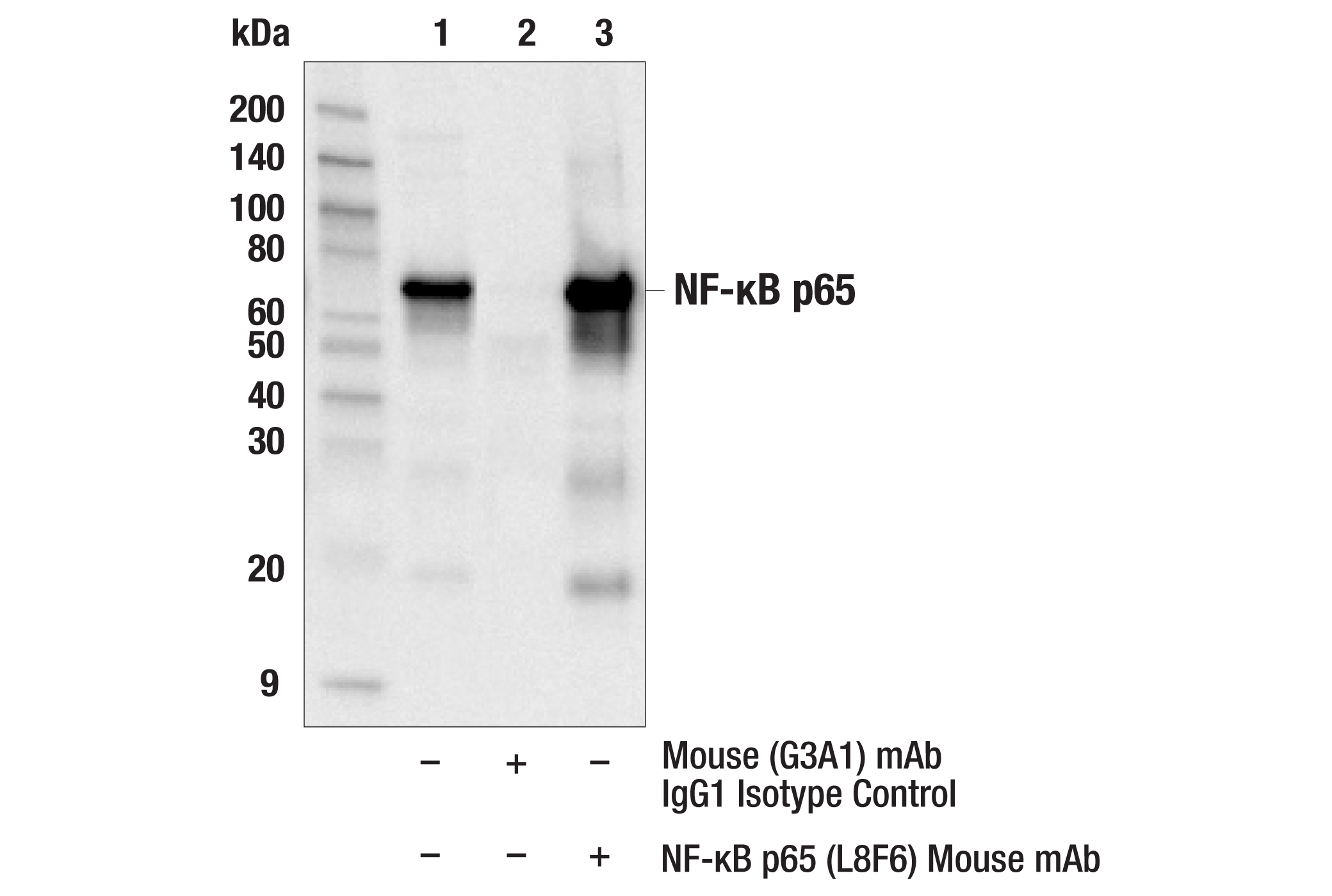
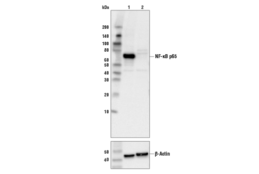
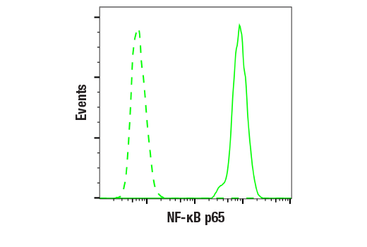
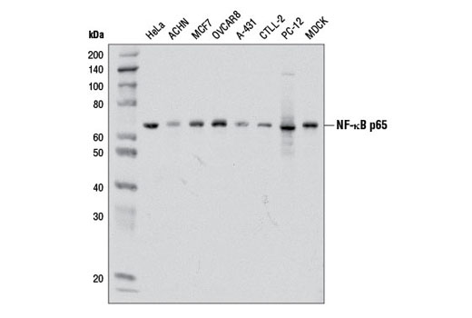
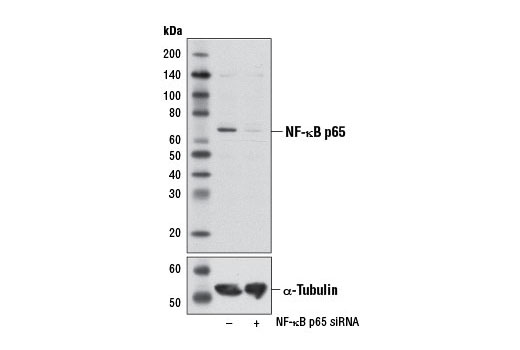
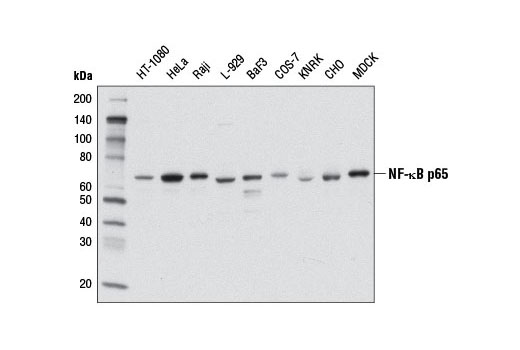
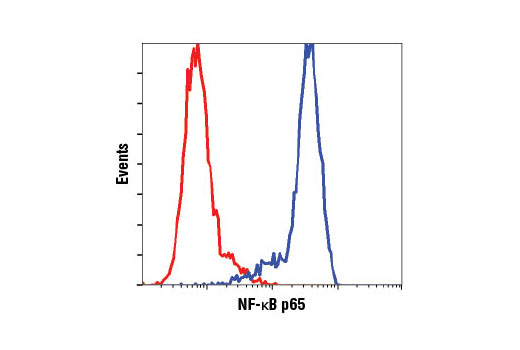
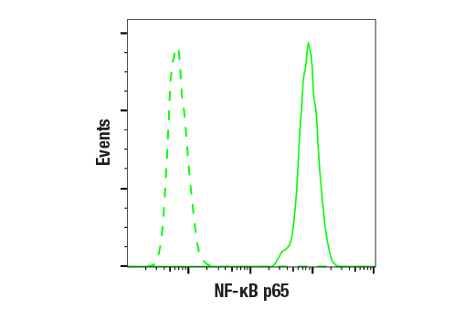
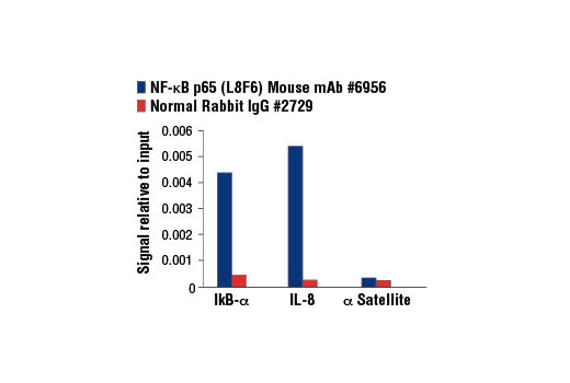
| NF-κB p65 (L8F6) Mouse mAb | 6956 | 20 µl | 65 kDa | Mouse IgG2b |
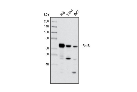
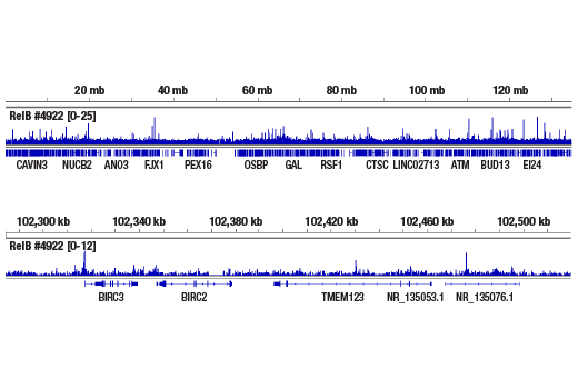
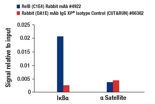
| RelB (C1E4) Rabbit mAb | 4922 | 20 µl | 70 kDa | Rabbit IgG |
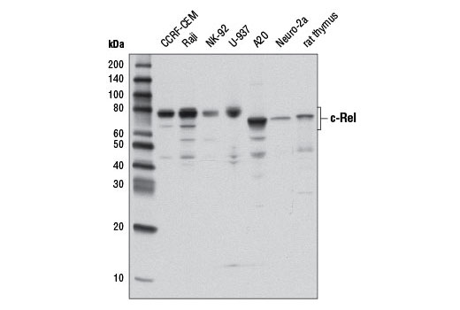
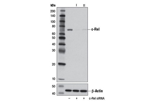
| c-Rel (D4Y6M) Rabbit mAb | 12707 | 20 µl | 68-78 kDa | Rabbit IgG |
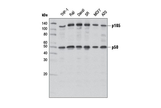
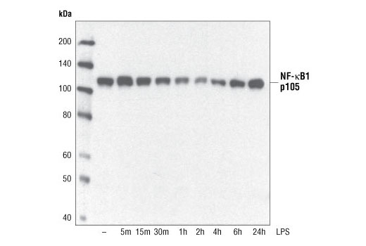
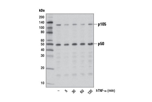
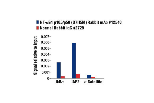
| NF-κB1 p105/p50 (D7H5M) Rabbit mAb | 12540 | 20 µl | 50 Active form. 120 Precursor kDa | Rabbit IgG |
| NF-κB1 p105 Antibody | 4717 | 20 µl | 120 kDa | Rabbit |
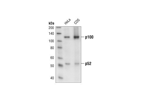
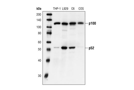
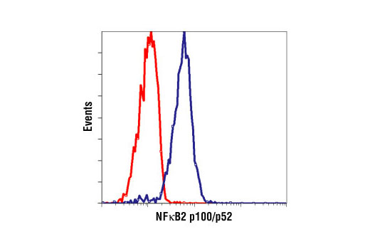
| NF-κB2 p100/p52 Antibody | 4882 | 20 µl | 52 (mature). 120 (precursor). kDa | Rabbit |
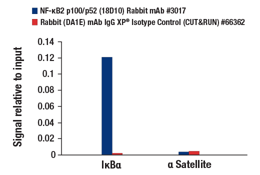
| NF-κB2 p100/p52 (18D10) Rabbit mAb | 3017 | 20 µl | 52 active form. 120 precursor. kDa | Rabbit IgG |
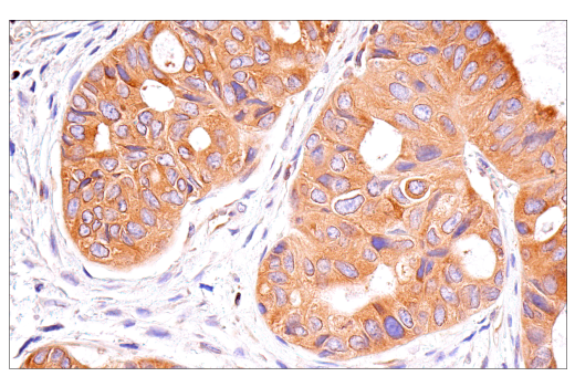
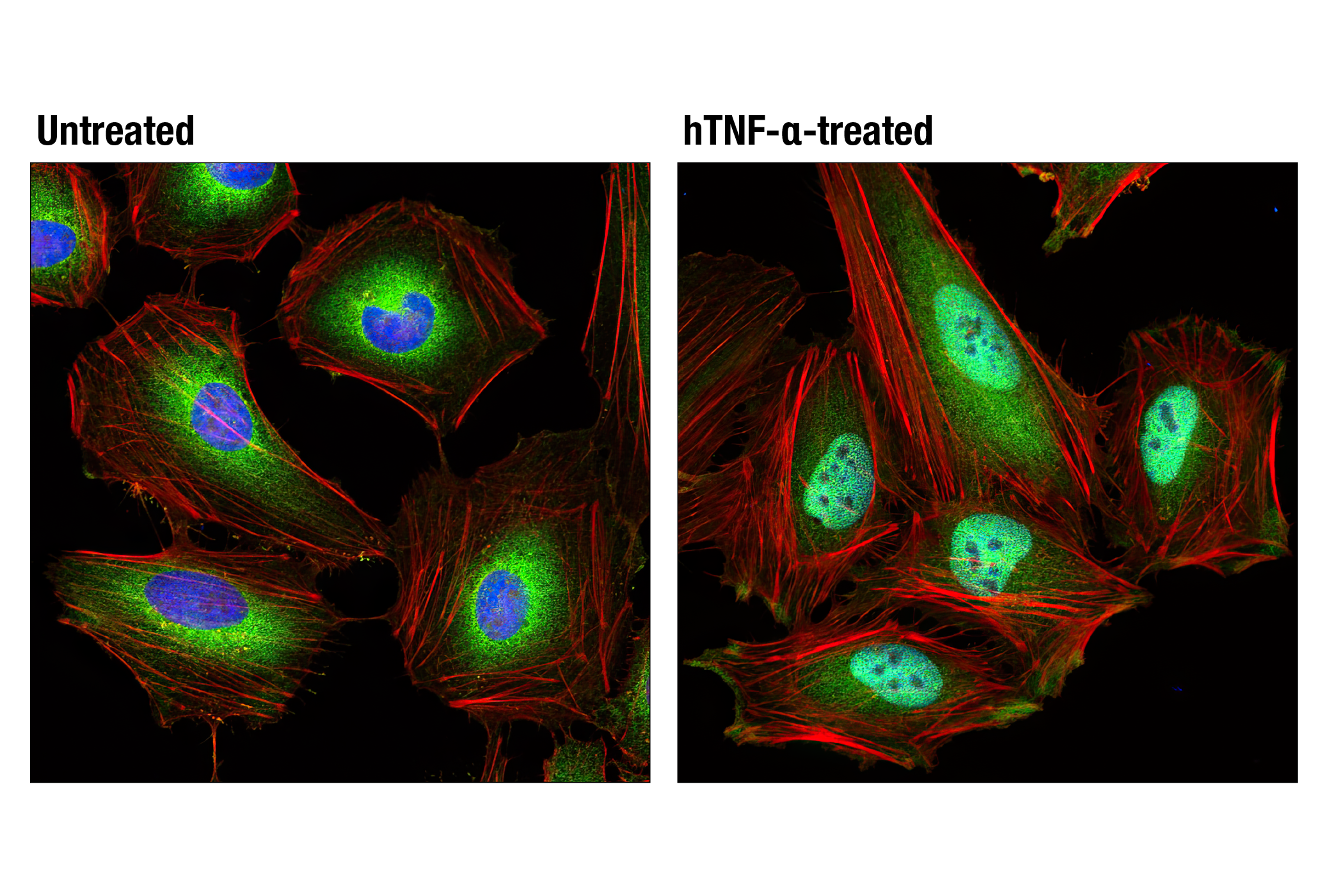
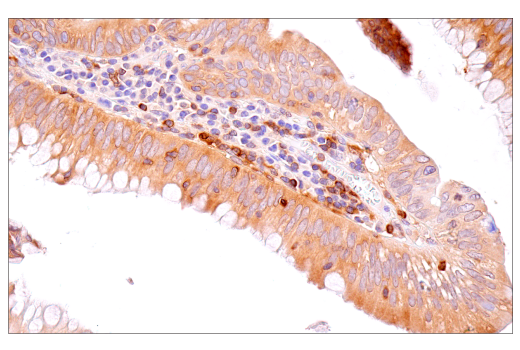
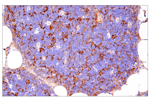
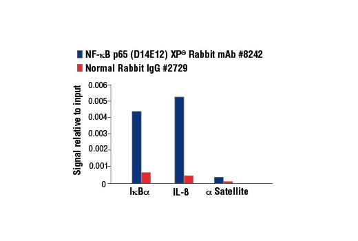
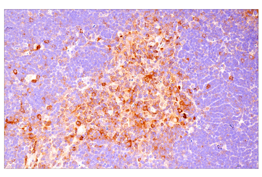
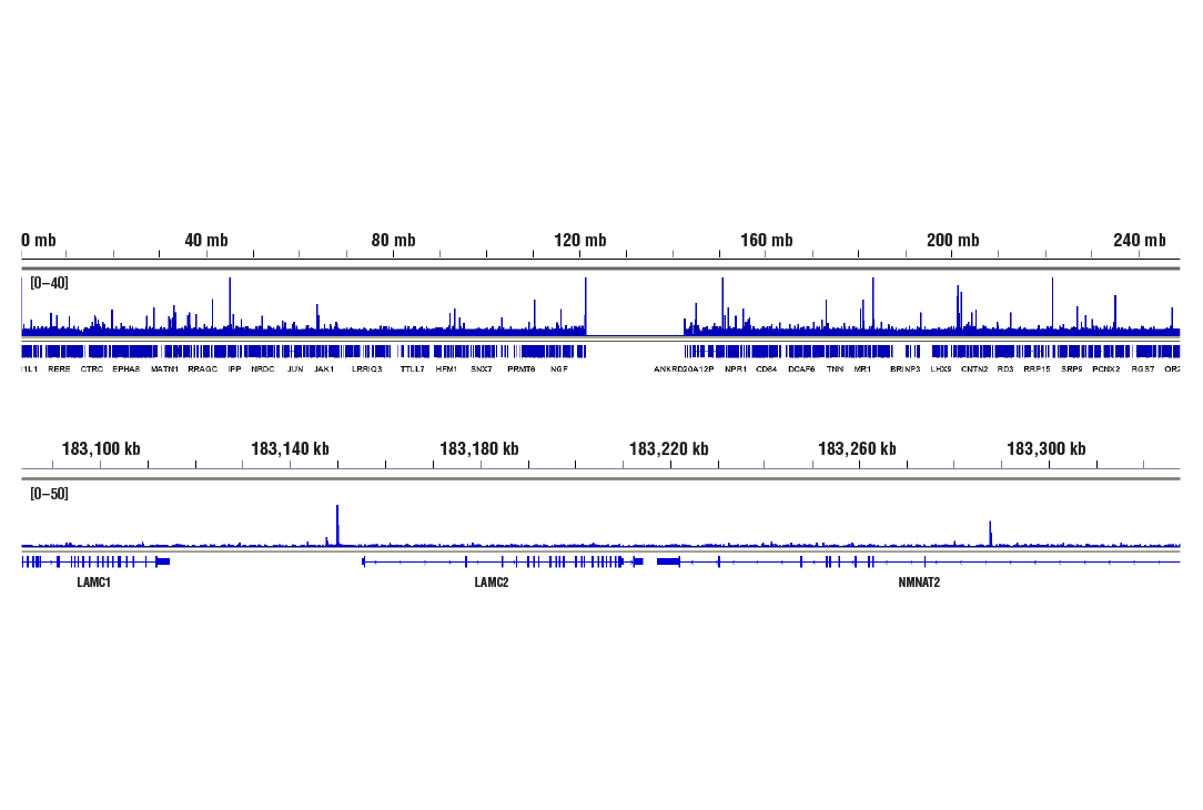
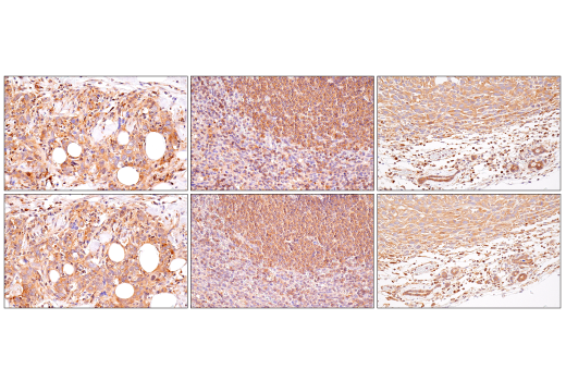
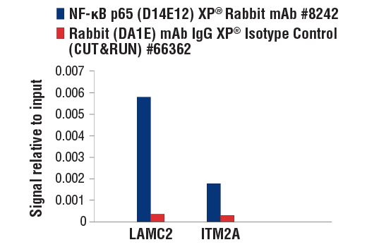
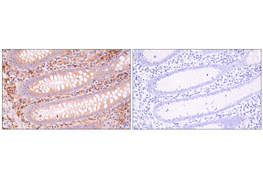
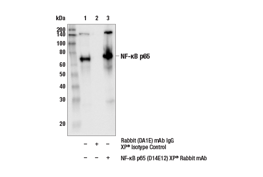
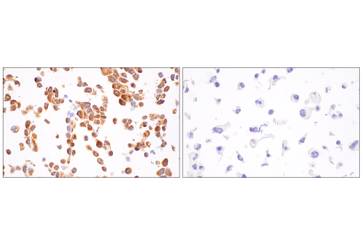
| NF-κB p65 (D14E12) XP® Rabbit mAb | 8242 | 20 µl | 65 kDa | Rabbit IgG |
| Anti-rabbit IgG, HRP-linked Antibody | 7074 | 100 µl | Goat | |
| Anti-mouse IgG, HRP-linked Antibody | 7076 | 100 µl | Horse |
Please visit cellsignal.com for individual component applications, species cross-reactivity, dilutions, protocols, and additional product information.
Description
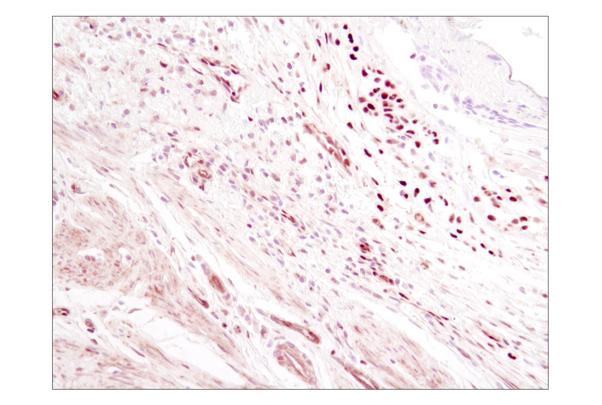
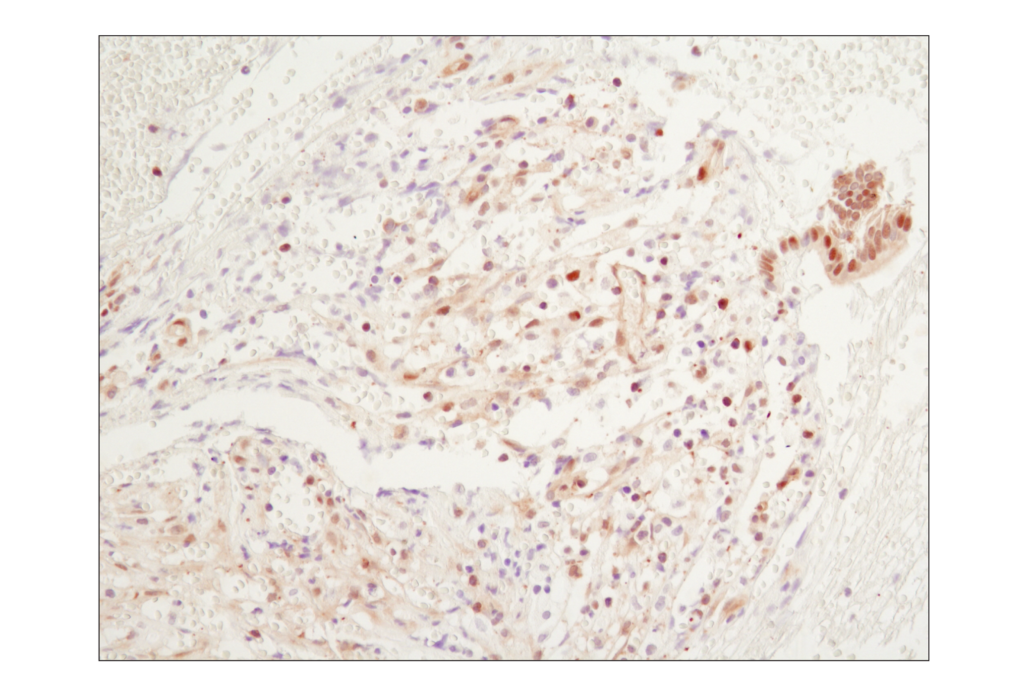
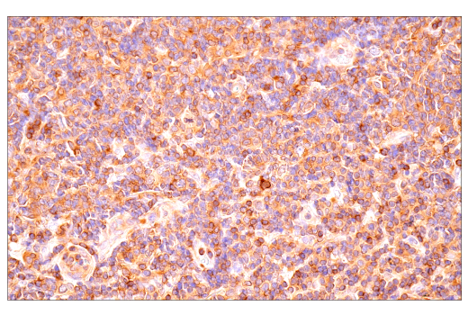
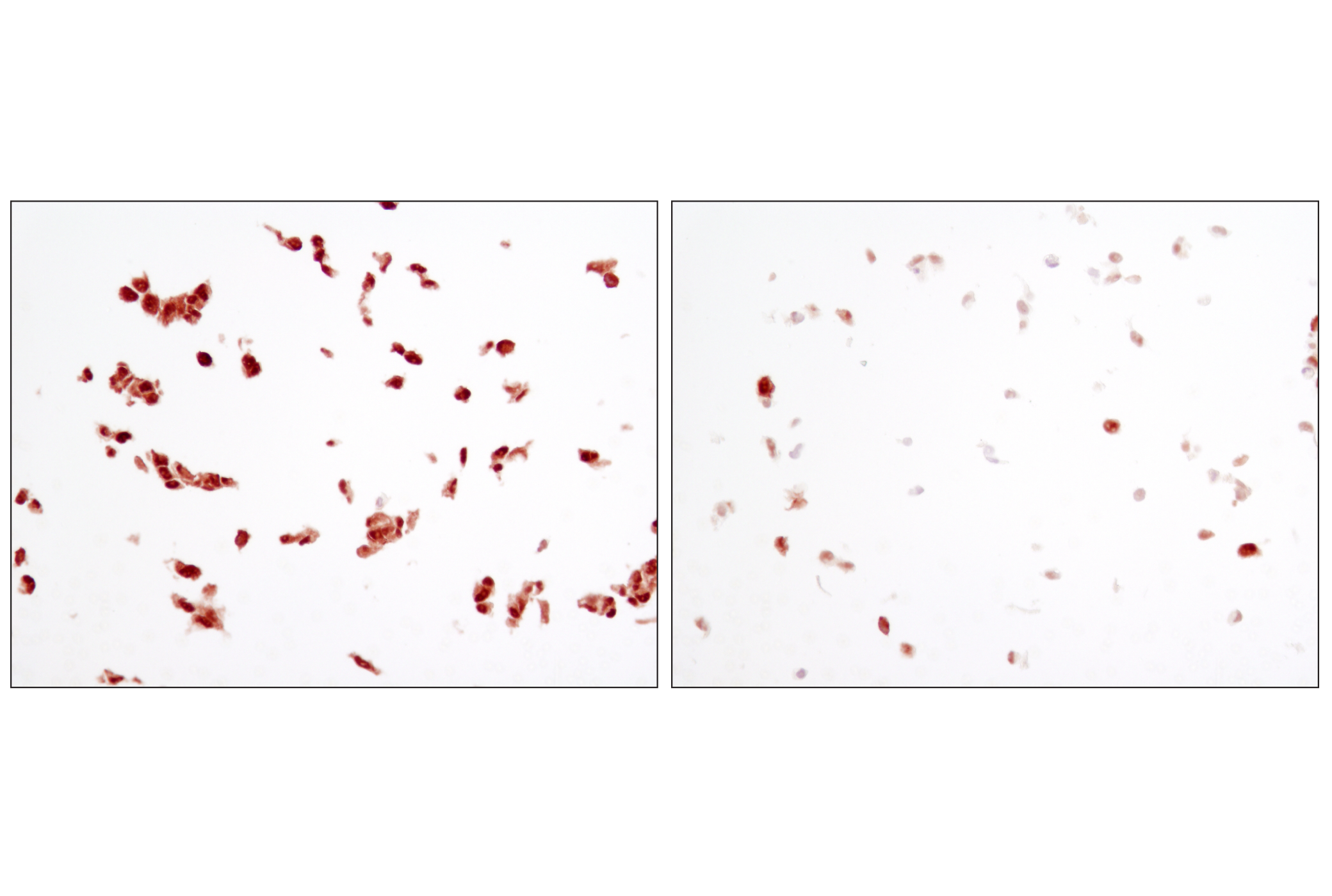
This kit contains reagents to examine total protein levels of the five NF-κB/Rel family members: p65/RelA, RelB, c-Rel, NF-κB1 (p105/p50) and NF-κB2 (p100/p52).
Storage
Background
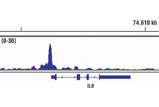
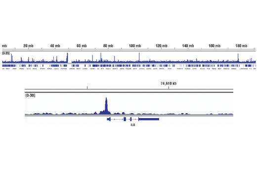
Transcription factors of the nuclear factor κB (NF-κB)/Rel family play a pivotal role in inflammatory and immune responses (1,2). There are five family members in mammals: RelA, c-Rel, RelB, NF-κB1 (p105/p50), and NF-κB2 (p100/p52). Both p105 and p100 are proteolytically processed by the proteasome to produce p50 and p52, respectively. Rel proteins bind p50 and p52 to form dimeric complexes that bind DNA and regulate transcription. In unstimulated cells, NF-κB is sequestered in the cytoplasm by IκB inhibitory proteins (3-5). NF-κB-activating agents can induce the phosphorylation of IκB proteins, targeting them for rapid degradation through the ubiquitin-proteasome pathway and releasing NF-κB to enter the nucleus where it regulates gene expression (6-8). NIK and IKKα (IKK1) regulate the phosphorylation and processing of NF-κB2 (p100) to produce p52, which translocates to the nucleus (9-11).
- Baeuerle, P.A. and Henkel, T. (1994) Annu Rev Immunol 12, 141-79.
- Baeuerle, P.A. and Baltimore, D. (1996) Cell 87, 13-20.
- Haskill, S. et al. (1991) Cell 65, 1281-9.
- Thompson, J.E. et al. (1995) Cell 80, 573-82.
- Whiteside, S.T. et al. (1997) EMBO J 16, 1413-26.
- Traenckner, E.B. et al. (1995) EMBO J 14, 2876-83.
- Scherer, D.C. et al. (1995) Proc Natl Acad Sci USA 92, 11259-63.
- Chen, Z.J. et al. (1996) Cell 84, 853-62.
- Senftleben, U. et al. (2001) Science 293, 1495-9.
- Coope, H.J. et al. (2002) EMBO J 21, 5375-85.
- Xiao, G. et al. (2001) Mol Cell 7, 401-9.
Background References
Trademarks and Patents
使用に関する制限
法的な権限を与えられたCSTの担当者が署名した書面によって別途明示的に合意された場合を除き、 CST、その関連会社または代理店が提供する製品には以下の条件が適用されます。お客様が定める条件でここに定められた条件に含まれるものを超えるもの、 または、ここに定められた条件と異なるものは、法的な権限を与えられたCSTの担当者が別途書面にて受諾した場合を除き、拒絶され、 いかなる効力も効果も有しません。
研究専用 (For Research Use Only) またはこれに類似する表示がされた製品は、 いかなる目的についても FDA または外国もしくは国内のその他の規制機関により承認、認可または許可を受けていません。 お客様は製品を診断もしくは治療目的で使用してはならず、また、製品に表示された内容に違反する方法で使用してはなりません。 CST が販売または使用許諾する製品は、エンドユーザーであるお客様に対し、使途を研究および開発のみに限定して提供されるものです。 診断、予防もしくは治療目的で製品を使用することまたは製品を再販売 (単独であるか他の製品等の一部であるかを問いません) もしくはその他の商業的利用の目的で購入することについては、CST から別途許諾を得る必要があります。 お客様は以下の事項を遵守しなければなりません。(a) CST の製品 (単独であるか他の資材と一緒であるかを問いません) を販売、使用許諾、貸与、寄付もしくはその他の態様で第三者に譲渡したり使用させたりしてはなりません。また、商用の製品を製造するために CST の製品を使用してはなりません。(b) 複製、改変、リバースエンジニアリング、逆コンパイル、 分解または他の方法により製品の構造または技術を解明しようとしてはなりません。また、 CST の製品またはサービスと競合する製品またはサービスを開発する目的で CST の製品を使用してはなりません。(c) CST の製品の商標、商号、ロゴ、特許または著作権に関する通知または表示を除去したり改変したりしてはなりません。(d) CST の製品をCST 製品販売条件(CST’s Product Terms of Sale) および該当する書面のみに従って使用しなければなりません。(e) CST の製品に関連してお客様が使用する第三者の製品またはサービスに関する使用許諾条件、 サービス提供条件またはこれに類する合意事項を遵守しなければなりません。