| Product Includes | Product # | Quantity | Mol. Wt | Isotype/Source |
|---|---|---|---|---|
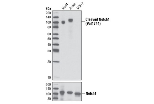
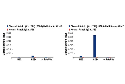
| Cleaved Notch1 (Val1744) (D3B8) Rabbit mAb | 4147 | 20 µl | 110 kDa | Rabbit IgG |
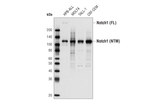
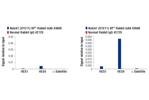
| Notch1 (D1E11) XP® Rabbit mAb | 3608 | 20 µl | 120, 300 kDa | Rabbit IgG |
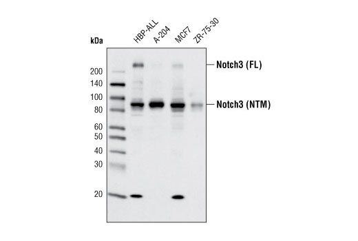
| Notch3 (D11B8) Rabbit mAb | 5276 | 20 µl | 90, 270 kDa | Rabbit IgG |
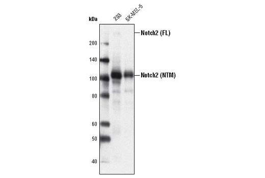
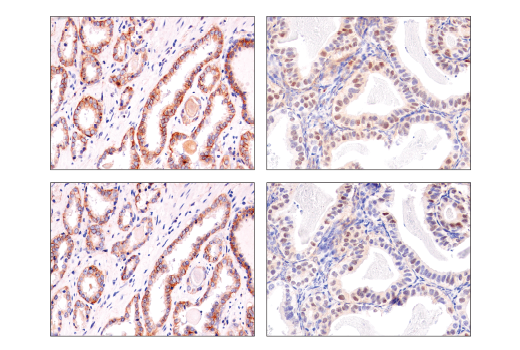
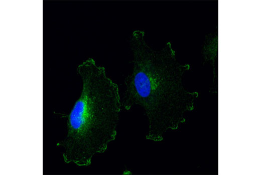
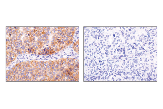
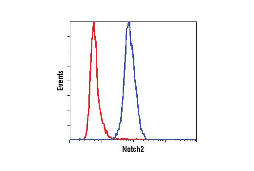
| Notch2 (D76A6) XP® Rabbit mAb | 5732 | 20 µl | 110, 300 kDa | Rabbit |
| Anti-rabbit IgG, HRP-linked Antibody | 7074 | 100 µl | Goat |
Please visit cellsignal.com for individual component applications, species cross-reactivity, dilutions, protocols, and additional product information.
Description
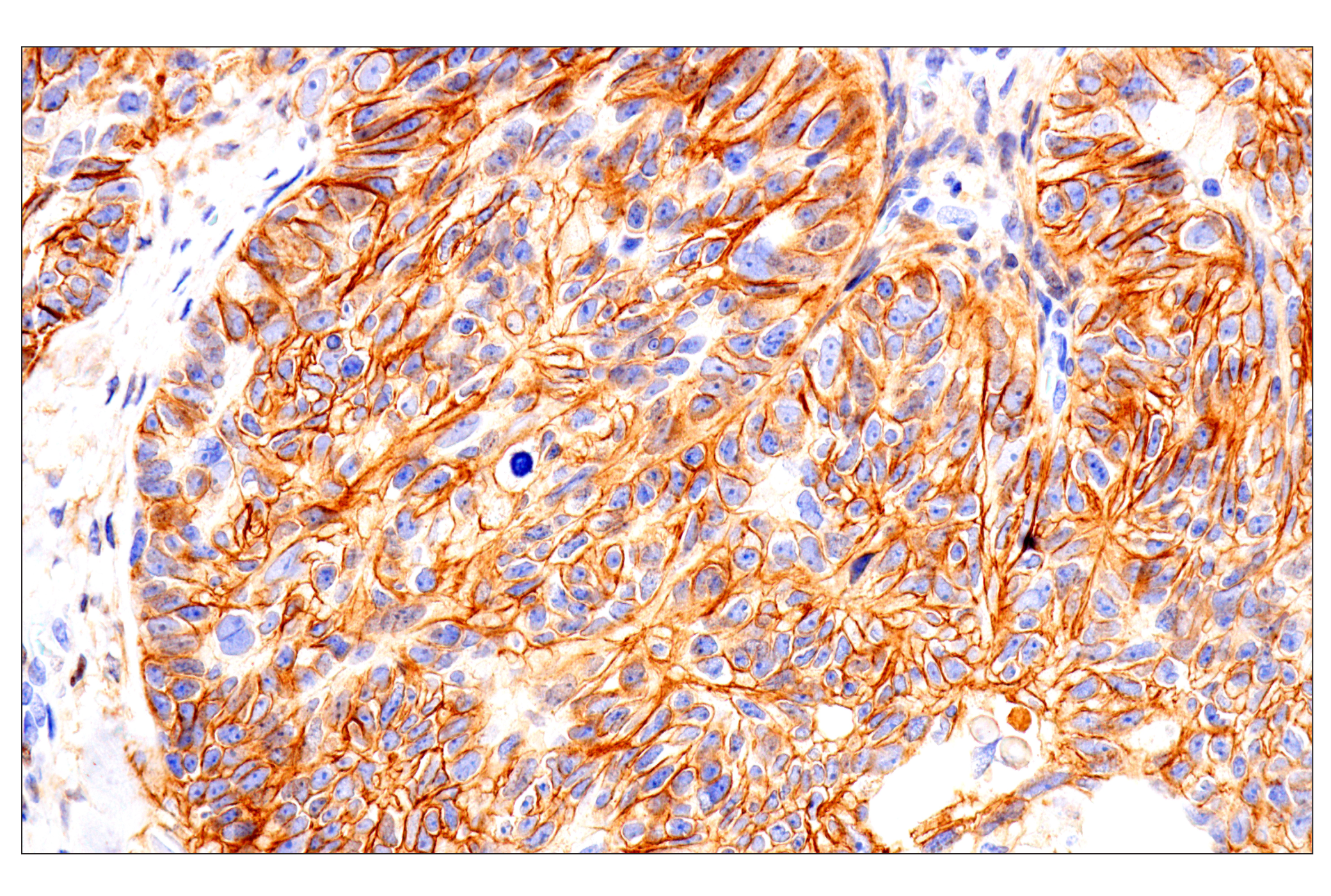
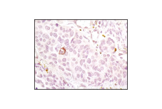
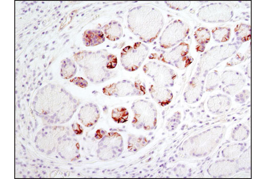
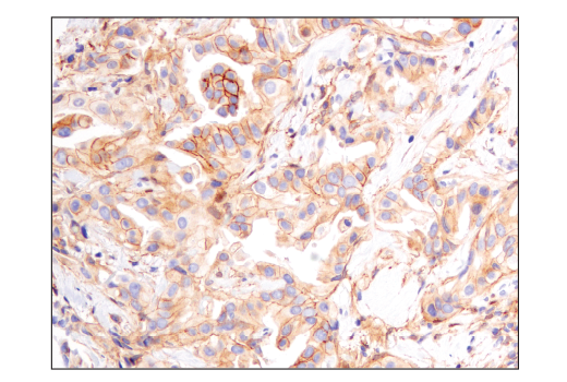
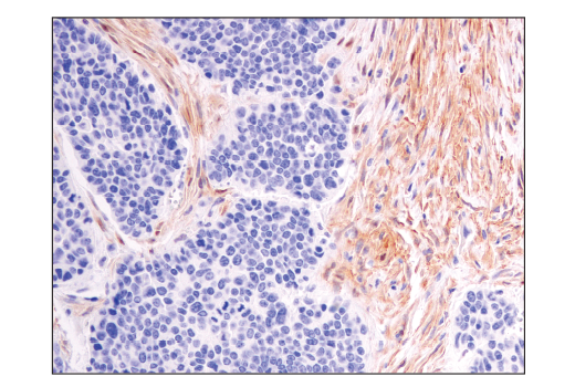
The Notch Isoform Antibody Sampler Kit provides an economical means to investigate Notch Signaling. The kit contains primary and secondary antibodies to perform two western mini-blots with each antibody.
Storage
Background
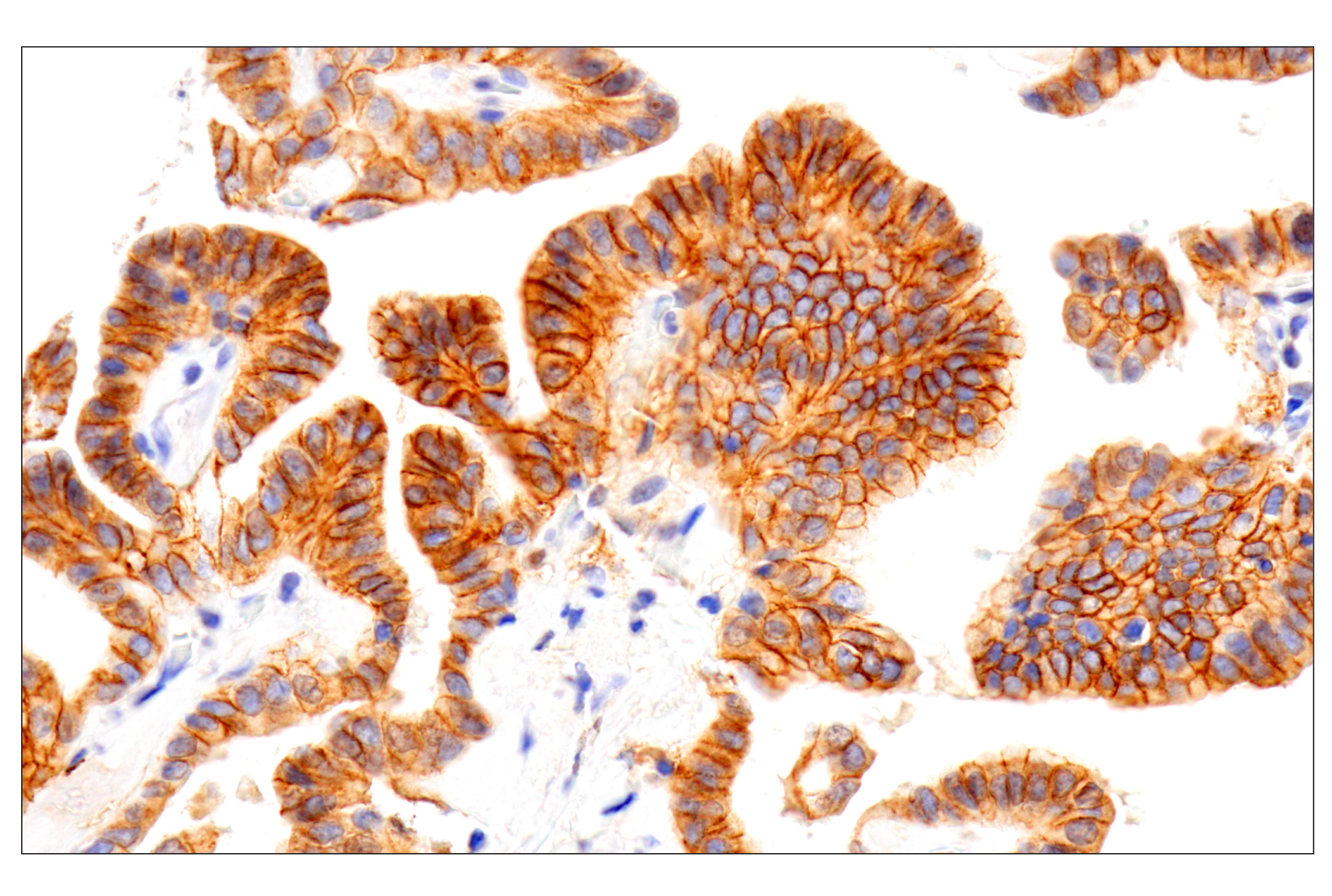
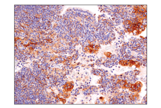
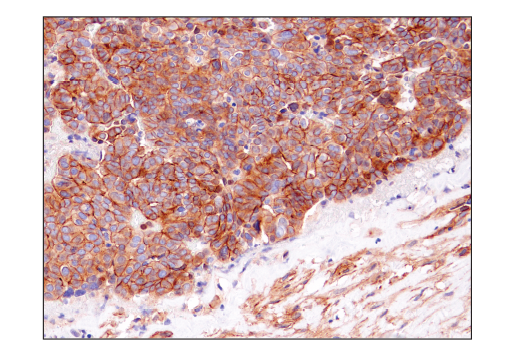
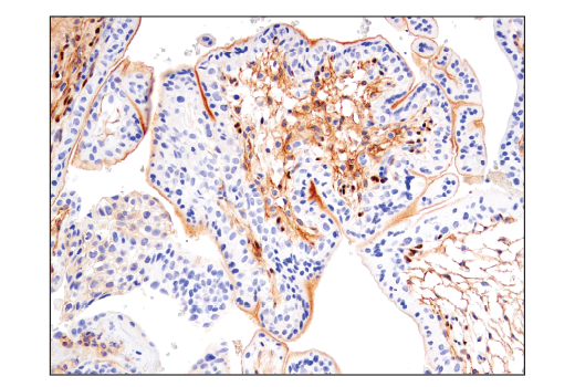
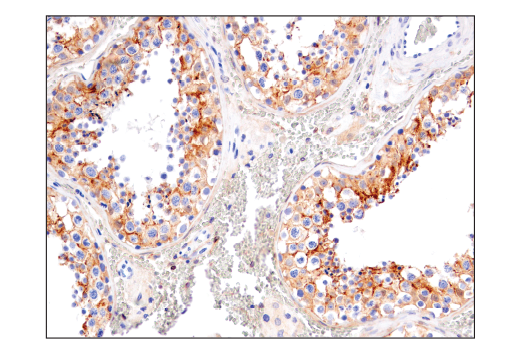
Notch proteins (Notch1-4) are a family of transmembrane receptors that play important roles in development and the determination of cell fate (1). Mature Notch receptors are processed and assembled as heterodimeric proteins, with each dimer comprised of a large extracellular ligand-binding domain, a single-pass transmembrane domain, and a smaller cytoplasmic subunit (Notch intracellular domain, NICD) (2). Binding of Notch receptors to ligands of the Delta-Serrate-Lag2 (DSL) family triggers heterodimer dissociation, exposing the receptors to proteolytic cleavages; these result in release of the NICD, which translocates to the nucleus and activates transcription of downstream target genes (3,4).
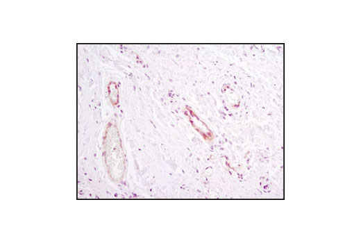
Constitutively activated Notch1 signaling is associated with the majority of cases of T cell acute lymphoblastic leukemia (T-ALL). The activation is either due to mutations in Notch1 itself or in the components of ubiquitin ligase complex, namely FBW7 (5-6). Notch2 is a member of the Notch family and mutation in Notch2 is associated with Alagille syndrome (7). Notch3 is a member of the Notch family and is processed similar to Notch1 (8). It is expressed primarily in arterial smooth muscle cells (SMC). Mutations altering the number of cysteine residues in the notch3 extracellular region are associated with cerebral autosomal dominant arteriopathy with subcortical infarcts and leukoencephalopathy (CADASIL), a hereditary angiopathy leading to strokes and dementia in adults (9-11). Recent studies indicate that Notch3 is overexpressed in many types of cancer (12-14).
- Artavanis-Tsakonas, S. et al. (1999) Science 284, 770-6.
- Chan, Y.M. and Jan, Y.N. (1998) Cell 94, 423-6.
- Schroeter, E.H. et al. (1998) Nature 393, 382-6.
- Rand, M.D. et al. (2000) Mol Cell Biol 20, 1825-35.
- Weng, A.P. et al. (2004) Science 306, 269-71.
- Thompson, B.J. et al. (2007) J Exp Med 204, 1825-35.
- McDaniell, R. et al. (2006) Am J Hum Genet 79, 169-73.
- Baron, M. (2003) Semin Cell Dev Biol 14, 113-9.
- Kalimo, H. et al. (2002) Brain Pathol 12, 371-84.
- Karlström, H. et al. (2002) Proc Natl Acad Sci U S A 99, 17119-24.
- Monet, M. et al. (2007) Hum Mol Genet 16, 982-92.
- Park, J.T. et al. (2006) Cancer Res 66, 6312-8.
- Gramantieri, L. et al. (2007) Liver Int 27, 997-1007.
- Yamaguchi, N. et al. (2008) Cancer Res 68, 1881-8.
Background References
Trademarks and Patents
使用に関する制限
法的な権限を与えられたCSTの担当者が署名した書面によって別途明示的に合意された場合を除き、 CST、その関連会社または代理店が提供する製品には以下の条件が適用されます。お客様が定める条件でここに定められた条件に含まれるものを超えるもの、 または、ここに定められた条件と異なるものは、法的な権限を与えられたCSTの担当者が別途書面にて受諾した場合を除き、拒絶され、 いかなる効力も効果も有しません。
研究専用 (For Research Use Only) またはこれに類似する表示がされた製品は、 いかなる目的についても FDA または外国もしくは国内のその他の規制機関により承認、認可または許可を受けていません。 お客様は製品を診断もしくは治療目的で使用してはならず、また、製品に表示された内容に違反する方法で使用してはなりません。 CST が販売または使用許諾する製品は、エンドユーザーであるお客様に対し、使途を研究および開発のみに限定して提供されるものです。 診断、予防もしくは治療目的で製品を使用することまたは製品を再販売 (単独であるか他の製品等の一部であるかを問いません) もしくはその他の商業的利用の目的で購入することについては、CST から別途許諾を得る必要があります。 お客様は以下の事項を遵守しなければなりません。(a) CST の製品 (単独であるか他の資材と一緒であるかを問いません) を販売、使用許諾、貸与、寄付もしくはその他の態様で第三者に譲渡したり使用させたりしてはなりません。また、商用の製品を製造するために CST の製品を使用してはなりません。(b) 複製、改変、リバースエンジニアリング、逆コンパイル、 分解または他の方法により製品の構造または技術を解明しようとしてはなりません。また、 CST の製品またはサービスと競合する製品またはサービスを開発する目的で CST の製品を使用してはなりません。(c) CST の製品の商標、商号、ロゴ、特許または著作権に関する通知または表示を除去したり改変したりしてはなりません。(d) CST の製品をCST 製品販売条件(CST’s Product Terms of Sale) および該当する書面のみに従って使用しなければなりません。(e) CST の製品に関連してお客様が使用する第三者の製品またはサービスに関する使用許諾条件、 サービス提供条件またはこれに類する合意事項を遵守しなければなりません。