View in English?
View in English?
View in English?

| Cat. # | Size | Qty. | Price | Inventory |
|---|---|---|---|---|
| 8595T | 1 Kit (8 x 20 microliters) |
|
| Product Includes | Quantity | Applications | Reactivity | MW(kDa) | Isotype |
|---|---|---|---|---|---|
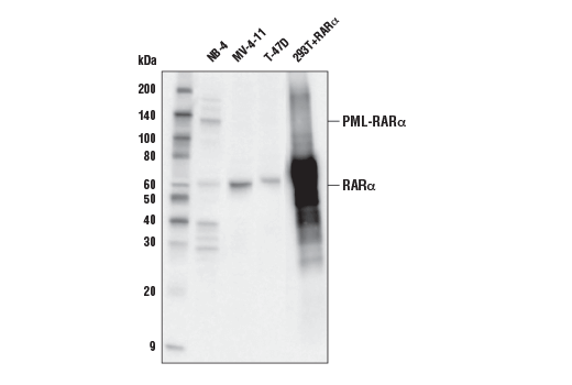
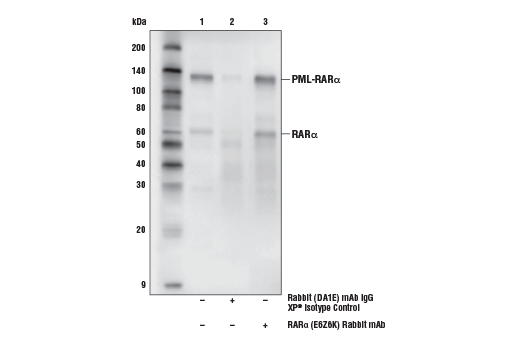
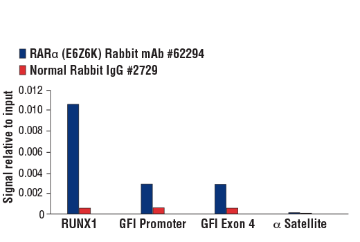
| RARα (E6Z6K) Rabbit mAb 62294 | 20 µl |
|
H M Mk | 60 | Rabbit IgG |
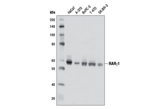
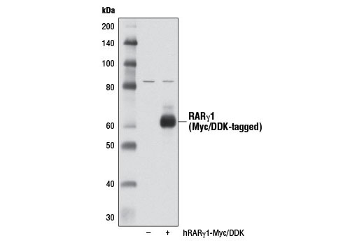
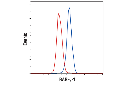
| RARγ1 (D3A4) XP® Rabbit mAb 8965 | 20 µl |
|
H M | 58 | Rabbit IgG |
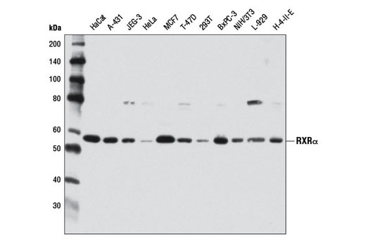
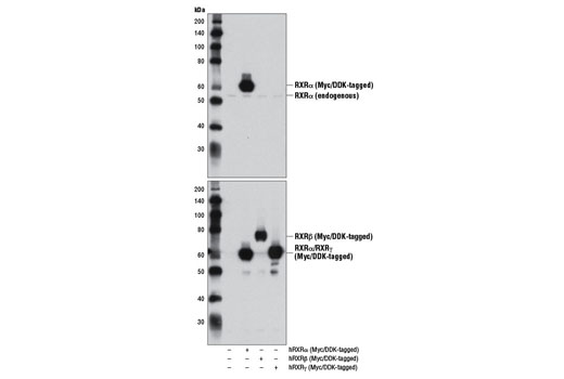
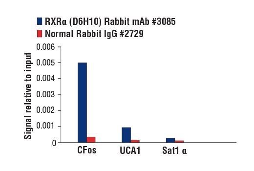
| RXRα (D6H10) Rabbit mAb 3085 | 20 µl |
|
H M R | 53 | Rabbit IgG |
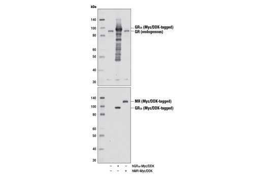
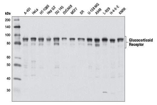
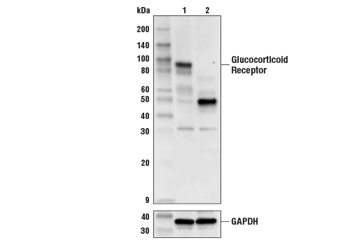
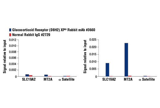
| Glucocorticoid Receptor (D8H2) XP® Rabbit mAb 3660 | 20 µl |
|
H M R Mk | 80, 91, 94 | Rabbit IgG |
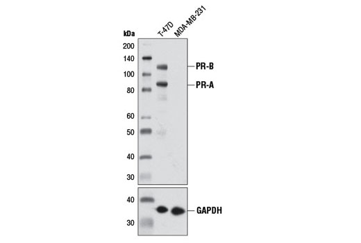
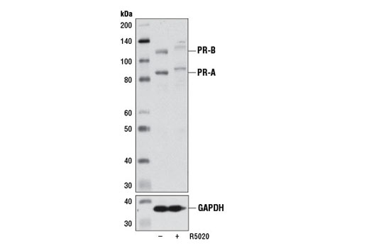
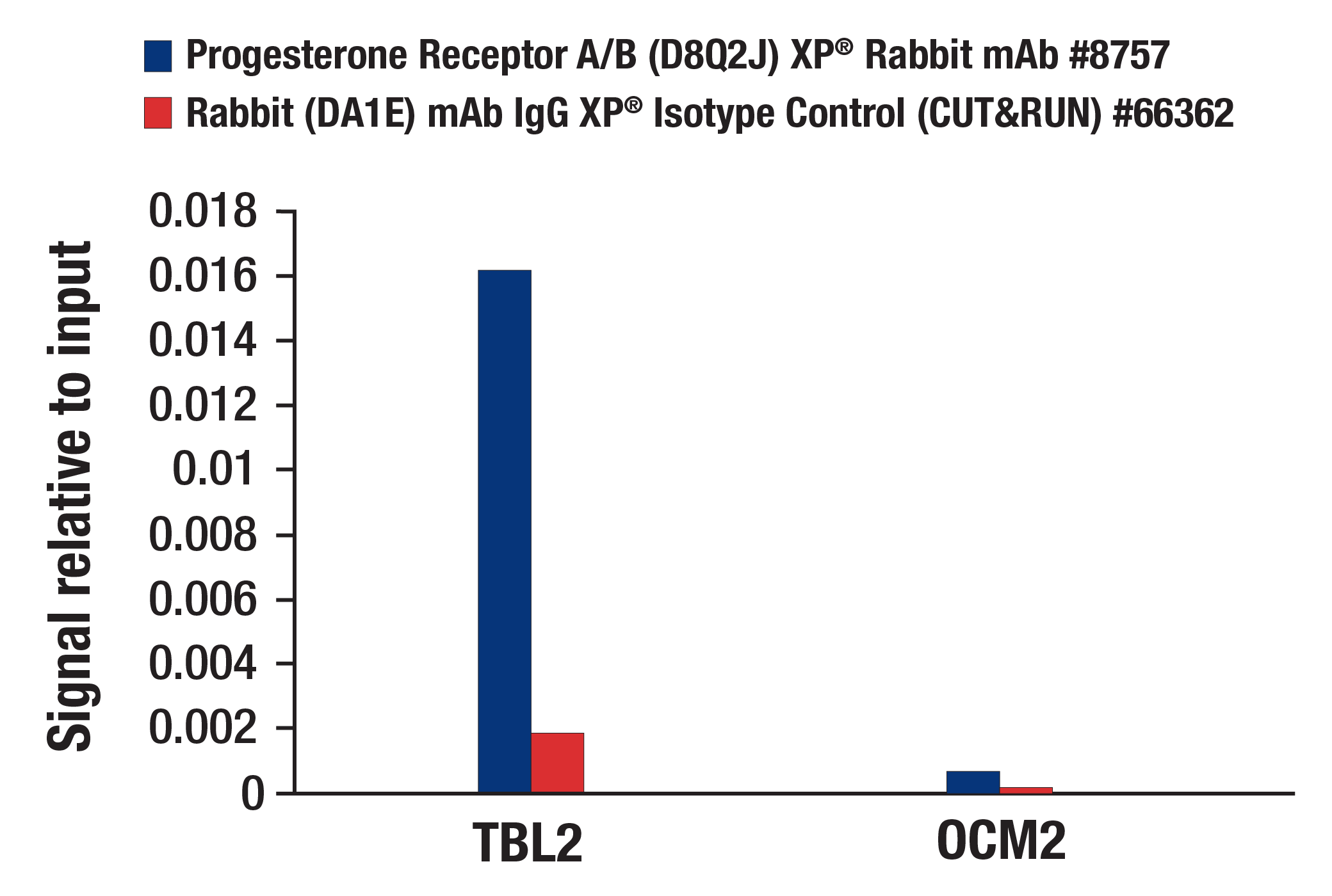
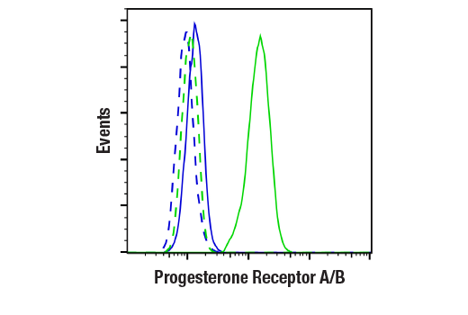
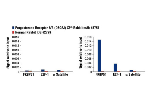
| Progesterone Receptor A/B (D8Q2J) XP® Rabbit mAb 8757 | 20 µl |
|
H | 90 (PR-A), 118 (PR-B) | Rabbit IgG |
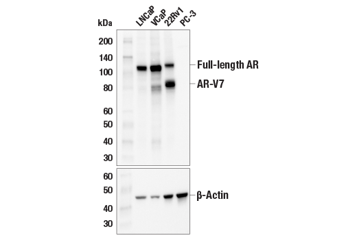
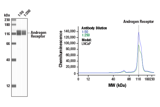
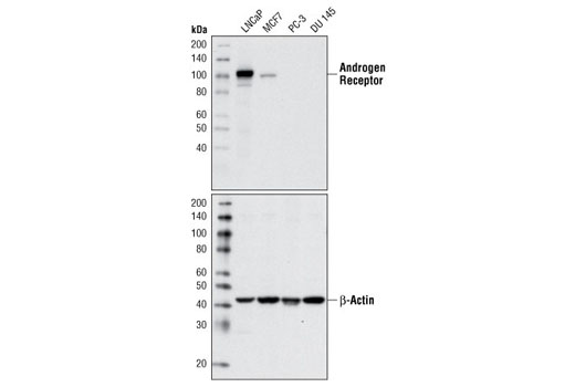
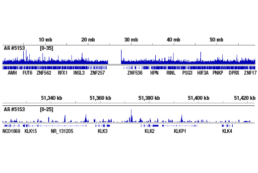
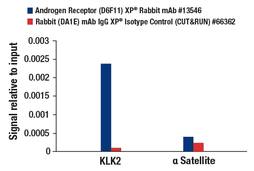
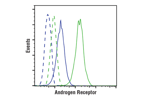
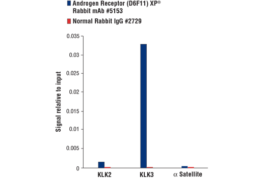
| Androgen Receptor (D6F11) XP® Rabbit mAb 5153 | 20 µl |
|
H | 110 | Rabbit IgG |
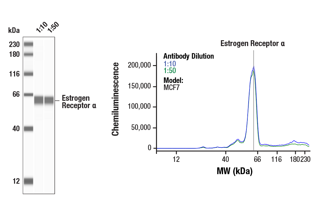
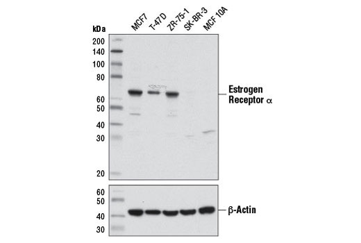
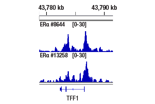
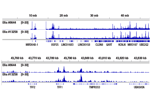
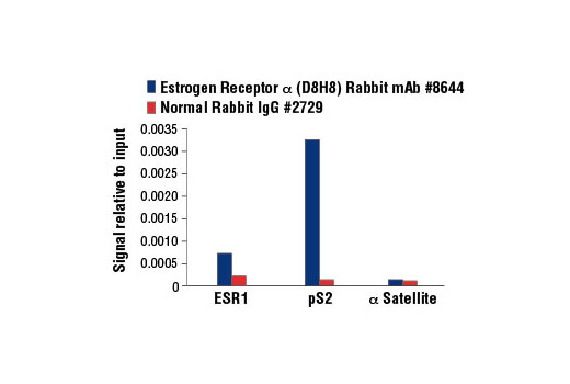
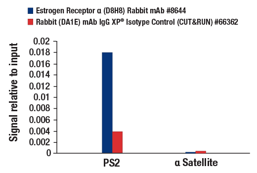
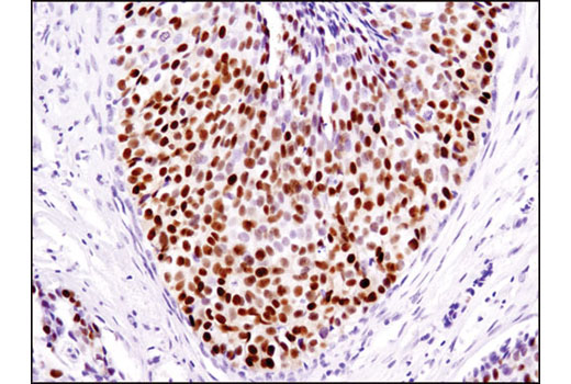
| Estrogen Receptor α (D8H8) Rabbit mAb 8644 | 20 µl |
|
H | 66 | Rabbit IgG |
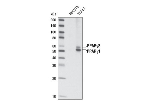
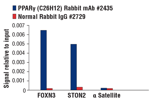
| PPARγ (C26H12) Rabbit mAb 2435 | 20 µl |
|
H M | 53, 57 | Rabbit IgG |
| Anti-rabbit IgG, HRP-linked Antibody 7074 | 100 µl |
|
Rab | Goat |
Product Information
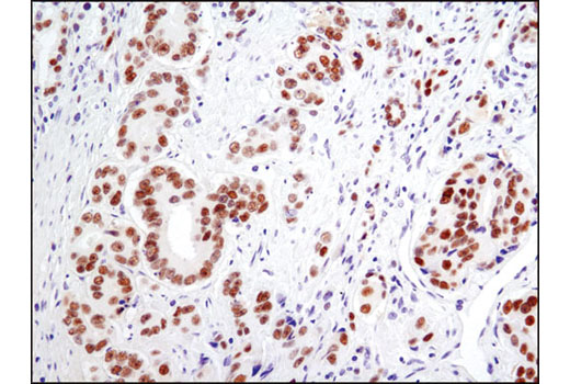
Monoclonal antibodies are produced by immunizing animals with a synthetic peptide corresponding to residues near the amino terminal region of human androgen receptor protein, residues in the carboxy terminus of human ERα protein, residues surrounding Leu378 of human glucocorticoid receptor protein, residues surrounding Asp69 of human PPARγ protein, residues surrounding Tyr541 of human progesterone receptor protein, residues near the amino terminus of human RARγ1 protein, residues near the amino terminus of human RXRα protein, or residues surrounding Leu220 of human RARα protein.
Nuclear Receptors are transcription factors responsible for sensing bioactive molecules, including steroid and thyroid hormones. They are regulated by multiple posttranslational modifications, which in turn impacts their ability to regulate the expression of specific genes involved in the control of reproduction, development, and metabolism.
Androgen receptor (AR), a zinc finger transcription factor belonging to the nuclear receptor superfamily, is activated by phosphorylation and dimerization upon ligand binding (1). This promotes nuclear localization and binding of AR to androgen response elements in androgen target genes. AR plays a crucial role in several stages of male development and the progression of prostate cancer (2,3).
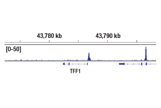
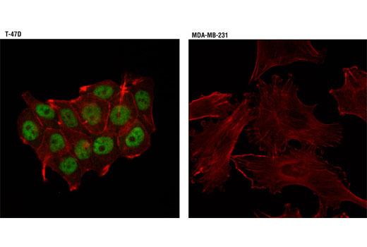
Estrogen receptor α (ERα), a member of the steroid receptor superfamily, contains highly conserved DNA binding and ligand binding domains (4). Through its estrogen-independent and estrogen-dependent activation domains (AF-1 and AF-2, respectively), ERα regulates transcription by recruiting coactivator proteins and interacting with general transcriptional machinery (5).
Glucocorticoid hormones control cellular proliferation, inflammation, and metabolism through their association with the glucocorticoid receptor (GR)/NR3C1, a member of the nuclear hormone receptor superfamily of transcription factors (6).
Peroxisome proliferator-activated receptor γ (PPARγ) is a member of the ligand-activated nuclear receptor superfamily and functions as a transcriptional activator (7). PPARγ is preferentially expressed in adipocytes, as well as in vascular smooth muscle cells and macrophages (8). Besides its role in mediating adipogenesis and lipid metabolism (8), PPARγ also modulates insulin sensitivity, cell proliferation, and inflammation (9).
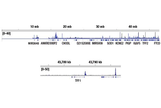
Human progesterone receptor (PR) is expressed as two forms: the full length PR B and the short form PR A. PR A lacks the first 164 amino acid residues of PR B (10,11). Both PR A and PR B are ligand activated, but differ in their relative ability to activate target gene transcription (12,13).
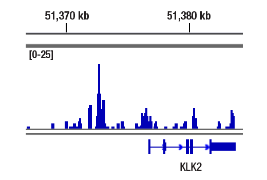
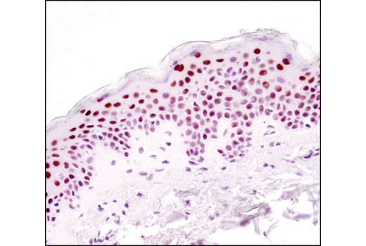
Nuclear retinoic acid receptors (RARs) consist of three subtypes encoded by separate genes: α (NR1B1), β (NR1B2), and γ (NR1B3). For each subtype, there are at least two isoforms, which are generated by differential promoter usage and alternative splicing and differ only in their N-terminal regions. Retinoids, which are metabolites of vitamin A, serve as ligands for RARs (14). RARs function as ligand-dependent transcriptional regulators and are found to be heterodimerized with retinoid X receptors (RXRs). These transcriptionally active dimers regulate the expression of genes involved in cellular differentiation, proliferation, and apoptosis (15,16).
The human retinoid X receptors are encoded by three distinct genes (RXRα, RXRβ, and RXRγ) and bind selectively and with high affinity to the vitamin A derivative, 9-cis-retinoic acid. RXRs are type-II nuclear hormone receptors that are largely localized to the nuclear compartment independent of ligand binding. Nuclear RXRs form heterodimers with nuclear hormone receptor subfamily 1 proteins, including thyroid hormone receptor, retinoic acid receptors, vitamin D receptor, peroxisome proliferator-activated receptors, liver X receptors, and farnesoid X receptor (17).
Except as otherwise expressly agreed in a writing signed by a legally authorized representative of CST, the following terms apply to Products provided by CST, its affiliates or its distributors. Any Customer's terms and conditions that are in addition to, or different from, those contained herein, unless separately accepted in writing by a legally authorized representative of CST, are rejected and are of no force or effect.
Products are labeled with For Research Use Only or a similar labeling statement and have not been approved, cleared, or licensed by the FDA or other regulatory foreign or domestic entity, for any purpose. Customer shall not use any Product for any diagnostic or therapeutic purpose, or otherwise in any manner that conflicts with its labeling statement. Products sold or licensed by CST are provided for Customer as the end-user and solely for research and development uses. Any use of Product for diagnostic, prophylactic or therapeutic purposes, or any purchase of Product for resale (alone or as a component) or other commercial purpose, requires a separate license from CST. Customer shall (a) not sell, license, loan, donate or otherwise transfer or make available any Product to any third party, whether alone or in combination with other materials, or use the Products to manufacture any commercial products, (b) not copy, modify, reverse engineer, decompile, disassemble or otherwise attempt to discover the underlying structure or technology of the Products, or use the Products for the purpose of developing any products or services that would compete with CST products or services, (c) not alter or remove from the Products any trademarks, trade names, logos, patent or copyright notices or markings, (d) use the Products solely in accordance with CST Product Terms of Sale and any applicable documentation, and (e) comply with any license, terms of service or similar agreement with respect to any third party products or services used by Customer in connection with the Products.
View in English?