Revision 1
Cell Signaling Technology

Orders: 877-616-CELL (2355) [email protected]

Support: 877-678-TECH (8324)

Web: [email protected] cellsignal.com

3 Trask LaneDanversMassachusetts01923USA
For Research Use Only. Not for Use in Diagnostic Procedures.
Applications:

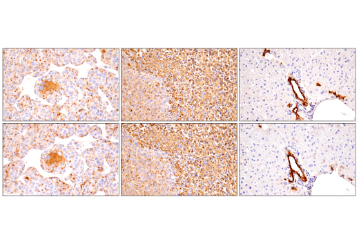
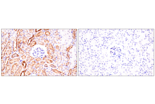
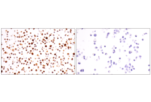
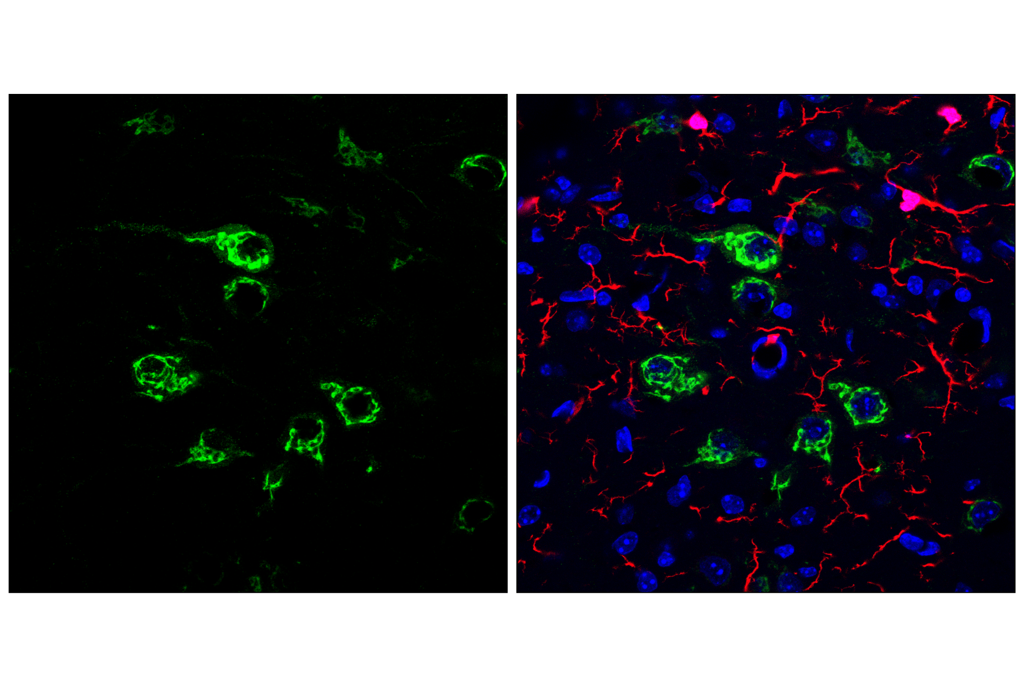
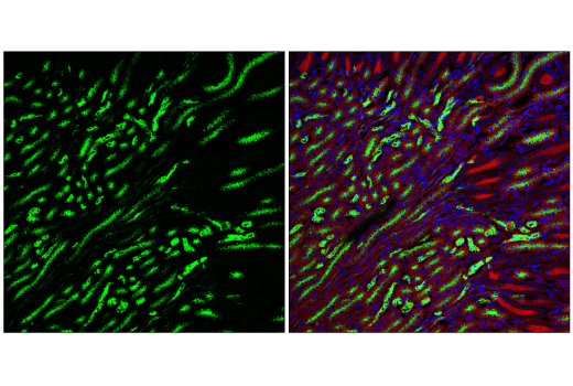
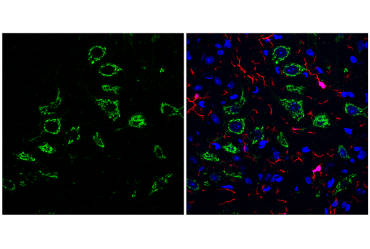
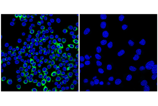
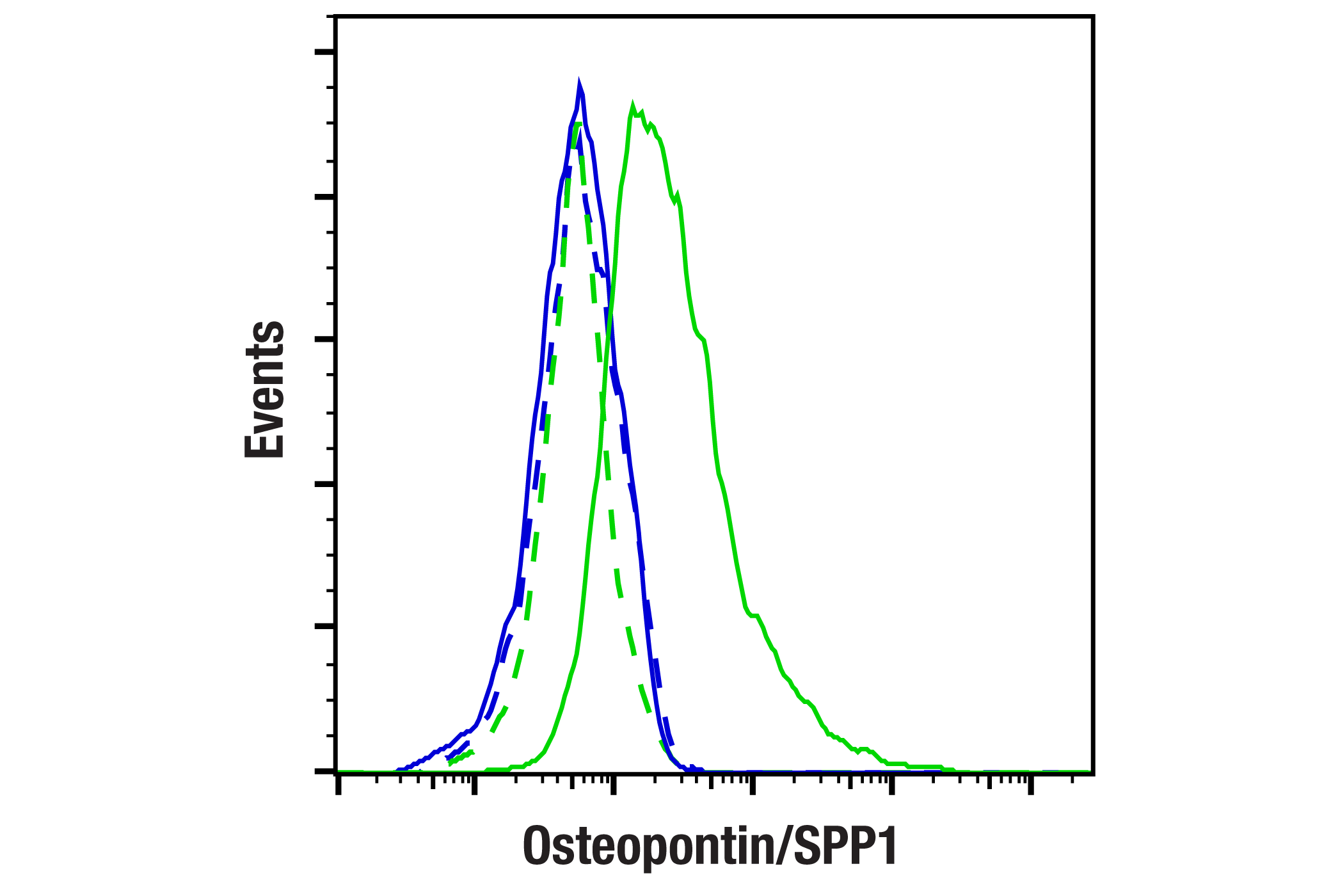
WB, IHC-P, IF-F, IF-IC, FC-FP

REACTIVITY:

M

SENSITIVITY:

Endogenous

MW (kDa):

58-75

Source/Isotype:

Rabbit IgG

UniProt ID:

#P10923

Entrez-Gene Id:

20750

Product Information

Product Usage Information

This product is the carrier free version of product #88742. All data were generated using the same antibody clone in the standard formulation which contains BSA and glycerol.

This formulation is ideal for use with technologies requiring specialized or custom antibody labeling, including fluorophores, metals, lanthanides, and oligonucleotides. It is not recommended for ChIP, ChIP-seq, CUT&RUN or CUT&Tag assays. If you require a carrier free formulation for chromatin profiling, please contact us. Optimal dilutions/concentrations should be determined by the end user.

Formulation

Supplied in 1X PBS, BSA and Azide Free.

For standard formulation of this product see product #88742

Storage

Store at -20°C. This product will freeze at -20°C so it is recommended to aliquot into single-use vials to avoid multiple freeze/thaw cycles. A slight precipitate may be present and can be dissolved by gently vortexing. This will not interfere with antibody performance.

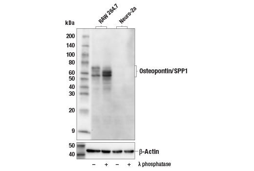
Specificity / Sensitivity

Osteopontin/SPP1 (E9Z1D) Rabbit mAb (BSA and Azide Free) recognizes endogenous levels of total osteopontin/SPP1 protein.

Species Reactivity:

Mouse

Source / Purification

Monoclonal antibody is produced by immunizing animals with a synthetic peptide corresponding to residues surrounding Ala149 of mouse osteopontin/SPP1 protein.

Background

Osteopontin is a highly phosphorylated secreted glycoprotein expressed in a variety of cell types. Osteopontin, encoded by the gene secreted phosphoprotein 1 (SPP1), was originally isolated and cloned from a rat osteosarcoma cell line (1). Osteopontin is a highly negatively charged protein that likely regulates extracellular matrix turnover and remodeling, both in bone and other tissue (2). Osteopontin also has three highly conserved receptor-binding domains, otherwise known as arginine-glycine-aspartic acid (RGD) domains. Binding of osteopontin to cell surface integrins likely activates cell signaling pathways that regulate cell proliferation, adhesion, invasion, migration, and fibrosis (3,4). Osteopontin may be regulated by posttranslational regulation, including phosphorylation, glycosylation, and proteolytic cleavage (5). Osteopontin is also linked to several human diseases. For example, cleaved osteopontin is higher in patients suffering with cirrhosis, a disease involving fibrosis of the liver (5). Its role in the brain is unclear, but SPP1 expression in microglia, the resident macrophages of the brain, is upregulated in mouse neurodegenerative disease models, including Alzheimer’s disease (6). Osteopontin’s role in disease likely involve its function as a key mediator of inflammatory responses (4).

  1. Oldberg, A. et al. (1986) Proc Natl Acad Sci U S A 83, 8819-23.
  2. Kazanecki, C.C. et al. (2007) J Cell Biochem 102, 912-24.
  3. Standal, T. et al. (2004) Exp Oncol 26, 179-84.
  4. Lamort, A.S. et al. (2019) Cells 8, 815. doi: 10.3390/cells8080815.
  5. Song, Z. et al. (2021) Hepatology 73, 1594-1608.
  6. Sala Frigerio, C. et al. (2019) Cell Rep 27, 1293-1306.e6.

Species Reactivity

Species reactivity is determined by testing in at least one approved application (e.g., western blot).

Applications Key

WB: Western Blotting IHC-P: Immunohistochemistry (Paraffin) IF-F: Immunofluorescence (Frozen) IF-IC: Immunofluorescence (Immunocytochemistry) FC-FP: Flow Cytometry (Fixed/Permeabilized)

Cross-Reactivity Key

H: human M: mouse R: rat Hm: hamster Mk: monkey Vir: virus Mi: mink C: chicken Dm: D. melanogaster X: Xenopus Z: zebrafish B: bovine Dg: dog Pg: pig Sc: S. cerevisiae Ce: C. elegans Hr: horse GP: Guinea Pig Rab: rabbit All: all species expected

Trademarks and Patents

Cell Signaling Technology is a trademark of Cell Signaling Technology, Inc.
All other trademarks are the property of their respective owners. Visit cellsignal.com/trademarks for more information.

使用に関する制限

法的な権限を与えられたCSTの担当者が署名した書面によって別途明示的に合意された場合を除き、 CST、その関連会社または代理店が提供する製品には以下の条件が適用されます。お客様が定める条件でここに定められた条件に含まれるものを超えるもの、 または、ここに定められた条件と異なるものは、法的な権限を与えられたCSTの担当者が別途書面にて受諾した場合を除き、拒絶され、 いかなる効力も効果も有しません。

研究専用 (For Research Use Only) またはこれに類似する表示がされた製品は、 いかなる目的についても FDA または外国もしくは国内のその他の規制機関により承認、認可または許可を受けていません。 お客様は製品を診断もしくは治療目的で使用してはならず、また、製品に表示された内容に違反する方法で使用してはなりません。 CST が販売または使用許諾する製品は、エンドユーザーであるお客様に対し、使途を研究および開発のみに限定して提供されるものです。 診断、予防もしくは治療目的で製品を使用することまたは製品を再販売 (単独であるか他の製品等の一部であるかを問いません) もしくはその他の商業的利用の目的で購入することについては、CST から別途許諾を得る必要があります。 お客様は以下の事項を遵守しなければなりません。(a) CST の製品 (単独であるか他の資材と一緒であるかを問いません) を販売、使用許諾、貸与、寄付もしくはその他の態様で第三者に譲渡したり使用させたりしてはなりません。また、商用の製品を製造するために CST の製品を使用してはなりません。(b) 複製、改変、リバースエンジニアリング、逆コンパイル、 分解または他の方法により製品の構造または技術を解明しようとしてはなりません。また、 CST の製品またはサービスと競合する製品またはサービスを開発する目的で CST の製品を使用してはなりません。(c) CST の製品の商標、商号、ロゴ、特許または著作権に関する通知または表示を除去したり改変したりしてはなりません。(d) CST の製品をCST 製品販売条件(CST’s Product Terms of Sale) および該当する書面のみに従って使用しなければなりません。(e) CST の製品に関連してお客様が使用する第三者の製品またはサービスに関する使用許諾条件、 サービス提供条件またはこれに類する合意事項を遵守しなければなりません。

Revision 1
#17090

Osteopontin/SPP1 (E9Z1D) Rabbit mAb (BSA and Azide Free)

Western Blotting Image 1: Osteopontin/SPP1 (E9Z1D) Rabbit mAb (BSA and Azide Free) Expand Image
Western blot analysis of extracts from RAW 264.7 and Neuro-2a cells, untreated (-) or treated with λ phosphatase (+), using Osteopontin/SPP1 (E9Z1D) Rabbit mAb (upper) or β-Actin (D6A8) Rabbit mAb #8457 (lower). Data were generated using the standard formulation of this product.
Immunohistochemistry Image 1: Osteopontin/SPP1 (E9Z1D) Rabbit mAb (BSA and Azide Free) Expand Image
Immunohistochemical analysis of paraffin-embedded mouse ovary using Osteopontin/SPP1 (E9Z1D) Rabbit mAb. Data were generated using the standard formulation of this product.
Immunohistochemistry Image 2: Osteopontin/SPP1 (E9Z1D) Rabbit mAb (BSA and Azide Free) Expand Image
Immunohistochemical analysis of paraffin-embedded mouse pancreas using Osteopontin/SPP1 (E9Z1D) Rabbit mAb. Data were generated using the standard formulation of this product.
Immunohistochemistry Image 3: Osteopontin/SPP1 (E9Z1D) Rabbit mAb (BSA and Azide Free) Expand Image
Immunohistochemical analysis of paraffin-embedded pons region of mouse brain using Osteopontin/SPP1 (E9Z1D) Rabbit mAb. Data were generated using the standard formulation of this product.
Immunohistochemistry Image 4: Osteopontin/SPP1 (E9Z1D) Rabbit mAb (BSA and Azide Free) Expand Image
Immunohistochemical analysis of paraffin-embedded renca syngeneic tumor (left), 4T1 syngeneic tumor (middle), or mouse liver (right) using Osteopontin/SPP1 (E9Z1D) Rabbit mAb (top) or an Osteopontin/SPP1 Rabbit mAb (bottom). These two antibodies detect unique, non-overlapping epitopes on mouse Osteopontin/SPP1. The similar staining patterns obtained with both antibodies help to confirm the specificity of the staining. Data were generated using the standard formulation of this product.
Immunohistochemistry Image 5: Osteopontin/SPP1 (E9Z1D) Rabbit mAb (BSA and Azide Free) Expand Image
Immunohistochemical analysis of paraffin-embedded mouse kidney using Osteopontin/SPP1 (E9Z1D) Rabbit mAb (left) compared to concentration-matched Rabbit (DA1E) mAb IgG XP® Isotype Control #3900 (right).  Data were generated using the standard formulation of this product.
Immunohistochemistry Image 6: Osteopontin/SPP1 (E9Z1D) Rabbit mAb (BSA and Azide Free) Expand Image
Immunohistochemical analysis of paraffin-embedded RAW 264.7 cell pellet (left, positive) or Neuro-2a cell pellet (right, negative) using Osteopontin/SPP1 (E9Z1D) Rabbit mAb. Data were generated using the standard formulation of this product.
Immunofluorescence Image 1: Osteopontin/SPP1 (E9Z1D) Rabbit mAb (BSA and Azide Free) Expand Image
Confocal immunofluorescent analysis of fixed frozen mouse pons labeled with Osteopontin/SPP1 (E4O2F) Rabbit mAb (left, green). Free secondary binding sites were then blocked with Rabbit (DA1E) mAb IgG XP® Isotype Control #3900 prior to colabeling with Iba1/AIF-1 (E4O4W) XP® Rabbit mAb (Alexa Fluor® 647 Conjugate) #78060 (right, red) and DAPI #4083 (right, blue). Data were generated using the standard formulation of this product.
Immunofluorescence Image 2: Osteopontin/SPP1 (E9Z1D) Rabbit mAb (BSA and Azide Free) Expand Image
Confocal immunofluorescent analysis of fixed frozen mouse kidney labeled with Osteopontin/SPP1 (E9Z1D) Rabbit mAb (left, green). Free secondary binding sites were then blocked with Rabbit (DA1E) mAb IgG XP® Isotype Control #3900 prior to co-labeling with Ras (E4K9L) Rabbit mAb (Alexa Fluor® 488 Conjugate) #51813 (right, red pseudocolor) and DAPI #4083 (right, blue). Data were generated using the standard formulation of this product.
Immunofluorescence Image 3: Osteopontin/SPP1 (E9Z1D) Rabbit mAb (BSA and Azide Free) Expand Image
Confocal immunofluorescent analysis of fixed frozen mouse thalamus labeled with Osteopontin/SPP1 (E9Z1D) Rabbit mAb (left, green). Free secondary binding sites were then blocked with Rabbit (DA1E) mAb IgG XP® Isotype Control #3900 prior to co-labeling with Iba1/AIF-1 (E4O4W) XP® Rabbit mAb (Alexa Fluor® 647 Conjugate) #78060 (right, red) and DAPI #4083 (right, blue). Data were generated using the standard formulation of this product.
Immunofluorescence Image 1: Osteopontin/SPP1 (E9Z1D) Rabbit mAb (BSA and Azide Free) Expand Image
Confocal immunofluorescent analysis of RAW 264.7 cells (left, positive) and Neuro-2a cells (right, negative) using Osteopontin/SPP1 (E9Z1D) Rabbit mAb (green) and DAPI #4083 (blue). Cells were treated with Brefeldin A #9972 (100 ng/mL, 90 min) to inhibit protein secretion. Data were generated using the standard formulation of this product.
Flow Cytometry Image 1: Osteopontin/SPP1 (E9Z1D) Rabbit mAb (BSA and Azide Free) Expand Image
Flow cytometric analysis of Neuro-2a cells (blue, negative) and RAW 264.7 cells (green, positive) using Osteopontin/SPP1 (E9Z1D) Rabbit mAb (solid lines) or concentration-matched Rabbit (DA1E) mAb IgG XP® Isotype Control #3900 (dashed lines). Anti-rabbit IgG (H+L), F(ab')2 Fragment (Alexa Fluor® 488 Conjugate) #4412 was used as a secondary antibody. Data were generated using the standard formulation of this product.