Revision 4
Cell Signaling Technology

Orders: 877-616-CELL (2355) [email protected]

Support: 877-678-TECH (8324)

Web: [email protected] cellsignal.com

3 Trask LaneDanversMassachusetts01923USA
For Research Use Only. Not for Use in Diagnostic Procedures.
Applications:

WB, IP, IHC-P, IF-F, IF-IC

REACTIVITY:

H M R Mk

SENSITIVITY:

Endogenous

MW (kDa):

70, 85

Source/Isotype:

Rabbit IgG

UniProt ID:

#P23443

Entrez-Gene Id:

6198

Product Information

Product Usage Information

Application Dilution
Western Blotting 1:1000
Immunoprecipitation 1:100
Immunohistochemistry (Paraffin) 1:50 - 1:200
Immunofluorescence (Frozen) 1:100 - 1:400
Immunofluorescence (Immunocytochemistry) 1:400 - 1:1600

Storage

Supplied in 10 mM sodium HEPES (pH 7.5), 150 mM NaCl, 100 µg/ml BSA, 50% glycerol and less than 0.02% sodium azide. Store at –20°C. Do not aliquot the antibody.

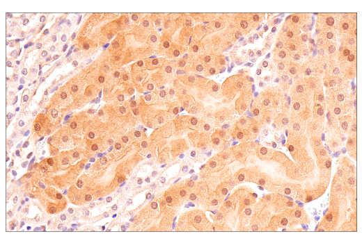
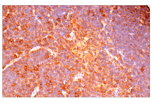
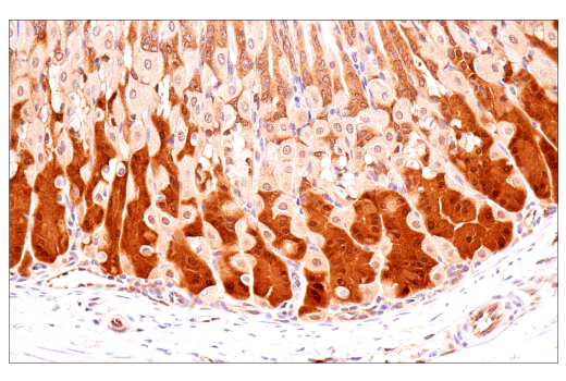
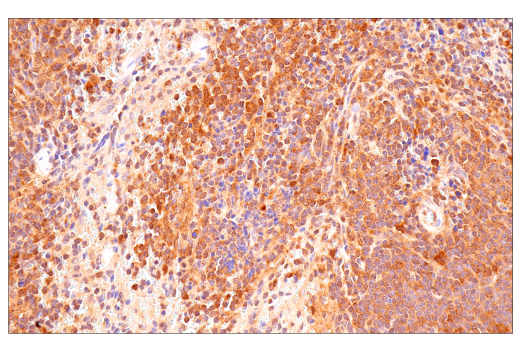
Specificity / Sensitivity

p70 S6 Kinase (E8K6T) XP® Rabbit mAb recognizes endogenous levels of total p70 S6 kinase protein. This antibody does not cross-react with p70 S6 kinase-2.

Species Reactivity:

Human, Mouse, Rat, Monkey

Source / Purification

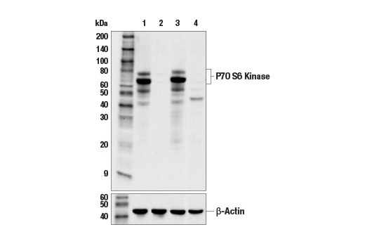
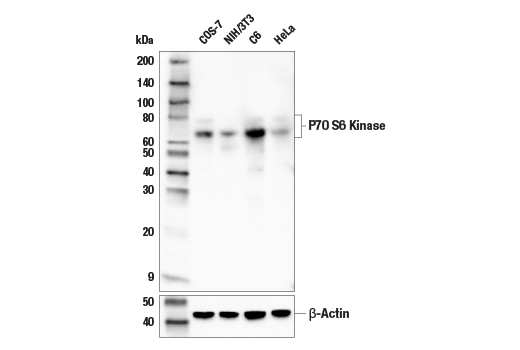
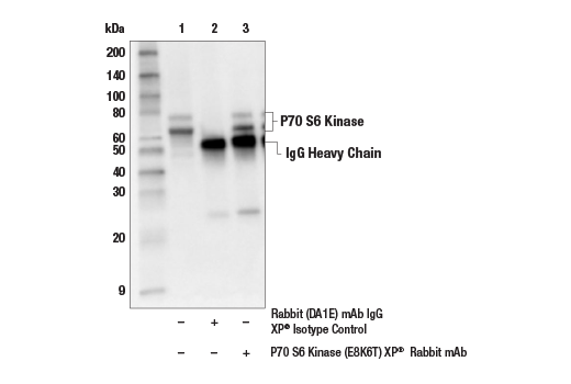
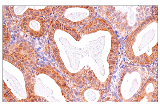
Monoclonal antibody is produced by immunizing animals with a synthetic peptide corresponding to residues surrounding Leu32 of human p70 S6 kinase protein.

Background

p70 S6 kinase is a mitogen activated Ser/Thr protein kinase that is required for cell growth and G1 cell cycle progression (1,2). p70 S6 kinase phosphorylates the S6 protein of the 40S ribosomal subunit and is involved in translational control of 5' oligopyrimidine tract mRNAs (1). A second isoform, p85 S6 kinase, is derived from the same gene and is identical to p70 S6 kinase except for 23 extra residues at the amino terminus, which encode a nuclear localizing signal (1). Both isoforms lie on a mitogen activated signaling pathway downstream of phosphoinositide-3 kinase (PI-3K) and the target of rapamycin, FRAP/mTOR, a pathway distinct from the Ras/MAP kinase cascade (1). The activity of p70 S6 kinase is controlled by multiple phosphorylation events located within the catalytic, linker and pseudosubstrate domains (1). Phosphorylation of Thr229 in the catalytic domain and Thr389 in the linker domain are most critical for kinase function (1). Phosphorylation of Thr389, however, most closely correlates with p70 kinase activity in vivo (3). Prior phosphorylation of Thr389 is required for the action of phosphoinositide 3-dependent protein kinase 1 (PDK1) on Thr229 (4,5). Phosphorylation of this site is stimulated by growth factors such as insulin, EGF and FGF, as well as by serum and some G-protein-coupled receptor ligands, and is blocked by wortmannin, LY294002 (PI-3K inhibitor) and rapamycin (FRAP/mTOR inhibitor) (1,6,7). Ser411, Thr421 and Ser424 lie within a Ser-Pro-rich region located in the pseudosubstrate region (1). Phosphorylation at these sites is thought to activate p70 S6 kinase via relief of pseudosubstrate suppression (1,2). Another LY294002 and rapamycin sensitive phosphorylation site, Ser371, is an in vitro substrate for mTOR and correlates well with the activity of a partially rapamycin resistant mutant p70 S6 kinase (8).

  1. Pullen, N. and Thomas, G. (1997) FEBS Lett 410, 78-82.
  2. Dufner, A. and Thomas, G. (1999) Exp Cell Res 253, 100-9.
  3. Weng, Q.P. et al. (1998) J Biol Chem 273, 16621-9.
  4. Pullen, N. et al. (1998) Science 279, 707-10.
  5. Alessi, D.R. et al. (1998) Curr Biol 8, 69-81.
  6. Polakiewicz, R.D. et al. (1998) J Biol Chem 273, 23534-41.
  7. Fingar, D.C. et al. (2002) Genes Dev 16, 1472-87.
  8. Saitoh, M. et al. (2002) J Biol Chem 277, 20104-12.

Species Reactivity

Species reactivity is determined by testing in at least one approved application (e.g., western blot).

Western Blot Buffer

IMPORTANT: For western blots, incubate membrane with diluted primary antibody in 5% w/v BSA, 1X TBS, 0.1% Tween® 20 at 4°C with gentle shaking, overnight.

Applications Key

WB: Western Blotting IP: Immunoprecipitation IHC-P: Immunohistochemistry (Paraffin) IF-F: Immunofluorescence (Frozen) IF-IC: Immunofluorescence (Immunocytochemistry)

Cross-Reactivity Key

H: human M: mouse R: rat Hm: hamster Mk: monkey Vir: virus Mi: mink C: chicken Dm: D. melanogaster X: Xenopus Z: zebrafish B: bovine Dg: dog Pg: pig Sc: S. cerevisiae Ce: C. elegans Hr: horse GP: Guinea Pig Rab: rabbit All: all species expected

Trademarks and Patents

Cell Signaling Technology is a trademark of Cell Signaling Technology, Inc.
SignalStain is a registered trademark of Cell Signaling Technology, Inc.
XP is a registered trademark of Cell Signaling Technology, Inc.
All other trademarks are the property of their respective owners. Visit cellsignal.com/trademarks for more information.

使用に関する制限

法的な権限を与えられたCSTの担当者が署名した書面によって別途明示的に合意された場合を除き、 CST、その関連会社または代理店が提供する製品には以下の条件が適用されます。お客様が定める条件でここに定められた条件に含まれるものを超えるもの、 または、ここに定められた条件と異なるものは、法的な権限を与えられたCSTの担当者が別途書面にて受諾した場合を除き、拒絶され、 いかなる効力も効果も有しません。

研究専用 (For Research Use Only) またはこれに類似する表示がされた製品は、 いかなる目的についても FDA または外国もしくは国内のその他の規制機関により承認、認可または許可を受けていません。 お客様は製品を診断もしくは治療目的で使用してはならず、また、製品に表示された内容に違反する方法で使用してはなりません。 CST が販売または使用許諾する製品は、エンドユーザーであるお客様に対し、使途を研究および開発のみに限定して提供されるものです。 診断、予防もしくは治療目的で製品を使用することまたは製品を再販売 (単独であるか他の製品等の一部であるかを問いません) もしくはその他の商業的利用の目的で購入することについては、CST から別途許諾を得る必要があります。 お客様は以下の事項を遵守しなければなりません。(a) CST の製品 (単独であるか他の資材と一緒であるかを問いません) を販売、使用許諾、貸与、寄付もしくはその他の態様で第三者に譲渡したり使用させたりしてはなりません。また、商用の製品を製造するために CST の製品を使用してはなりません。(b) 複製、改変、リバースエンジニアリング、逆コンパイル、 分解または他の方法により製品の構造または技術を解明しようとしてはなりません。また、 CST の製品またはサービスと競合する製品またはサービスを開発する目的で CST の製品を使用してはなりません。(c) CST の製品の商標、商号、ロゴ、特許または著作権に関する通知または表示を除去したり改変したりしてはなりません。(d) CST の製品をCST 製品販売条件(CST’s Product Terms of Sale) および該当する書面のみに従って使用しなければなりません。(e) CST の製品に関連してお客様が使用する第三者の製品またはサービスに関する使用許諾条件、 サービス提供条件またはこれに類する合意事項を遵守しなければなりません。

Revision 4
#34475

p70 S6 Kinase (E8K6T) XP® Rabbit mAb

Western Blotting Image 1: p70 S6 Kinase (E8K6T) XP® Rabbit mAb Expand Image
Western blot analysis of extracts from control mIMCD-3 cells (lane 1) or p70 S6 kinase-1 knockout mIMCD-3 cells (lane 2), and control wild-type immortalized MEF cells (lane3) or p70 S6 Kinase-1/p70 S6 kinase-2 double knockout MEF cells (lane 4), using p70 S6 Kinase (E8K6T) Rabbit mAb (upper) and β-Actin (D6A8) Rabbit mAb #8457 (lower). The absence of signal in the two knockout models confirms specificity of the antibody for p70 S6 kinase-1. Knockout cells were kindly provided by Dr. Mario Pende, INSERM, Paris, France.
Western Blotting Image 2: p70 S6 Kinase (E8K6T) XP® Rabbit mAb Expand Image
Western blot analysis of extracts from various cell lines using p70 S6 Kinase (E8K6T) XP® Rabbit mAb (upper) and β-Actin (D6A8) Rabbit mAb #8457 (lower).
Immunoprecipitation Image 1: p70 S6 Kinase (E8K6T) XP® Rabbit mAb Expand Image
Immunoprecipitation of p70 S6 kinase protein from HeLa extracts. Lane 1 is 10% input, lane 2 is Rabbit (DA1E) mAb IgG XP® Isotype Control #3900, and lane 3 is p70 S6 Kinase (E8K6T) XP® Rabbit mAb. Western blot analysis was performed using p70 S6 Kinase (E8K6T) XP® Rabbit mAb. Anti-rabbit IgG, HRP-linked Antibody #7074 was used as the secondary antibody.
Immunohistochemistry Image 1: p70 S6 Kinase (E8K6T) XP® Rabbit mAb Expand Image
Immunohistochemical analysis of paraffin-embedded human urothelial carcinoma using p70 S6 Kinase (E8K6T) XP® Rabbit mAb.
Immunohistochemistry Image 2: p70 S6 Kinase (E8K6T) XP® Rabbit mAb Expand Image
Immunohistochemical analysis of paraffin-embedded human endometrioid adenocarcinoma using p70 S6 Kinase (E8K6T) XP® Rabbit mAb.
Immunohistochemistry Image 3: p70 S6 Kinase (E8K6T) XP® Rabbit mAb Expand Image
Immunohistochemical analysis of paraffin-embedded human prostate carcinoma using p70 S6 Kinase (E8K6T) XP® Rabbit mAb.
Immunohistochemistry Image 4: p70 S6 Kinase (E8K6T) XP® Rabbit mAb Expand Image
Immunohistochemical analysis of paraffin-embedded normal human pancreas using p70 S6 Kinase (E8K6T) XP® Rabbit mAb.
Immunohistochemistry Image 5: p70 S6 Kinase (E8K6T) XP® Rabbit mAb Expand Image
Immunohistochemical analysis of paraffin-embedded normal human parathyroid using p70 S6 Kinase (E8K6T) XP® Rabbit mAb.
Immunohistochemistry Image 6: p70 S6 Kinase (E8K6T) XP® Rabbit mAb Expand Image
Immunohistochemical analysis of paraffin-embedded mouse testis using p70 S6 Kinase (E8K6T) XP® Rabbit mAb.
Immunohistochemistry Image 7: p70 S6 Kinase (E8K6T) XP® Rabbit mAb Expand Image
Immunohistochemical analysis of paraffin-embedded mouse kidney using p70 S6 Kinase (E8K6T) XP® Rabbit mAb.
Immunohistochemistry Image 8: p70 S6 Kinase (E8K6T) XP® Rabbit mAb Expand Image
Immunohistochemical analysis of paraffin-embedded mouse thymus using p70 S6 Kinase (E8K6T) XP® Rabbit mAb.
Immunohistochemistry Image 9: p70 S6 Kinase (E8K6T) XP® Rabbit mAb Expand Image
Immunohistochemical analysis of paraffin-embedded mouse stomach using p70 S6 Kinase (E8K6T) XP® Rabbit mAb.
Immunohistochemistry Image 10: p70 S6 Kinase (E8K6T) XP® Rabbit mAb Expand Image
Immunohistochemical analysis of paraffin-embedded mouse spleen using p70 S6 Kinase (E8K6T) XP® Rabbit mAb.
Immunohistochemistry Image 11: p70 S6 Kinase (E8K6T) XP® Rabbit mAb Expand Image
Immunohistochemical analysis of paraffin-embedded mouse cerebellum using p70 S6 Kinase (E8K6T) XP® Rabbit mAb.
Immunohistochemistry Image 12: p70 S6 Kinase (E8K6T) XP® Rabbit mAb Expand Image
Immunohistochemical analysis of paraffin-embedded human prostate carcinoma (left), colon carcinoma (middle), or normal breast (right) using p70 S6 Kinase (E8K6T) XP® Rabbit mAb (top) or p70 S6 Kinase Rabbit mAb (bottom). These two antibodies detect unique, non-overlapping epitopes on p70 S6 kinase protein. The similar staining patterns obtained with both antibodies help to confirm the specificity of the staining.
Immunohistochemistry Image 13: p70 S6 Kinase (E8K6T) XP® Rabbit mAb Expand Image
Immunohistochemical analysis of paraffin-embedded human small cell carcinoma of the salivary gland using p70 S6 Kinase (E8K6T) XP® Rabbit mAb (left) compared to concentration-matched Rabbit (DA1E) mAb IgG XP® Isotype Control #3900 (right).
Immunohistochemistry Image 14: p70 S6 Kinase (E8K6T) XP® Rabbit mAb Expand Image
Immunohistochemical analysis of paraffin-embedded MEF cell pellet, wild-type (left, positive) or p70 S6 Kinase-1/ p70 S6 Kinase-2 double knockout (right, negative) using p70 S6 Kinase (E8K6T) XP® Rabbit mAb. Cell lines courtesy of Dr. Mario Pende, INSERM, Paris, France.
Immunofluorescence Image 1: p70 S6 Kinase (E8K6T) XP® Rabbit mAb Expand Image
Confocal immunofluorescent analysis of fixed frozen mouse spleen labeled with p70 S6 Kinase (E8K6T)XP(R) Rabbit mAb (left, green) and colabeled with CD45R/B220 (RA3-6B2) Rat mAb (redFluor 710 Conjugate) #82984 (center, red). Sections were mounted in ProLong® Gold Antifade Reagent with DAPI #8961 (right, blue).
Immunofluorescence Image 2: p70 S6 Kinase (E8K6T) XP® Rabbit mAb Expand Image
Confocal immunofluorescent analysis of fixed frozen mouse kidney, labeled with p70 S6 Kinase (E8K6T)XP® Rabbit mAb (left, green) and DyLight 554 Phalloidin #13054 (right, red). Sections were mounted in ProLong® Gold Antifade Reagent with DAPI #8961 (right, blue).
Immunofluorescence Image 1: p70 S6 Kinase (E8K6T) XP® Rabbit mAb Expand Image
Confocal immunofluorescent analysis of MCF7 cells, wild-type (left, positive) or p70 S6 Kinase-1/ p70 S6 Kinase-2 double knockout (right, negative) using p70 S6 Kinase (E6L9T) XP® Rabbit mAb (green) and DAPI #4083 (blue). Cell lines courtesy of Dr. Mario Pende, INSERM, Paris, France.