| Product Includes | Product # | Quantity | Mol. Wt | Isotype/Source |
|---|---|---|---|---|
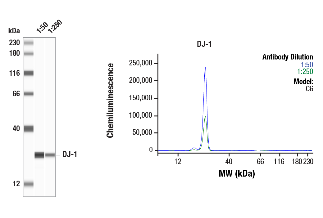
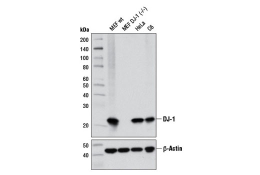
| DJ-1 (D29E5) XP® Rabbit mAb | 5933 | 20 µl | 22 kDa | Rabbit IgG |
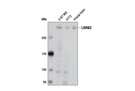
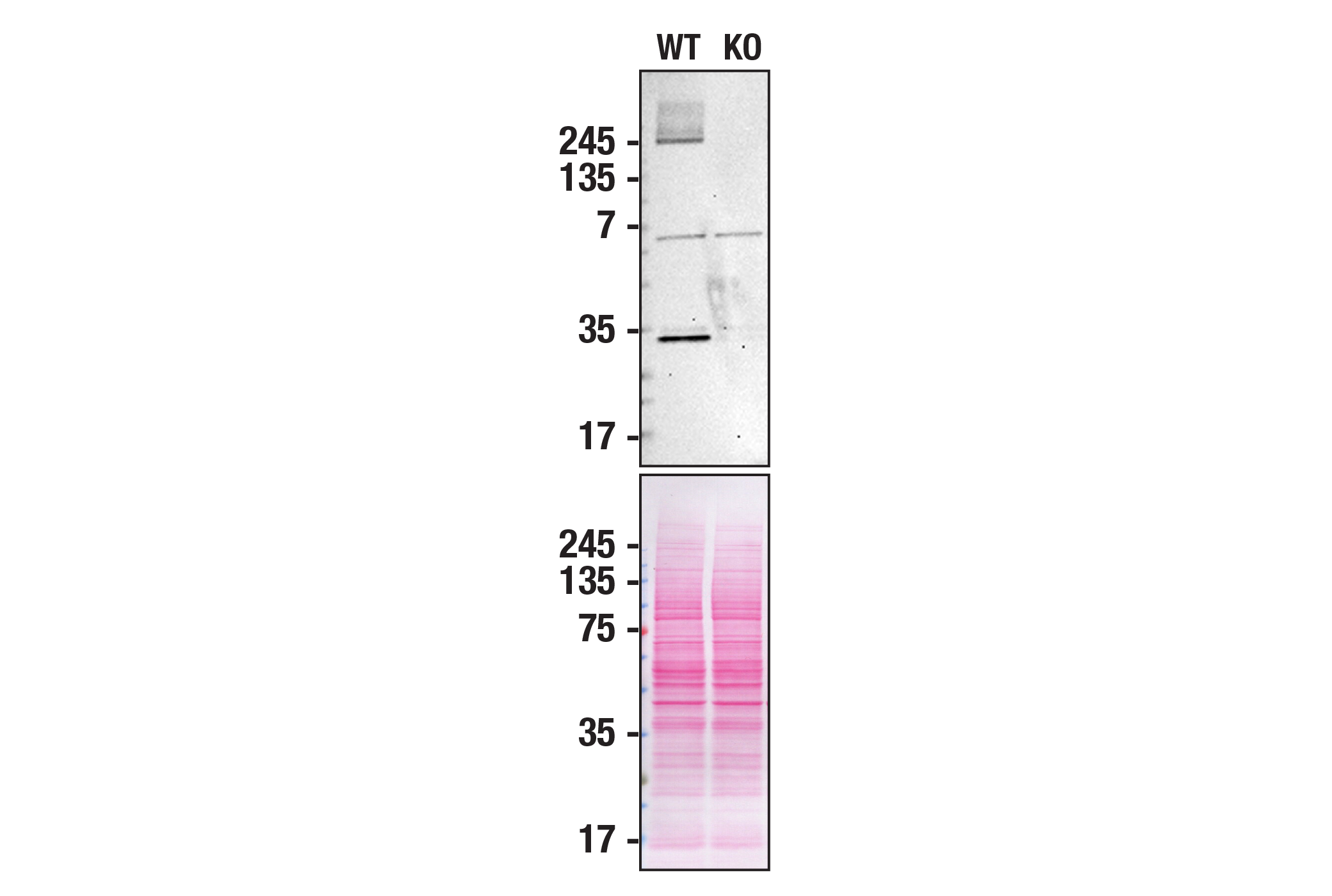
| LRRK2 (D18E12) Rabbit mAb | 13046 | 20 µl | 290 kDa | Rabbit IgG |
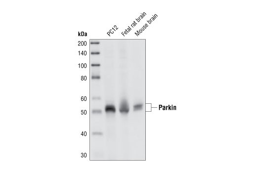
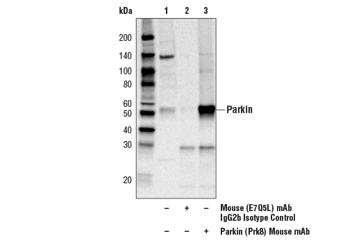
| Parkin (Prk8) Mouse mAb | 4211 | 20 µl | 50 kDa | Mouse IgG2b |
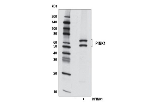
| PINK1 (D8G3) Rabbit mAb | 6946 | 20 µl | 60, 50 kDa | Rabbit IgG |
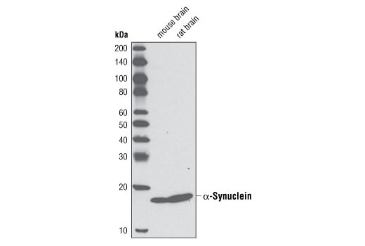
| α-Synuclein (D37A6) Rabbit mAb | 4179 | 20 µl | 18 kDa | Rabbit IgG |
| Anti-rabbit IgG, HRP-linked Antibody | 7074 | 100 µl | Goat | |
| Anti-mouse IgG, HRP-linked Antibody | 7076 | 100 µl | Horse |
Please visit cellsignal.com for individual component applications, species cross-reactivity, dilutions, protocols, and additional product information.
Description
The Parkinson's Research Antibody Sampler Kit provides an economical means of detecting target proteins related to Parkinson's disease. The kit contains enough primary and secondary antibody to perform two western blots per primary.
Storage
Background
Parkinson’s disease (PD), the second most common neurodegenerative disease after Alzheimer’s, is a progressive movement disorder characterized by rigidity, tremors, and postural instability. The pathological hallmark of PD is progressive loss of dopaminergic neurons in the substantia nigra of the ventral midbrain and the presence of intracellular Lewy bodies in surviving neurons of the brain stem (1). Research studies have shown that various genes and loci (α-synuclein/PARK1 and 4, parkin/PARK2, UCH-L1/PARK5, PINK1/PARK6, DJ-1/PARK7, LRRK2/PARK8, synphilin-1, and NR4A2) are genetically linked to PD (2).
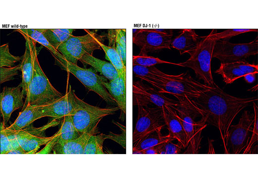
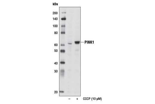
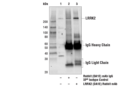
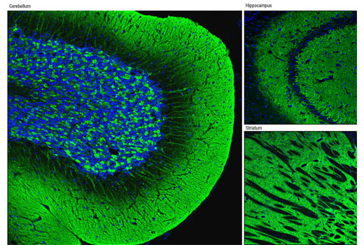
α-Synuclein, a 140 amino acid protein expressed abundantly in the brain, is a major component of aggregates found in Lewy bodies (3). Parkin is involved in protein degradation through the ubiquitin-proteasome pathway, and investigators have shown that mutations in Parkin cause early onset of PD (4). In the case of autosomal recessive juvenile Parkinsonism (AR-JP), deletions have been found on chromosome 6 in the Parkin gene (5). PTEN induced putative kinase 1 (PINK1) is a mitochondrial serine/threonine kinase involved in the normal function and integrity of mitochondria, as well as a reduction of cytochrome c release from mitochondria (6-8). PINK1 phosphorylates Parkin and promotes its translocation to mitochondria (7). Mutations of PINK1 are associated with loss of protective function, mitrochondrial dysfunction, aggregation of α-synuclein, and proteasome dysfunction (6,8). DJ-1 is involved in multiple cellular functions; it has been shown to cooperate with Ras to increase cell transformation, to regulate transcription of the androgen receptor, and may function as an indicator of oxidative stress, while loss-of-function mutations in DJ-1 cause early onset of PD (9-12). Dopamine D2 receptor-mediated functions are greatly impaired in DJ-1 (-/-) mice, resulting in reduced long-term depression (13). Leucine-rich repeat kinase 2 (LRRK2) contains amino-terminal leucine-rich repeats (LRR), a Ras-like small GTP binding protein-like (ROC) domain, an MLK protein kinase domain, and a carboxy-terminal WD40-repeat. At least 20 LRRK2 mutations have been linked to PD (14). The most prevalent mutation, G2019S, causes increased LRRK2 kinase activity, leading to progressive neurite loss and decreased neuronal survival (15).
- Fahn, S. (2003) Ann N Y Acad Sci 991, 1-14.
- Moore, D.J. et al. (2005) Annu Rev Neurosci 28, 57-87.
- Goldberg, M.S. and Lansbury, P.T. (2000) Nat Cell Biol 2, E115-9.
- Borrelli, E. (2005) Neuron 45, 479-81.
- Polymeropoulos, M.H. et al. (1997) Science 276, 2045-7.
- Liu, W. et al. (2009) PLoS One 4, e4597.
- Kim, Y. et al. (2008) Biochem Biophys Res Commun 377, 975-80.
- Petit, A. et al. (2005) J Biol Chem 280, 34025-32.
- Bonifati, V. et al. (2003) Science 299, 256-9.
- Nagakubo, D. et al. (1997) Biochem Biophys Res Commun 231, 509-13.
- Takahashi, K. et al. (2001) J Biol Chem 276, 37556-63.
- Mitsumoto, A. and Nakagawa, Y. (2001) Free Radic Res 35, 885-93.
- Goldberg, M.S. et al. (2005) Neuron 45, 489-96.
- Mata, I.F. et al. (2006) Trends Neurosci 29, 286-93.
- MacLeod, D. et al. (2006) Neuron 52, 587-93.
Background References
Trademarks and Patents
使用に関する制限
法的な権限を与えられたCSTの担当者が署名した書面によって別途明示的に合意された場合を除き、 CST、その関連会社または代理店が提供する製品には以下の条件が適用されます。お客様が定める条件でここに定められた条件に含まれるものを超えるもの、 または、ここに定められた条件と異なるものは、法的な権限を与えられたCSTの担当者が別途書面にて受諾した場合を除き、拒絶され、 いかなる効力も効果も有しません。
研究専用 (For Research Use Only) またはこれに類似する表示がされた製品は、 いかなる目的についても FDA または外国もしくは国内のその他の規制機関により承認、認可または許可を受けていません。 お客様は製品を診断もしくは治療目的で使用してはならず、また、製品に表示された内容に違反する方法で使用してはなりません。 CST が販売または使用許諾する製品は、エンドユーザーであるお客様に対し、使途を研究および開発のみに限定して提供されるものです。 診断、予防もしくは治療目的で製品を使用することまたは製品を再販売 (単独であるか他の製品等の一部であるかを問いません) もしくはその他の商業的利用の目的で購入することについては、CST から別途許諾を得る必要があります。 お客様は以下の事項を遵守しなければなりません。(a) CST の製品 (単独であるか他の資材と一緒であるかを問いません) を販売、使用許諾、貸与、寄付もしくはその他の態様で第三者に譲渡したり使用させたりしてはなりません。また、商用の製品を製造するために CST の製品を使用してはなりません。(b) 複製、改変、リバースエンジニアリング、逆コンパイル、 分解または他の方法により製品の構造または技術を解明しようとしてはなりません。また、 CST の製品またはサービスと競合する製品またはサービスを開発する目的で CST の製品を使用してはなりません。(c) CST の製品の商標、商号、ロゴ、特許または著作権に関する通知または表示を除去したり改変したりしてはなりません。(d) CST の製品をCST 製品販売条件(CST’s Product Terms of Sale) および該当する書面のみに従って使用しなければなりません。(e) CST の製品に関連してお客様が使用する第三者の製品またはサービスに関する使用許諾条件、 サービス提供条件またはこれに類する合意事項を遵守しなければなりません。