| Product Includes | Product # | Quantity | Mol. Wt | Isotype/Source |
|---|---|---|---|---|
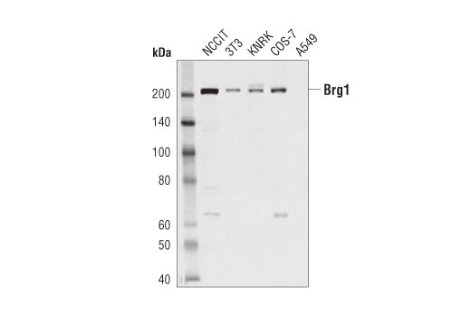
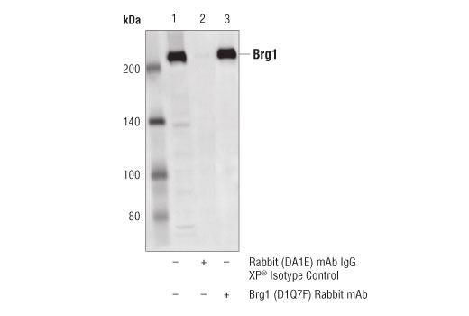
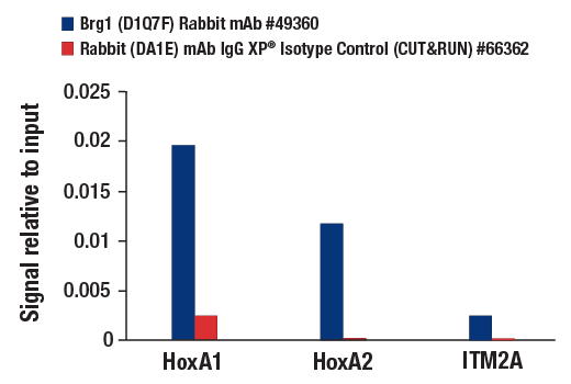
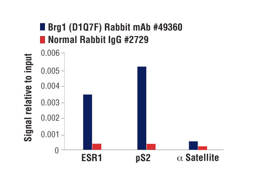
| Brg1 (D1Q7F) Rabbit mAb | 49360 | 20 µl | 220 kDa | Rabbit IgG |
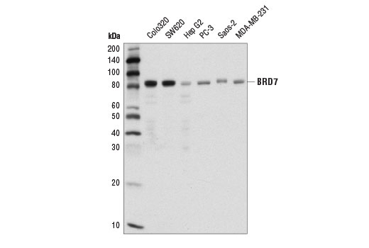
| BRD7 (D9K2T) Rabbit mAb | 15125 | 20 µl | 85 kDa | Rabbit IgG |
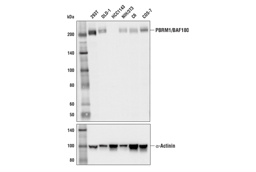
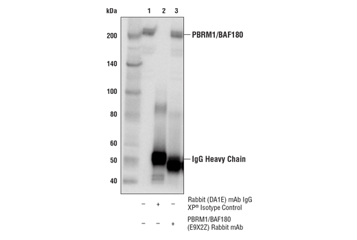
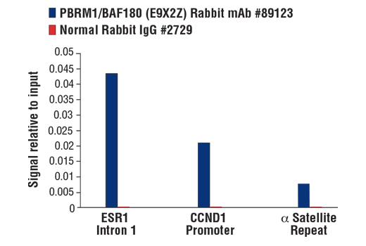
| PBRM1/BAF180 (E9X2Z) Rabbit mAb | 89123 | 20 µl | 205 kDa | Rabbit IgG |
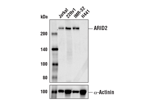
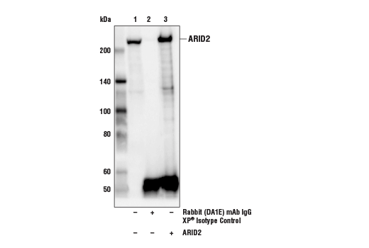
| ARID2 (D8D8U) Rabbit mAb | 82342 | 20 µl | 220 kDa | Rabbit IgG |
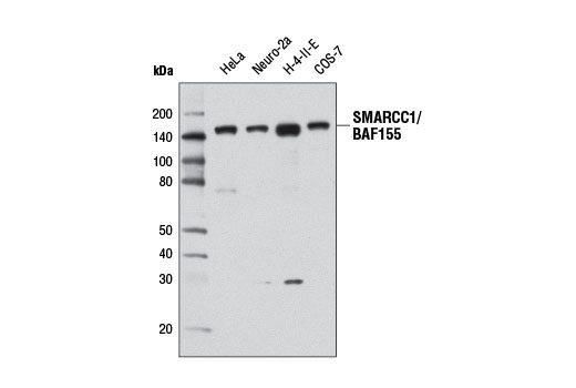
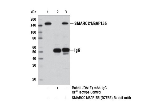
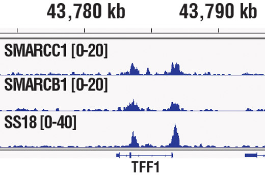
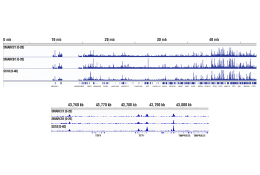
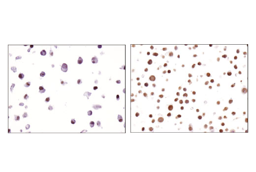
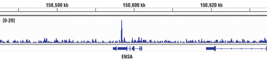
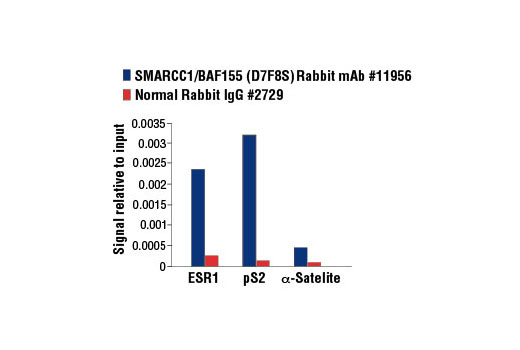
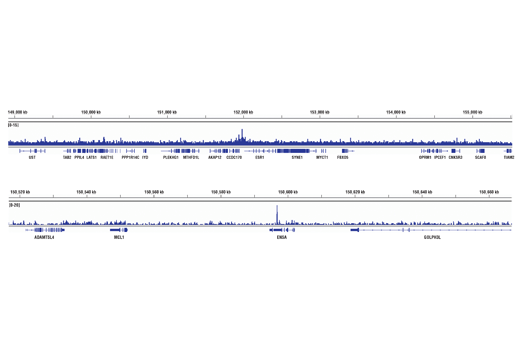
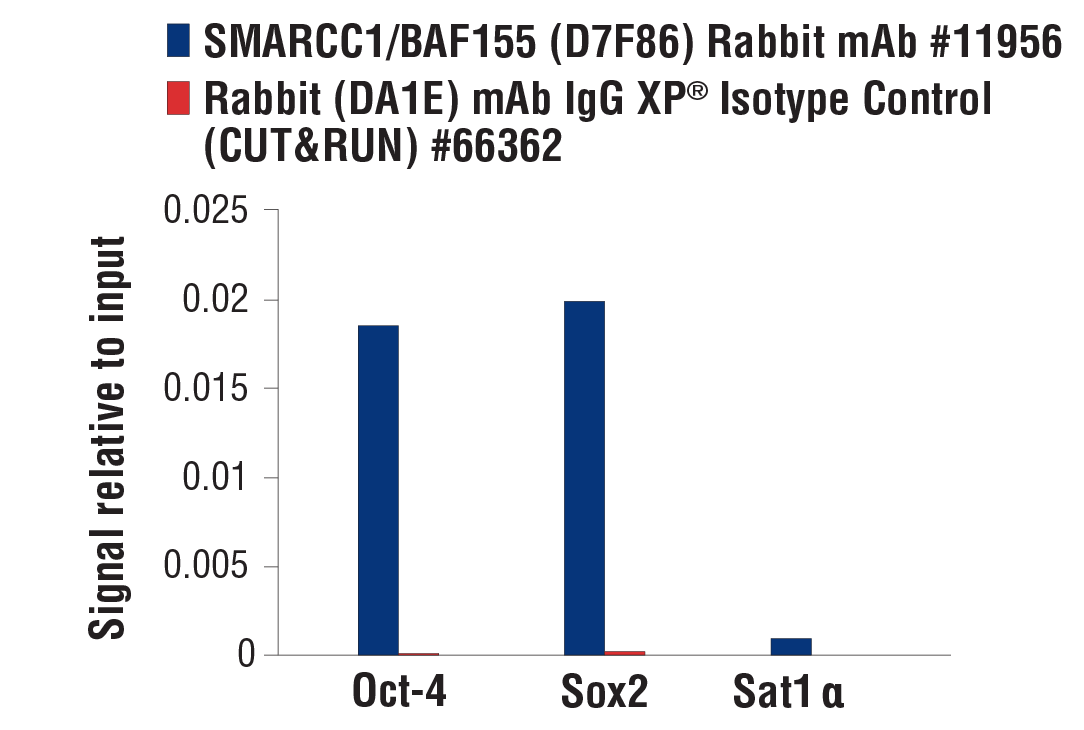
| SMARCC1/BAF155 (D7F8S) Rabbit mAb | 11956 | 20 µl | 155 kDa | Rabbit IgG |
| Anti-rabbit IgG, HRP-linked Antibody | 7074 | 100 µl | Goat |
Please visit cellsignal.com for individual component applications, species cross-reactivity, dilutions, protocols, and additional product information.
Description
The PBAF Complex Antibody Sampler Kit provides an economical means of detecting members of the PBAF chromatin remodeling complex. ARID2, BRD7, and PBRM1/BAF180 are unique members of the PBAF complex, while Brg1 and SMARCC1/BAF155 are shared with BAF and non-canonical BAF complexes. The kit includes enough antibody to perform two western blot experiments with each primary antibody.
Storage
Background
ATP-dependent chromatin remodeling complexes play an essential role in the regulation of various nuclear processes, such as gene expression, DNA replication, and repair (1,2). The SWI/SNF chromatin remodeling complex consists of more than 10 subunits with a single molecule of the ATPase catalytic subunit BRM or BRG1, but not both. The activities of these two subunits drive the disruption of histone-DNA contacts that lead to changes in accessibility of crucial regulatory elements within chromatin (2-5). The BRM/BRG1 containing SWI/SNF complexes are recruited to target promoters by transcription factors, such as nuclear receptors, p53, RB, and BRCA1 to regulate gene activation, cell growth, the cell cycle, and differentiation processes (1,6-9).
PBRM1/BAF180, ARID2, and BRD7 are unique members of the SWI/SNF complex PBAF, which binds to kinetochores in mitotic chromatin (10,11). PBAF is involved in nuclear receptor-mediated transcription and retinoic acid driven gene activation (12,13). PBRM1/BAF180 has been shown to be a potent tumor suppressor, as it is the second-most mutated gene in renal carcinomas (14). ARID2 is the targeting subunit of the PBAF complex and critical for complex stability (15). Bromodomain-containing BRD7 interacts with acetylated histones to regulate gene transcription (16,17). SMARCC1/BAF155 is a core subunit of all BAF complexes including PBAF, and is necessary for efficient nucleosome remodeling by BRG1 in vitro (18).
- Ho, L. and Crabtree, G.R. (2010) Nature 463, 474-84.
- Becker, P.B. and Hörz, W. (2002) Annu Rev Biochem 71, 247-73.
- Eberharter, A. and Becker, P.B. (2004) J Cell Sci 117, 3707-11.
- Bowman, G.D. (2010) Curr Opin Struct Biol 20, 73-81.
- Gangaraju, V.K. and Bartholomew, B. (2007) Mutat Res 618, 3-17.
- Lessard, J.A. and Crabtree, G.R. (2010) Annu Rev Cell Dev Biol 26, 503-32.
- Morettini, S. et al. (2008) Front Biosci 13, 5522-32.
- Wolf, I.M. et al. (2008) J Cell Biochem 104, 1580-6.
- Simone, C. (2006) J Cell Physiol 207, 309-14.
- Nie, Z. et al. (2000) Mol Cell Biol 20, 8879-88.
- Xue, Y. et al. (2000) Proc Natl Acad Sci U S A 97, 13015-20.
- Lemon, B. et al. (2001) Nature 414, 924-8.
- Wang, Z. et al. (2004) Genes Dev 18, 3106-16.
- Varela, I. et al. (2011) Nature 469, 539-42.
- Yan, Z. et al. (2005) Genes Dev 19, 1662-7.
- Peng, C. et al. (2006) J Cell Biochem 97, 882-92.
- Kaeser, M.D. et al. (2008) J Biol Chem 283, 32254-63.
- Phelan, M.L. et al. (1999) Mol Cell 3, 247-53.
Background References
Trademarks and Patents
使用に関する制限
法的な権限を与えられたCSTの担当者が署名した書面によって別途明示的に合意された場合を除き、 CST、その関連会社または代理店が提供する製品には以下の条件が適用されます。お客様が定める条件でここに定められた条件に含まれるものを超えるもの、 または、ここに定められた条件と異なるものは、法的な権限を与えられたCSTの担当者が別途書面にて受諾した場合を除き、拒絶され、 いかなる効力も効果も有しません。
研究専用 (For Research Use Only) またはこれに類似する表示がされた製品は、 いかなる目的についても FDA または外国もしくは国内のその他の規制機関により承認、認可または許可を受けていません。 お客様は製品を診断もしくは治療目的で使用してはならず、また、製品に表示された内容に違反する方法で使用してはなりません。 CST が販売または使用許諾する製品は、エンドユーザーであるお客様に対し、使途を研究および開発のみに限定して提供されるものです。 診断、予防もしくは治療目的で製品を使用することまたは製品を再販売 (単独であるか他の製品等の一部であるかを問いません) もしくはその他の商業的利用の目的で購入することについては、CST から別途許諾を得る必要があります。 お客様は以下の事項を遵守しなければなりません。(a) CST の製品 (単独であるか他の資材と一緒であるかを問いません) を販売、使用許諾、貸与、寄付もしくはその他の態様で第三者に譲渡したり使用させたりしてはなりません。また、商用の製品を製造するために CST の製品を使用してはなりません。(b) 複製、改変、リバースエンジニアリング、逆コンパイル、 分解または他の方法により製品の構造または技術を解明しようとしてはなりません。また、 CST の製品またはサービスと競合する製品またはサービスを開発する目的で CST の製品を使用してはなりません。(c) CST の製品の商標、商号、ロゴ、特許または著作権に関する通知または表示を除去したり改変したりしてはなりません。(d) CST の製品をCST 製品販売条件(CST’s Product Terms of Sale) および該当する書面のみに従って使用しなければなりません。(e) CST の製品に関連してお客様が使用する第三者の製品またはサービスに関する使用許諾条件、 サービス提供条件またはこれに類する合意事項を遵守しなければなりません。