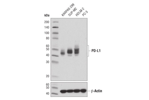
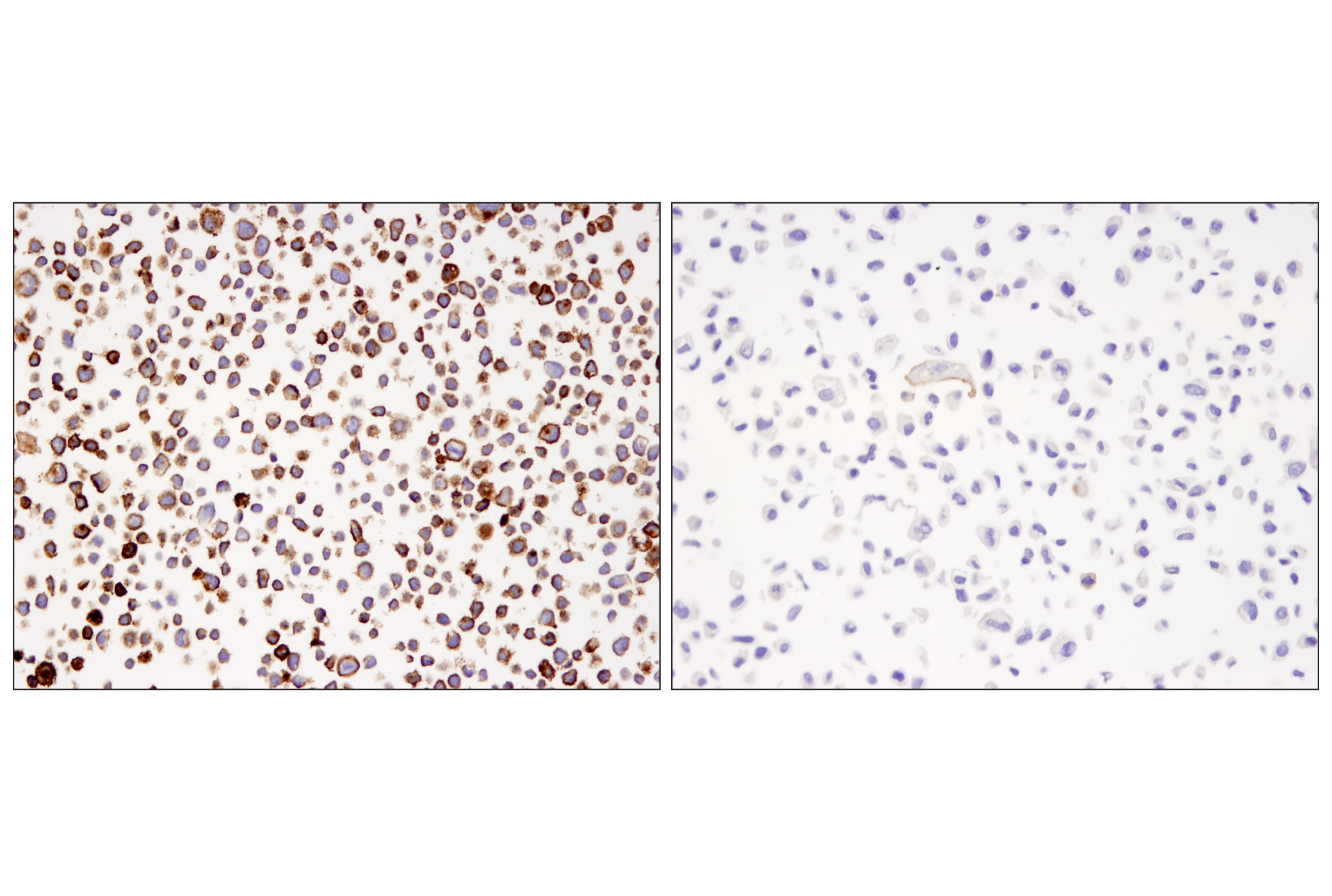
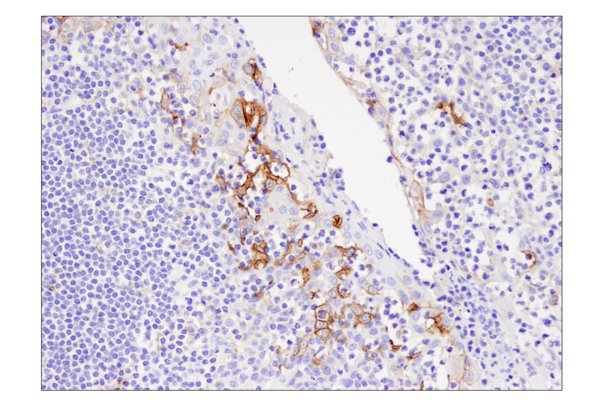
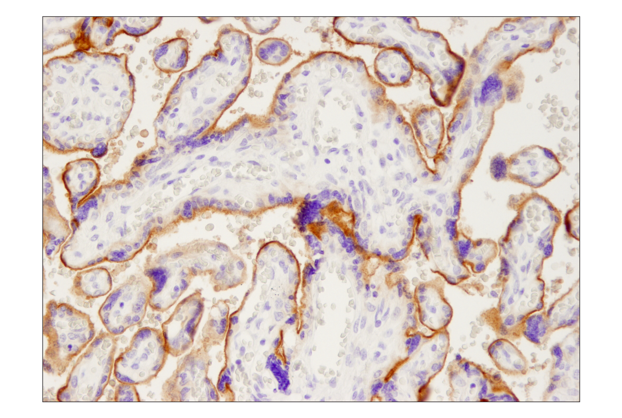
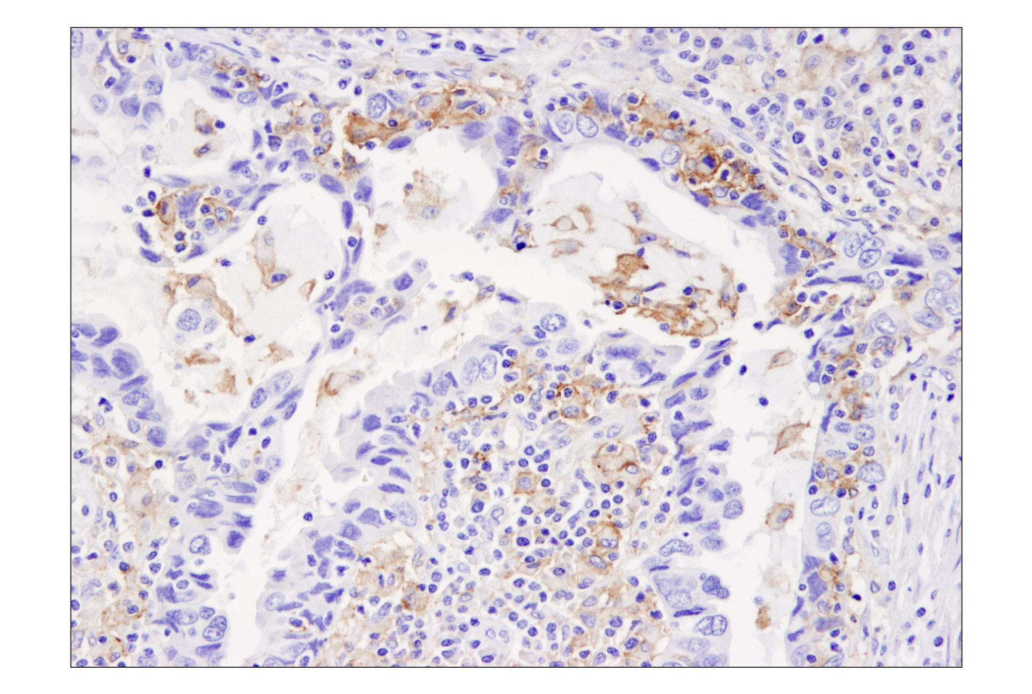
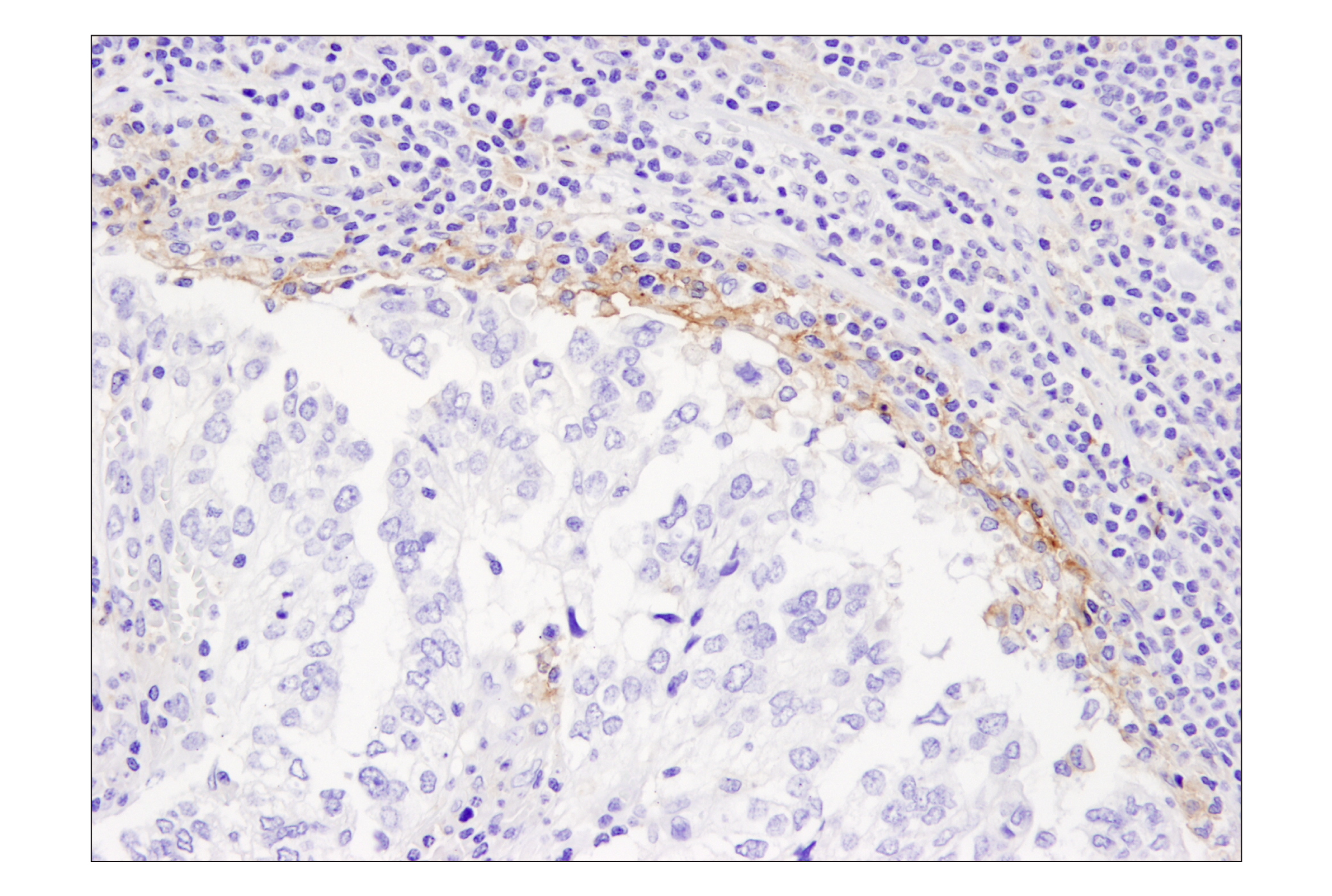
WB, IHC-P
H
Endogenous
40-50
Mouse IgG1
#Q9NZQ7
29126
Product Information
Product Usage Information
This product is the carrier free version of product #29122. All data were generated using the same antibody clone in the standard formulation which contains BSA and glycerol.
This formulation is ideal for use with technologies requiring specialized or custom antibody labeling, including fluorophores, metals, lanthanides, and oligonucleotides. It is not recommended for ChIP, ChIP-Seq, CUT&RUN, or CUT&Tag assays. If you require a carrier free formulation for chromatin profiling, please contact us. Optimal dilutions/concentrations should be determined by the end user.
Formulation
Supplied in 1X PBS, BSA and Azide Free.
For standard formulation of this product see product #29122.
Storage
Specificity / Sensitivity
Species Reactivity:
Human
Source / Purification
Monoclonal antibody is produced by immunizing animals with a synthetic peptide corresponding to residues near the carboxy terminus of human PD-L1 protein.
Background
Programmed cell death 1 ligand 1 (PD-L1, B7-H1, CD274) is a member of the B7 family of cell surface ligands that regulate T cell activation and immune responses. The PD-L1 ligand binds the PD-1 transmembrane receptor and inhibits T cell activation. PD-L1 was discovered following a search for novel B7 protein homologs and was later shown to be expressed by antigen presenting cells, activated T cells, and tissues including placenta, heart, and lung (1-3). Similar in structure to related B7 family members, PD-L1 protein contains extracellular IgV and IgC domains and a short, cytoplasmic region. Research studies demonstrate that PD-L1 is expressed in several tumor types, including melanoma, ovary, colon, lung, breast, and renal cell carcinomas (4-6). Expression of PD-L1 in cancer is associated with tumor-infiltrating lymphocytes, which mediate PD-L1 expression through the release of interferon gamma (7). Additional research links PD-L1 expression to cancers associated with viral infections (8,9).
- Dong, H. et al. (1999) Nat Med 5, 1365-9.
- Freeman, G.J. et al. (2000) J Exp Med 192, 1027-34.
- Liang, S.C. et al. (2003) Eur J Immunol 33, 2706-16.
- Dong, H. et al. (2002) Nat Med 8, 793-800.
- Thompson, R.H. et al. (2006) Cancer Res 66, 3381-5.
- Pardoll, D.M. (2012) Nat Rev Cancer 12, 252-64.
- Taube, J.M. et al. (2012) Sci Transl Med 4, 127ra37.
- Lyford-Pike, S. et al. (2013) Cancer Res 73, 1733-41.
- Chen, B.J. et al. (2013) Clin Cancer Res 19, 3462-73.
Species Reactivity
Species reactivity is determined by testing in at least one approved application (e.g., western blot).
Applications Key
WB: Western Blotting IHC-P: Immunohistochemistry (Paraffin)
Cross-Reactivity Key
H: human M: mouse R: rat Hm: hamster Mk: monkey Vir: virus Mi: mink C: chicken Dm: D. melanogaster X: Xenopus Z: zebrafish B: bovine Dg: dog Pg: pig Sc: S. cerevisiae Ce: C. elegans Hr: horse GP: Guinea Pig Rab: rabbit All: all species expected
Trademarks and Patents
使用に関する制限
法的な権限を与えられたCSTの担当者が署名した書面によって別途明示的に合意された場合を除き、 CST、その関連会社または代理店が提供する製品には以下の条件が適用されます。お客様が定める条件でここに定められた条件に含まれるものを超えるもの、 または、ここに定められた条件と異なるものは、法的な権限を与えられたCSTの担当者が別途書面にて受諾した場合を除き、拒絶され、 いかなる効力も効果も有しません。
研究専用 (For Research Use Only) またはこれに類似する表示がされた製品は、 いかなる目的についても FDA または外国もしくは国内のその他の規制機関により承認、認可または許可を受けていません。 お客様は製品を診断もしくは治療目的で使用してはならず、また、製品に表示された内容に違反する方法で使用してはなりません。 CST が販売または使用許諾する製品は、エンドユーザーであるお客様に対し、使途を研究および開発のみに限定して提供されるものです。 診断、予防もしくは治療目的で製品を使用することまたは製品を再販売 (単独であるか他の製品等の一部であるかを問いません) もしくはその他の商業的利用の目的で購入することについては、CST から別途許諾を得る必要があります。 お客様は以下の事項を遵守しなければなりません。(a) CST の製品 (単独であるか他の資材と一緒であるかを問いません) を販売、使用許諾、貸与、寄付もしくはその他の態様で第三者に譲渡したり使用させたりしてはなりません。また、商用の製品を製造するために CST の製品を使用してはなりません。(b) 複製、改変、リバースエンジニアリング、逆コンパイル、 分解または他の方法により製品の構造または技術を解明しようとしてはなりません。また、 CST の製品またはサービスと競合する製品またはサービスを開発する目的で CST の製品を使用してはなりません。(c) CST の製品の商標、商号、ロゴ、特許または著作権に関する通知または表示を除去したり改変したりしてはなりません。(d) CST の製品をCST 製品販売条件(CST’s Product Terms of Sale) および該当する書面のみに従って使用しなければなりません。(e) CST の製品に関連してお客様が使用する第三者の製品またはサービスに関する使用許諾条件、 サービス提供条件またはこれに類する合意事項を遵守しなければなりません。