Revision 4

#9958Store at -20C

1 Kit

(3 x 20 microliters)

Cell Signaling Technology

Orders: 877-616-CELL (2355) [email protected]

Support: 877-678-TECH (8324)

Web: [email protected] cellsignal.com

3 Trask LaneDanversMassachusetts01923USA
For Research Use Only. Not for Use in Diagnostic Procedures.
Product Includes Product # Quantity Mol. Wt Isotype/Source
Phospho-IKKα (Ser176)/IKKβ (Ser177) (C84E11) Rabbit mAb 2078 20 µl 85 (IKKalpha), 87 (IKKbeta) kDa Rabbit 
Phospho-IKKα/β (Ser176/180) (16A6) Rabbit mAb 2697 20 µl 85 IKK-alpha 87 IKK-beta kDa Rabbit IgG
Phospho-IKKα/β (Ser176/180) Antibody II 2694 20 µl 85 IKK-alpha 87 IKK-beta kDa Rabbit 
Anti-rabbit IgG, HRP-linked Antibody 7074 100 µl Goat 

Please visit cellsignal.com for individual component applications, species cross-reactivity, dilutions, protocols, and additional product information.

Description

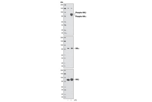
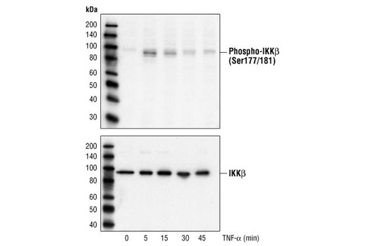
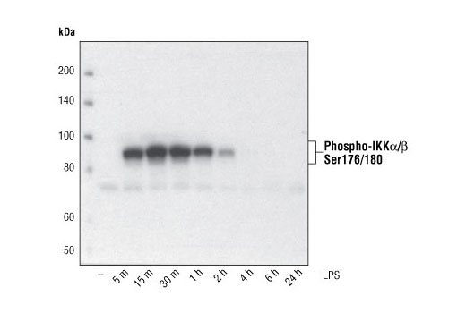
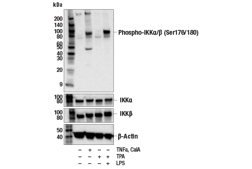
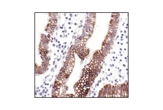
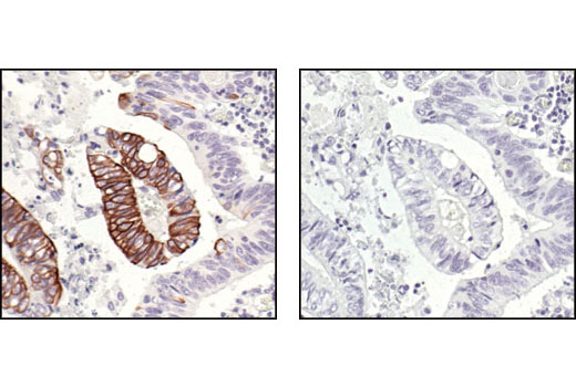
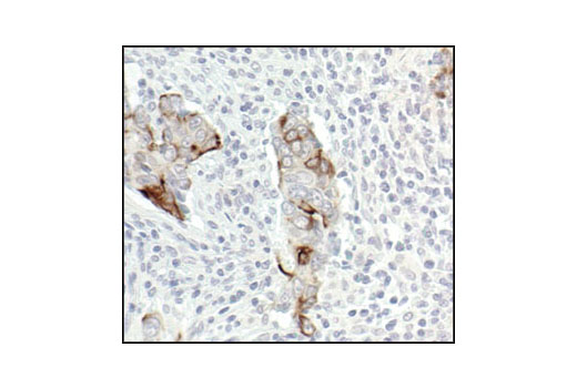
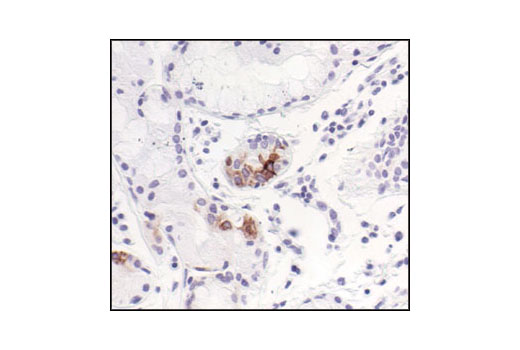
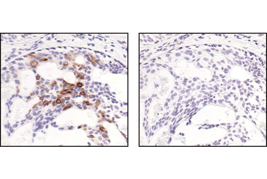
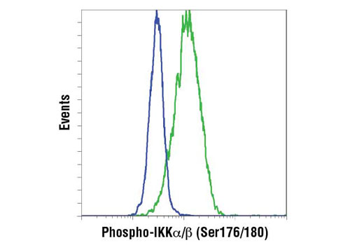
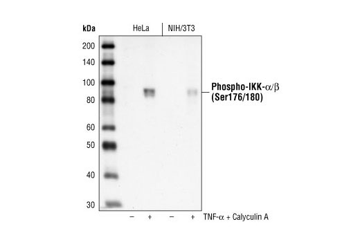
The Phospho-IKKalpha/beta (Ser176/180) Antibody Sampler Kit contains reagents to examine protein levels of IKKalpha when phosphorylated at Ser176/180 and IKKbeta when phosphorylated at Ser177/181. The kit contains primary and secondary antibodies to perform two Western blots with each antibody.

Storage

Supplied in 10 mM sodium HEPES (pH 7.5), 150 mM NaCl, 100 µg/ml BSA, 50% glycerol and less than 0.02% sodium azide. Store at –20°C. Do not aliquot the antibody.

Background

The NF-κB/Rel transcription factors are present in the cytosol in an inactive state, complexed with the inhibitory IκB proteins (1-3). Most agents that activate NF-κB do so through a common pathway based on phosphorylation-induced, proteasome-mediated degradation of IκB (3-7). The key regulatory step in this pathway involves activation of a high molecular weight IκB kinase (IKK) complex whose catalysis is generally carried out by three tightly associated IKK subunits. IKKα and IKKβ serve as the catalytic subunits of the kinase and IKKγ serves as the regulatory subunit (8,9). Activation of IKK depends upon phosphorylation at Ser177 and Ser181 in the activation loop of IKKβ (Ser176 and Ser180 in IKKα), which causes conformational changes, resulting in kinase activation (10-13).

  1. Baeuerle, P.A. and Baltimore, D. (1988) Science 242, 540-6.
  2. Beg, A.A. and Baldwin, A.S. (1993) Genes Dev 7, 2064-70.
  3. Finco, T.S. et al. (1994) Proc Natl Acad Sci USA 91, 11884-8.
  4. Brown, K. et al. (1995) Science 267, 1485-8.
  5. Brockman, J.A. et al. (1995) Mol Cell Biol 15, 2809-18.
  6. Traenckner, E.B. et al. (1995) EMBO J 14, 2876-83.
  7. Chen, Z.J. et al. (1996) Cell 84, 853-62.
  8. Zandi, E. et al. (1997) Cell 91, 243-52.
  9. Karin, M. (1999) Oncogene 18, 6867-74.
  10. DiDonato, J.A. et al. (1997) Nature 388, 548-54.
  11. Mercurio, F. et al. (1997) Science 278, 860-6.
  12. Johnson, L.N. et al. (1996) Cell 85, 149-58.
  13. Delhase, M. et al. (1999) Science 284, 309-13.

Background References

    Trademarks and Patents

    Cell Signaling Technology is a trademark of Cell Signaling Technology, Inc.
    All other trademarks are the property of their respective owners. Visit cellsignal.com/trademarks for more information.

    使用に関する制限

    法的な権限を与えられたCSTの担当者が署名した書面によって別途明示的に合意された場合を除き、 CST、その関連会社または代理店が提供する製品には以下の条件が適用されます。お客様が定める条件でここに定められた条件に含まれるものを超えるもの、 または、ここに定められた条件と異なるものは、法的な権限を与えられたCSTの担当者が別途書面にて受諾した場合を除き、拒絶され、 いかなる効力も効果も有しません。

    研究専用 (For Research Use Only) またはこれに類似する表示がされた製品は、 いかなる目的についても FDA または外国もしくは国内のその他の規制機関により承認、認可または許可を受けていません。 お客様は製品を診断もしくは治療目的で使用してはならず、また、製品に表示された内容に違反する方法で使用してはなりません。 CST が販売または使用許諾する製品は、エンドユーザーであるお客様に対し、使途を研究および開発のみに限定して提供されるものです。 診断、予防もしくは治療目的で製品を使用することまたは製品を再販売 (単独であるか他の製品等の一部であるかを問いません) もしくはその他の商業的利用の目的で購入することについては、CST から別途許諾を得る必要があります。 お客様は以下の事項を遵守しなければなりません。(a) CST の製品 (単独であるか他の資材と一緒であるかを問いません) を販売、使用許諾、貸与、寄付もしくはその他の態様で第三者に譲渡したり使用させたりしてはなりません。また、商用の製品を製造するために CST の製品を使用してはなりません。(b) 複製、改変、リバースエンジニアリング、逆コンパイル、 分解または他の方法により製品の構造または技術を解明しようとしてはなりません。また、 CST の製品またはサービスと競合する製品またはサービスを開発する目的で CST の製品を使用してはなりません。(c) CST の製品の商標、商号、ロゴ、特許または著作権に関する通知または表示を除去したり改変したりしてはなりません。(d) CST の製品をCST 製品販売条件(CST’s Product Terms of Sale) および該当する書面のみに従って使用しなければなりません。(e) CST の製品に関連してお客様が使用する第三者の製品またはサービスに関する使用許諾条件、 サービス提供条件またはこれに類する合意事項を遵守しなければなりません。

    Revision 4
    #9958

    Phospho-IKKα/β (Ser176/180) Antibody Sampler Kit

    Phospho-IKKα/β (Ser176/180) Antibody Sampler Kit: Image 1 Expand Image
    Western blot analysis of extracts from differentiated THP-1 cells, untreated or LPS-treated for 15 minutes, using Phospho-IKKα (Ser176)/IKKβ (Ser177) (C84E11) Rabbit mAb (upper), IKKα Antibody #2682 (middle) and IKKβ (2C8) Rabbit mAb #2370 (lower).
    Phospho-IKKα/β (Ser176/180) Antibody Sampler Kit: Image 2 Expand Image
    Western blot analysis of extracts from HeLa cells, treated with TNF-α for the indicated times, using Phospho-IKKα/β (Ser176/180) Antibody II (upper) or IKKβ Antibody #2684 (lower).
    Phospho-IKKα/β (Ser176/180) Antibody Sampler Kit: Image 3 Expand Image
    Western blot analysis of extracts from THP-1 cells, differentiated with TPA (#9905, 80 nM for 24h) and treated with 1 μg/ml LPS for the indicated times, using Phospho-IKKα/β (Ser176/180) (16A6) Rabbit mAb.
    Phospho-IKKα/β (Ser176/180) Antibody Sampler Kit: Image 4 Expand Image
    After the primary antibody is bound to the target protein, a complex with HRP-linked secondary antibody is formed. The LumiGLO® is added and emits light during enzyme catalyzed decomposition.
    Phospho-IKKα/β (Ser176/180) Antibody Sampler Kit: Image 5 Expand Image
    Western blot analysis of extracts from HeLa cells, untreated or treated with TNFa (#16789, 20 ng/mL, 10 min) and Calyculin A (#9902, 100 nM, 10 min); THP-1 cells differentiated with TPA (#4174, 80 nM for 16hr) untreated and treated with LPS (#14011, 1 μg/ml for 1hr) using #2697 (upper) or IKKα (D3W6N) Rabbit mAb #61294, IKKβ (D30C6) Rabbit mAb #8943 (middle), and β-Actin (D6A8) Rabbit mAb #8457 (lower).
    Phospho-IKKα/β (Ser176/180) Antibody Sampler Kit: Image 6 Expand Image
    Immunohistochemical analysis of paraffin-embedded human gall bladder (chronic cholecystitis), using Phospho-IKKα/β (Ser176/180) (16A6) Rabbit mAb.
    Phospho-IKKα/β (Ser176/180) Antibody Sampler Kit: Image 7 Expand Image
    Immunohistochemical analysis of paraffin-embedded human colon carcinoma, untreated (left) or λ phosphatase-treated (right), using Phospho-IKKα/β (Ser176/180) (16A6) Rabbit mAb.
    Phospho-IKKα/β (Ser176/180) Antibody Sampler Kit: Image 8 Expand Image
    Immunohistochemical analysis of paraffin-embedded human colon carcinoma, showing cytoplasmic localization, using Phospho-IKKα/β (Ser176/180) (16A6) Rabbit mAb.
    Phospho-IKKα/β (Ser176/180) Antibody Sampler Kit: Image 9 Expand Image
    Immunohistochemical analysis of paraffin-embedded human lung (chronic bronchitis), using Phospho-IKKα/β (Ser176/180) (16A6) Rabbit mAb.
    Phospho-IKKα/β (Ser176/180) Antibody Sampler Kit: Image 10 Expand Image
    Immunohistochemical analysis of paraffin-embedded human breast carcinoma, using Phospho-IKKα/β (Ser176/180) (16A6) Rabbit mAb in the presence of control peptide (left) or Phospho-IKK-alpha/beta (Ser176/180) Blocking Peptide #1023 (right).
    Phospho-IKKα/β (Ser176/180) Antibody Sampler Kit: Image 11 Expand Image
    Flow cytometric analysis of THP-1 cells, untreated (blue) and with TPA and LPS (green) using IKK-α (Ser176/Ser180) phosphate Rabbit mAb. Anti-rabbit IgG (H+L), F(ab')2 Fragment (PE Conjugate) #8885 was used as a secondary antibody.
    Phospho-IKKα/β (Ser176/180) Antibody Sampler Kit: Image 12 Expand Image
    Western blot analysis of extracts from TNF-alpha and Calyculin A treated HeLa and NIH/3T3 cells, using Phospho-IKKα/β (Ser176/180) (16A6) Rabbit mAb.