WB, IHC-P, IF-F, IF-IC, FC-FP
H M R Mk
Endogenous
46, 48
Rabbit IgG
#Q92597
10397
Product Information
Product Usage Information
| Application | Dilution |
|---|---|
| Western Blotting | 1:1000 |
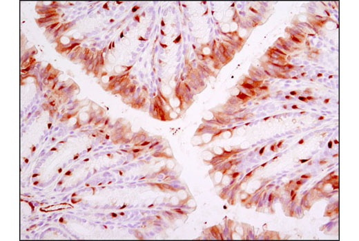
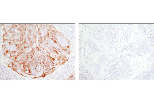
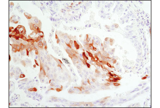
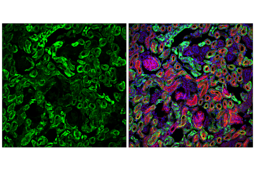
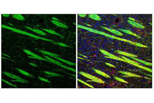
| Immunohistochemistry (Paraffin) | 1:250 - 1:1000 |
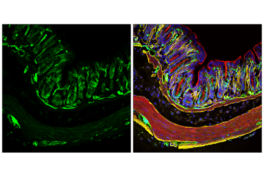
| Immunofluorescence (Frozen) | 1:50 - 1:100 |
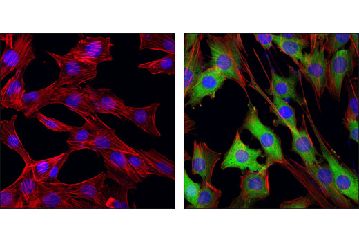
| Immunofluorescence (Immunocytochemistry) | 1:200 - 1:800 |
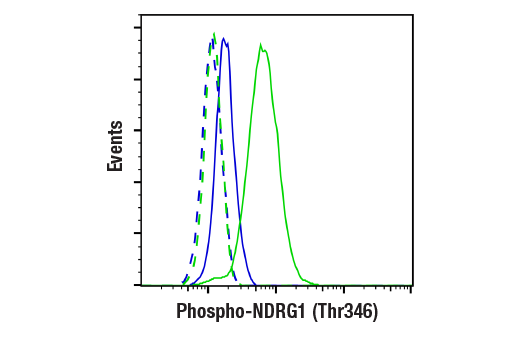
| Flow Cytometry (Fixed/Permeabilized) | 1:200 |
Storage
Supplied in 10 mM sodium HEPES (pH 7.5), 150 mM NaCl, 100 µg/ml BSA, 50% glycerol and less than 0.02% sodium azide. Store at –20°C. Do not aliquot the antibody.
For a carrier-free (BSA and azide free) version of this product see product #89166.
Specificity / Sensitivity
Species Reactivity:
Human, Mouse, Rat, Monkey
Source / Purification
Monoclonal antibody is produced by immunizing animals with a synthetic phosphopeptide corresponding to residues surrounding Thr346 of mouse NDRG1 protein.
Background
N-myc downstream-regulated gene 1 (NDRG1), also termed Cap43, Drg1, RTP/rit42, and Proxy-1, is a member of the NDRG family, which is composed of four members (NDRG1-4) that function in growth, differentiation, and cell survival (1-5). NDRG1 is ubiquitously expressed and highly responsive to a variety of stress signals, including DNA damage (4), hypoxia (5), and elevated levels of nickel and calcium (2). Expression of NDRG1 is elevated in N-myc defective mice and is negatively regulated by N- and c-myc (1,6). During DNA damage, NDRG1 is induced in a p53-dependent fashion and is necessary for p53-mediated apoptosis (4,7). Research studies have shown that NDRG1 may also play a role in cancer progression by promoting differentiation, inhibiting growth, and modulating metastasis and angiogenesis (3,4,6,8,9). Nonsense mutation of the NDRG1 gene has been shown to cause hereditary motor and sensory neuropathy-Lom (HMSNL), which is supported by studies demonstrating the role of NDRG1 in maintaining myelin sheaths and axonal survival (10,11). NDRG1 is upregulated during mast cell maturation and its deletion leads to attenuated allergic responses (12). Both NDRG1 and NDRG2 are substrates of SGK1, although the precise physiological role of SGK1-mediated phosphorylation is not known (13). NDRG1 is phosphorylated by SGK1 at Thr328, Ser330, Thr346, Thr356, and Thr366. Phosphorylation by SGK1 primes NDRG1 for phosphorylation by GSK-3.
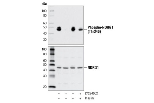
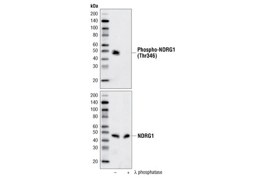
Phospho-NDRG1 (Thr346) (D98G11) XP® Rabbit mAb is directed at a site that was identified at Cell Signaling Technology (CST) using PhosphoScan®, CST's LC-MS/MS platform for modification site discovery. Phosphorylation at Thr346 was discovered using an Akt substrate antibody and was shown to be induced by insulin treatment in multiple cell lines. Please visit PhosphoSitePlus®, CST's modification site knowledgebase, at www.phosphosite.org for more information.
- Shimono, A. et al. (1999) Mech Dev 83, 39-52.
- Zhou, D. et al. (1998) Cancer Res 58, 2182-9.
- van Belzen, N. et al. (1997) Lab Invest 77, 85-92.
- Kurdistani, S.K. et al. (1998) Cancer Res 58, 4439-44.
- Park, H. et al. (2000) Biochem Biophys Res Commun 276, 321-8.
- Li, J. and Kretzner, L. (2003) Mol Cell Biochem 250, 91-105.
- Stein, S. et al. (2004) J Biol Chem 279, 48930-40.
- Maruyama, Y. et al. (2006) Cancer Res 66, 6233-42.
- Nishio, S. et al. (2008) Cancer Lett 264, 36-43.
- Kalaydjieva, L. et al. (2000) Am J Hum Genet 67, 47-58.
- Okuda, T. et al. (2004) Mol Cell Biol 24, 3949-56.
- Taketomi, Y. et al. (2007) J Immunol 178, 7042-53.
- Murray, J.T. et al. (2004) Biochem J 384, 477-88.
Species Reactivity
Species reactivity is determined by testing in at least one approved application (e.g., western blot).
Western Blot Buffer
IMPORTANT: For western blots, incubate membrane with diluted primary antibody in 5% w/v nonfat dry milk, 1X TBS, 0.1% Tween® 20 at 4°C with gentle shaking, overnight.
Applications Key
WB: Western Blotting IHC-P: Immunohistochemistry (Paraffin) IF-F: Immunofluorescence (Frozen) IF-IC: Immunofluorescence (Immunocytochemistry) FC-FP: Flow Cytometry (Fixed/Permeabilized)
Cross-Reactivity Key
H: human M: mouse R: rat Hm: hamster Mk: monkey Vir: virus Mi: mink C: chicken Dm: D. melanogaster X: Xenopus Z: zebrafish B: bovine Dg: dog Pg: pig Sc: S. cerevisiae Ce: C. elegans Hr: horse GP: Guinea Pig Rab: rabbit All: all species expected
Trademarks and Patents
使用に関する制限
法的な権限を与えられたCSTの担当者が署名した書面によって別途明示的に合意された場合を除き、 CST、その関連会社または代理店が提供する製品には以下の条件が適用されます。お客様が定める条件でここに定められた条件に含まれるものを超えるもの、 または、ここに定められた条件と異なるものは、法的な権限を与えられたCSTの担当者が別途書面にて受諾した場合を除き、拒絶され、 いかなる効力も効果も有しません。
研究専用 (For Research Use Only) またはこれに類似する表示がされた製品は、 いかなる目的についても FDA または外国もしくは国内のその他の規制機関により承認、認可または許可を受けていません。 お客様は製品を診断もしくは治療目的で使用してはならず、また、製品に表示された内容に違反する方法で使用してはなりません。 CST が販売または使用許諾する製品は、エンドユーザーであるお客様に対し、使途を研究および開発のみに限定して提供されるものです。 診断、予防もしくは治療目的で製品を使用することまたは製品を再販売 (単独であるか他の製品等の一部であるかを問いません) もしくはその他の商業的利用の目的で購入することについては、CST から別途許諾を得る必要があります。 お客様は以下の事項を遵守しなければなりません。(a) CST の製品 (単独であるか他の資材と一緒であるかを問いません) を販売、使用許諾、貸与、寄付もしくはその他の態様で第三者に譲渡したり使用させたりしてはなりません。また、商用の製品を製造するために CST の製品を使用してはなりません。(b) 複製、改変、リバースエンジニアリング、逆コンパイル、 分解または他の方法により製品の構造または技術を解明しようとしてはなりません。また、 CST の製品またはサービスと競合する製品またはサービスを開発する目的で CST の製品を使用してはなりません。(c) CST の製品の商標、商号、ロゴ、特許または著作権に関する通知または表示を除去したり改変したりしてはなりません。(d) CST の製品をCST 製品販売条件(CST’s Product Terms of Sale) および該当する書面のみに従って使用しなければなりません。(e) CST の製品に関連してお客様が使用する第三者の製品またはサービスに関する使用許諾条件、 サービス提供条件またはこれに類する合意事項を遵守しなければなりません。