| Product Includes | Product # | Quantity | Mol. Wt | Isotype/Source |
|---|---|---|---|---|
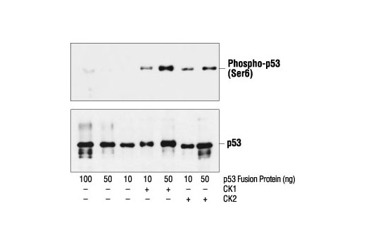
| Phospho-p53 (Ser6) Antibody | 9285 | 20 µl | 53 kDa | Rabbit |
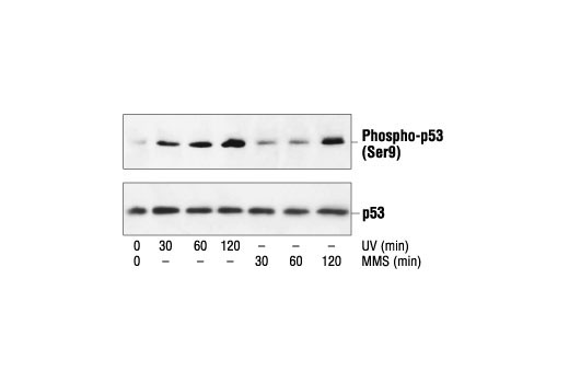
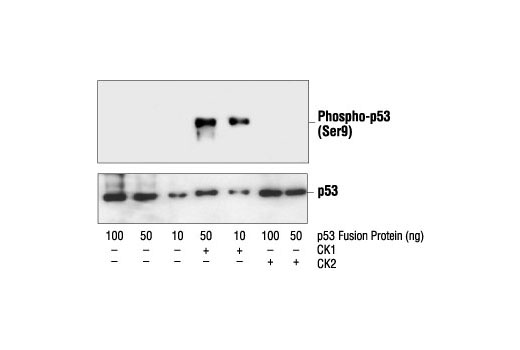
| Phospho-p53 (Ser9) Antibody | 9288 | 20 µl | 53 kDa | Rabbit |
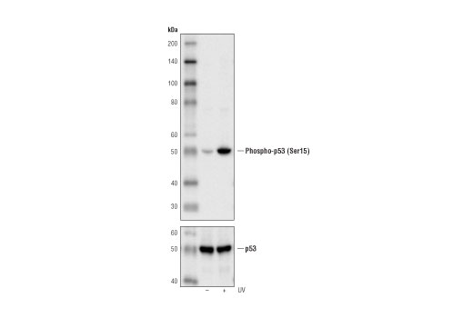
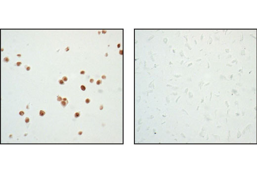
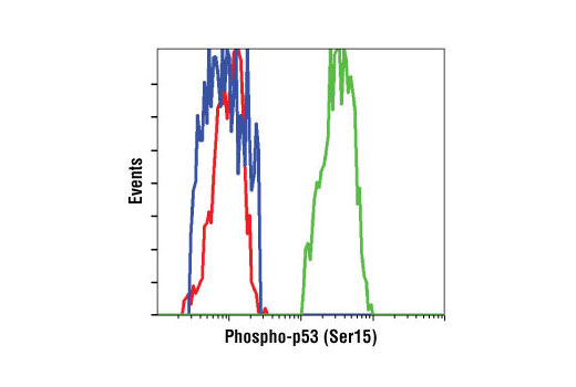
| Phospho-p53 (Ser15) (16G8) Mouse mAb | 9286 | 20 µl | 53 kDa | Mouse IgG1 |
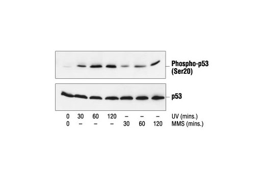
| Phospho-p53 (Ser20) Antibody | 9287 | 20 µl | 53 kDa | Rabbit |
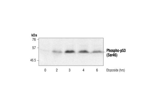
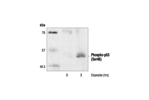
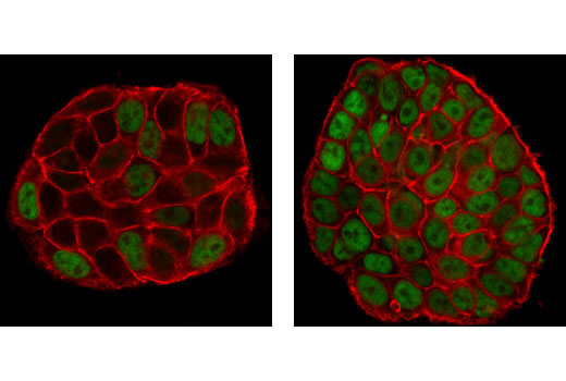
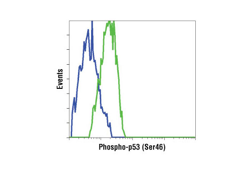
| Phospho-p53 (Ser46) Antibody | 2521 | 20 µl | 53 kDa | Rabbit |
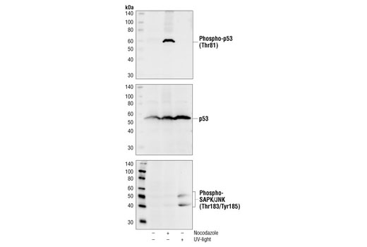
| Phospho-p53 (Thr81) Antibody | 2676 | 20 µl | 53 kDa | Rabbit |
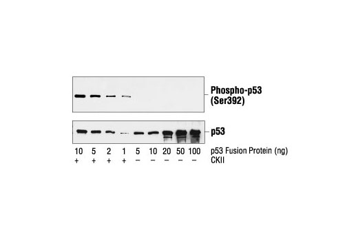
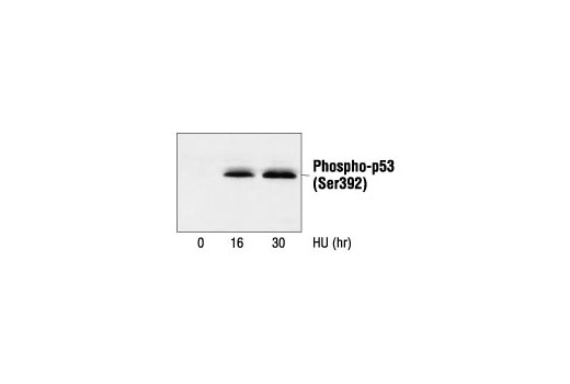
| Phospho-p53 (Ser392) Antibody | 9281 | 20 µl | 53 kDa | Rabbit |
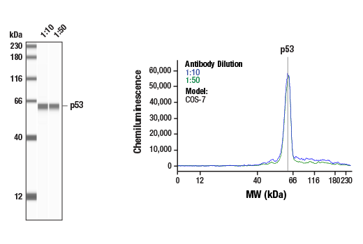
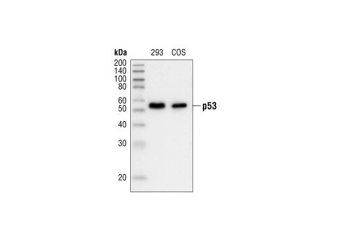
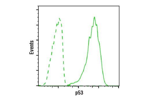
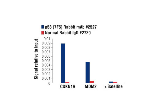
| p53 (7F5) Rabbit mAb | 2527 | 20 µl | 53 kDa | Rabbit IgG |
| Anti-rabbit IgG, HRP-linked Antibody | 7074 | 100 µl | Goat |
Please visit cellsignal.com for individual component applications, species cross-reactivity, dilutions, protocols, and additional product information.
Description
The Phospho-p53 Antibody Sampler Kit provides a fast and economical means of evaluating multiple phosphorylation sites of p53 protein. The kit includes enough antibodies to perform two western blot experiments with each primary antibody.
Storage
Background
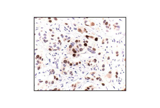
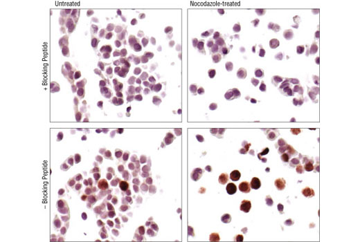
The p53 tumor suppressor protein plays a major role in cellular response to DNA damage and other genomic aberrations. Activation of p53 can lead to either cell cycle arrest and DNA repair or apoptosis (1). p53 is phosphorylated at multiple sites in vivo and by several different protein kinases in vitro (2,3). DNA damage induces phosphorylation of p53 at Ser15 and Ser20 and leads to a reduced interaction between p53 and its negative regulator, the oncoprotein MDM2 (4). MDM2 inhibits p53 accumulation by targeting it for ubiquitination and proteasomal degradation (5,6). p53 can be phosphorylated by ATM, ATR, and DNA-PK at Ser15 and Ser37. Phosphorylation impairs the ability of MDM2 to bind p53, promoting both the accumulation and activation of p53 in response to DNA damage (4,7). Chk2 and Chk1 can phosphorylate p53 at Ser20, enhancing its tetramerization, stability, and activity (8,9). p53 is phosphorylated at Ser392 in vivo (10,11) and by CAK in vitro (11). Phosphorylation of p53 at Ser392 is increased in human tumors (12) and has been reported to influence the growth suppressor function, DNA binding, and transcriptional activation of p53 (10,13,14). p53 is phosphorylated at Ser6 and Ser9 by CK1δ and CK1ε both in vitro and in vivo (13,15). Phosphorylation of p53 at Ser46 regulates the ability of p53 to induce apoptosis (16). Acetylation of p53 is mediated by p300 and CBP acetyltransferases. Inhibition of deacetylation suppressing MDM2 from recruiting HDAC1 complex by p19 (ARF) stabilizes p53. Acetylation appears to play a positive role in the accumulation of p53 protein in stress response (17). Following DNA damage, human p53 becomes acetylated at Lys382 (Lys379 in mouse) in vivo to enhance p53-DNA binding (18). Deacetylation of p53 occurs through interaction with the SIRT1 protein, a deacetylase that may be involved in cellular aging and the DNA damage response (19).
- Levine, A.J. (1997) Cell 88, 323-31.
- Meek, D.W. (1994) Semin Cancer Biol 5, 203-10.
- Milczarek, G.J. et al. (1997) Life Sci 60, 1-11.
- Shieh, S.Y. et al. (1997) Cell 91, 325-34.
- Chehab, N.H. et al. (1999) Proc Natl Acad Sci U S A 96, 13777-82.
- Honda, R. et al. (1997) FEBS Lett 420, 25-7.
- Tibbetts, R.S. et al. (1999) Genes Dev 13, 152-7.
- Shieh, S.Y. et al. (1999) EMBO J 18, 1815-23.
- Hirao, A. et al. (2000) Science 287, 1824-7.
- Hao, M. et al. (1996) J Biol Chem 271, 29380-5.
- Lu, H. et al. (1997) Mol Cell Biol 17, 5923-34.
- Ullrich, S.J. et al. (1993) Proc Natl Acad Sci U S A 90, 5954-8.
- Kohn, K.W. (1999) Mol Biol Cell 10, 2703-34.
- Lohrum, M. and Scheidtmann, K.H. (1996) Oncogene 13, 2527-39.
- Knippschild, U. et al. (1997) Oncogene 15, 1727-36.
- Oda, K. et al. (2000) Cell 102, 849-62.
- Ito, A. et al. (2001) EMBO J 20, 1331-40.
- Sakaguchi, K. et al. (1998) Genes Dev 12, 2831-41.
- Solomon, J.M. et al. (2006) Mol Cell Biol 26, 28-38.
Background References
Trademarks and Patents
使用に関する制限
法的な権限を与えられたCSTの担当者が署名した書面によって別途明示的に合意された場合を除き、 CST、その関連会社または代理店が提供する製品には以下の条件が適用されます。お客様が定める条件でここに定められた条件に含まれるものを超えるもの、 または、ここに定められた条件と異なるものは、法的な権限を与えられたCSTの担当者が別途書面にて受諾した場合を除き、拒絶され、 いかなる効力も効果も有しません。
研究専用 (For Research Use Only) またはこれに類似する表示がされた製品は、 いかなる目的についても FDA または外国もしくは国内のその他の規制機関により承認、認可または許可を受けていません。 お客様は製品を診断もしくは治療目的で使用してはならず、また、製品に表示された内容に違反する方法で使用してはなりません。 CST が販売または使用許諾する製品は、エンドユーザーであるお客様に対し、使途を研究および開発のみに限定して提供されるものです。 診断、予防もしくは治療目的で製品を使用することまたは製品を再販売 (単独であるか他の製品等の一部であるかを問いません) もしくはその他の商業的利用の目的で購入することについては、CST から別途許諾を得る必要があります。 お客様は以下の事項を遵守しなければなりません。(a) CST の製品 (単独であるか他の資材と一緒であるかを問いません) を販売、使用許諾、貸与、寄付もしくはその他の態様で第三者に譲渡したり使用させたりしてはなりません。また、商用の製品を製造するために CST の製品を使用してはなりません。(b) 複製、改変、リバースエンジニアリング、逆コンパイル、 分解または他の方法により製品の構造または技術を解明しようとしてはなりません。また、 CST の製品またはサービスと競合する製品またはサービスを開発する目的で CST の製品を使用してはなりません。(c) CST の製品の商標、商号、ロゴ、特許または著作権に関する通知または表示を除去したり改変したりしてはなりません。(d) CST の製品をCST 製品販売条件(CST’s Product Terms of Sale) および該当する書面のみに従って使用しなければなりません。(e) CST の製品に関連してお客様が使用する第三者の製品またはサービスに関する使用許諾条件、 サービス提供条件またはこれに類する合意事項を遵守しなければなりません。