Revision 1

#9920Store at -20C

1 Kit

(6 x 20 microliters)

Cell Signaling Technology

Orders: 877-616-CELL (2355) [email protected]

Support: 877-678-TECH (8324)

Web: [email protected] cellsignal.com

3 Trask LaneDanversMassachusetts01923USA
For Research Use Only. Not for Use in Diagnostic Procedures.
Product Includes Product # Quantity Mol. Wt Isotype/Source
Phospho-AMPK Substrate Motif [LXRXX(pS/pT) MultiMab® Rabbit mAb mix 5759 20 µl Rabbit 
Phospho-Akt Substrate (RXXS*/T*) (110B7E) Rabbit mAb 9614 20 µl Rabbit IgG
Phospho-PKA Substrate (RRXS*/T*) (100G7E) Rabbit mAb 9624 20 µl Rabbit IgG
Phospho-ATM/ATR Substrate Motif [(pS/pT) QG] MultiMab® Rabbit mAb mix 6966 20 µl Rabbit IgG
Phospho-PKC Substrate Motif [(R/K)XpSX(R/K)] MultiMab® Rabbit mAb mix 6967 20 µl Rabbit IgG
Phospho-CDK Substrate Motif [(K/H)pSP] MultiMab® Rabbit mAb mix 9477 20 µl Rabbit IgG
Anti-rabbit IgG, HRP-linked Antibody 7074 100 µl Goat 

Please visit cellsignal.com for individual component applications, species cross-reactivity, dilutions, protocols, and additional product information.

Description

The Phospho-(Ser/Thr) Kinase Substrate Antibody Sampler Kit provides an economical means to investigate the downstream activity of select serine/threonine kinases. The kit contains enough primary antibody to perform two western blot experiments with each antibody.

Storage

Supplied in 10 mM sodium HEPES (pH 7.5), 150 mM NaCl, 100 µg/ml BSA, 50% glycerol and less than 0.02% sodium azide. Store at –20°C. Do not aliquot the antibody.

Background

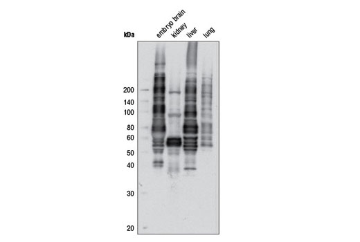
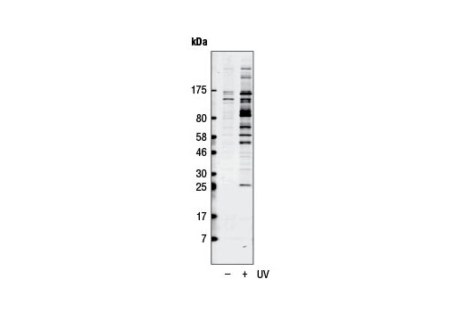
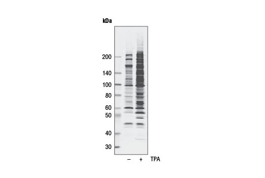
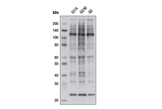
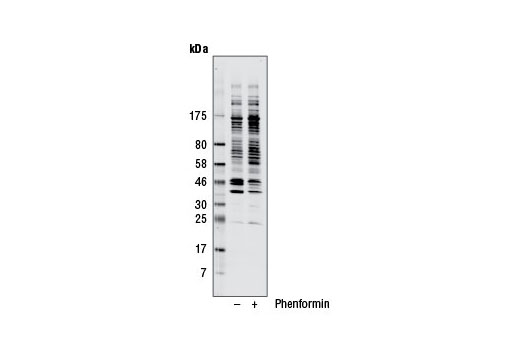
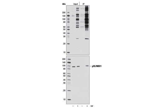
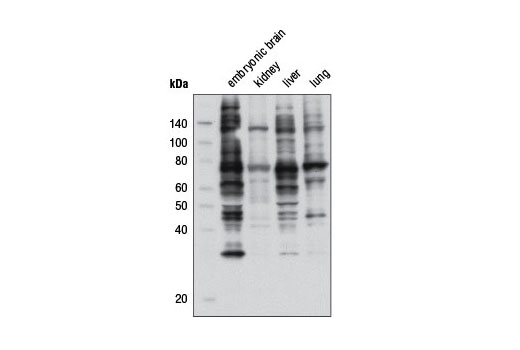
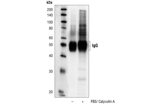
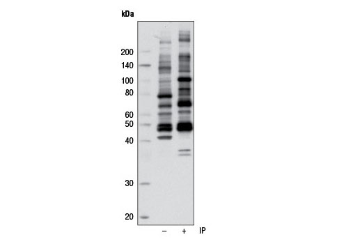
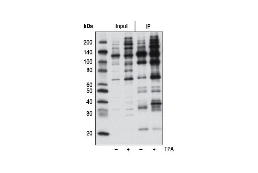
Phospho-(Ser/Thr) kinases and phosphatases play critical roles in a wide range of biological processes. Each phospho-(Ser/Thr) kinase phosphorylates serine or threonine within a specific motif. Akt phosphorylates substrates at a serine or threonine only in a conserved motif characterized by arginine at positions -5 and -3 (1). Conventional PKC isozymes phosphorylate substrates containing serine or threonine, with arginine or lysine at the -3, -2 and +2 positions, and a hydrophobic amino acid at position +1 (2,3). A consensus phosphorylation site of PKA is serine or threonine with arginine at the -2 and -3 positions (3). AMPK phosphorylates consensus motif (L/M)XRXX(S/T)XXXL (6). Antibodies recognizing the LXRXX(S/T) motif are very useful in the identification of AMPK substrates. The consensus amino acid sequence for CDK substrate is (K/R)(S*)PX(K/R), where denotes any one of the 20 amino acids and S* is the phosphorylation site (4-6). ATM and the related kinase ATR phosphorylate serine or threonine in an S*/T*Q motif (7,8).
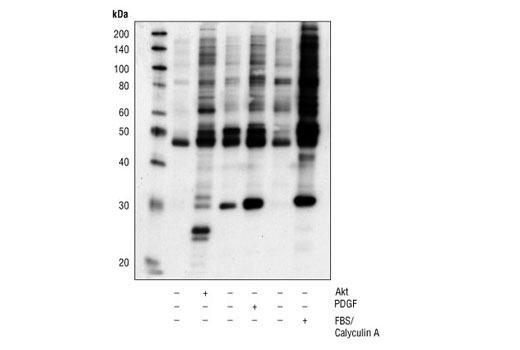
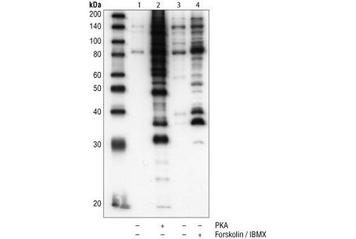
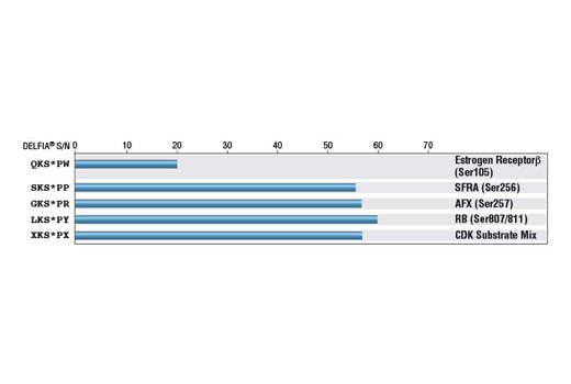
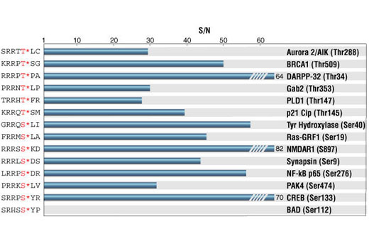
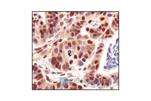
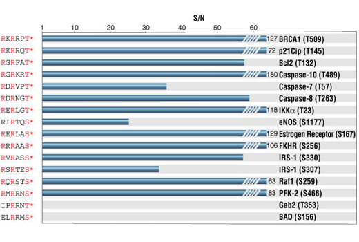
Antibodies specific to particular kinase substrates are invaluable reagents in determining kinase activity and identifying potential new kinase substrates. CST has developed antibodies that recognize phosphorylated serine or threonine within the context of a protein motif that is phosphorylated by Akt, PKC, PKA, MAPK/CDK, CDKs or ATM/ATR. As shown by peptide pairing ELISA, each phospho-(Ser/Thr) kinase substrate antibody in this sampler kit is specific to its kinase substrate motif.

  1. Alessi, D.R. et al. (1996) FEBS Lett 399, 333-8.
  2. Nishikawa, K. et al. (1997) J Biol Chem 272, 952-60.
  3. Pearson, R.B. and Kemp, B.E. (1991) Methods Enzymol 200, 62-81.
  4. Songyang, Z. et al. (1996) Mol Cell Biol 16, 6486-93.
  5. Songyang, Z. (1999) Prog Biophys Mol Biol 71, 359-72.
  6. Holmes, J.K. and Solomon, M.J. (1996) J Biol Chem 271, 25240-6.
  7. Kastan, M.B. and Lim, D.S. (2000) Nat Rev Mol Cell Biol 1, 179-86.
  8. Zhao, H. and Piwnica-Worms, H. (2001) Mol Cell Biol 21, 4129-39.

Background References

    Trademarks and Patents

    Cell Signaling Technology is a trademark of Cell Signaling Technology, Inc.
    All other trademarks are the property of their respective owners. Visit cellsignal.com/trademarks for more information.

    使用に関する制限

    法的な権限を与えられたCSTの担当者が署名した書面によって別途明示的に合意された場合を除き、 CST、その関連会社または代理店が提供する製品には以下の条件が適用されます。お客様が定める条件でここに定められた条件に含まれるものを超えるもの、 または、ここに定められた条件と異なるものは、法的な権限を与えられたCSTの担当者が別途書面にて受諾した場合を除き、拒絶され、 いかなる効力も効果も有しません。

    研究専用 (For Research Use Only) またはこれに類似する表示がされた製品は、 いかなる目的についても FDA または外国もしくは国内のその他の規制機関により承認、認可または許可を受けていません。 お客様は製品を診断もしくは治療目的で使用してはならず、また、製品に表示された内容に違反する方法で使用してはなりません。 CST が販売または使用許諾する製品は、エンドユーザーであるお客様に対し、使途を研究および開発のみに限定して提供されるものです。 診断、予防もしくは治療目的で製品を使用することまたは製品を再販売 (単独であるか他の製品等の一部であるかを問いません) もしくはその他の商業的利用の目的で購入することについては、CST から別途許諾を得る必要があります。 お客様は以下の事項を遵守しなければなりません。(a) CST の製品 (単独であるか他の資材と一緒であるかを問いません) を販売、使用許諾、貸与、寄付もしくはその他の態様で第三者に譲渡したり使用させたりしてはなりません。また、商用の製品を製造するために CST の製品を使用してはなりません。(b) 複製、改変、リバースエンジニアリング、逆コンパイル、 分解または他の方法により製品の構造または技術を解明しようとしてはなりません。また、 CST の製品またはサービスと競合する製品またはサービスを開発する目的で CST の製品を使用してはなりません。(c) CST の製品の商標、商号、ロゴ、特許または著作権に関する通知または表示を除去したり改変したりしてはなりません。(d) CST の製品をCST 製品販売条件(CST’s Product Terms of Sale) および該当する書面のみに従って使用しなければなりません。(e) CST の製品に関連してお客様が使用する第三者の製品またはサービスに関する使用許諾条件、 サービス提供条件またはこれに類する合意事項を遵守しなければなりません。

    Revision 1
    #9920

    Phospho-(Ser/Thr) Kinase Substrate Antibody Sampler Kit

    Phospho-(Ser/Thr) Kinase Substrate Antibody Sampler Kit: Image 1 Expand Image
    Western blot analysis of extracts from various mouse tissues using Phospho-AMPK Substrate Motif [LXRXX(pS/pT) MultiMab® Rabbit mAb mix
    Phospho-(Ser/Thr) Kinase Substrate Antibody Sampler Kit: Image 2 Expand Image
    Western blot analysis of extracts from HeLa cells, untreated (-) or UV-treated (+, 50 mJ/cm2, 2 hr), usingPhospho-ATM/ATR Substrate Motif [(pS/pT) QG] MultiMab® Rabbit mAb mix. Western blot was imaged using Odyssey® Infrared Imaging System (LI-COR® Biotechnologies).
    Phospho-(Ser/Thr) Kinase Substrate Antibody Sampler Kit: Image 3 Expand Image
    Western blot analysis of extracts from Jurkat cells, untreated (-) or TPA-treated (200 nM, 30 min; +), using Phospho-PKC Substrate Motif [(R/K)XpSX(R/K)] MultiMab® Rabbit mAb mix. Western blot was imaged using Odyssey® Infrared Imaging System (LI-COR® Biotechnologies).
    Phospho-(Ser/Thr) Kinase Substrate Antibody Sampler Kit: Image 4 Expand Image
    After the primary antibody is bound to the target protein, a complex with HRP-linked secondary antibody is formed. The LumiGLO® is added and emits light during enzyme catalyzed decomposition.
    Phospho-(Ser/Thr) Kinase Substrate Antibody Sampler Kit: Image 5 Expand Image
    Western blot analysis of extracts from COS-7 cells, synchronized in G1/S, G2/M, and G0 phase of the cell cycle, usingPhospho-CDK Substrate Motif [(K/H)pSP] MultiMab® Rabbit mAb mix.
    Phospho-(Ser/Thr) Kinase Substrate Antibody Sampler Kit: Image 6 Expand Image
    Western blot analysis of extracts from serum-starved NIH/3T3 cells, phosphorylated in vitro with Akt kinase, or treated in culture with PDGF or FBS/Calyculin A, using Phospho-Akt Substrate (RXXS*/T*) (110B7E) Rabbit mAb.
    Phospho-(Ser/Thr) Kinase Substrate Antibody Sampler Kit: Image 7 Expand Image
    Western blot analysis of extracts from serum-starved A431 cells, phosphorylated in vitro with PKA kinase or treated in culture with forskolin/IBMX, using Phospho-PKA Substrate (RRXS*/T*) (100G7E) Rabbit mAb. Lysis buffer: 1.0% Triton X-100 (lanes 1 and 2), 2.0% SDS (lanes 3 and 4).
    Phospho-(Ser/Thr) Kinase Substrate Antibody Sampler Kit: Image 8 Expand Image
    Western blot analysis of extracts from H1650 cells, untreated or treated with phenformin (5 mM, 1 hr), usingPhospho-AMPK Substrate Motif [LXRXX(pS/pT) MultiMab® Rabbit mAb mix. Western blot was imaged using Odyssey® Infrared Imaging System (LI-COR® Biosciences).
    Phospho-(Ser/Thr) Kinase Substrate Antibody Sampler Kit: Image 9 Expand Image
    Immunoprecipitation of Hela cells, untreated (-) or UV-treated (+, 50 mJ/cm2, 2 hr), usingPhospho-ATM/ATR Substrate Motif [(pS/pT) QG] MultiMab® Rabbit mAb mix (lanes 3 and 4). Lanes 1 and 2 show 10% input. Western blot detection was performed using the same antibody (upper) and p95/NBS1 Antibody #3002 (lower).
    Phospho-(Ser/Thr) Kinase Substrate Antibody Sampler Kit: Image 10 Expand Image
    Western blot analysis of extracts from various mouse tissues using Phospho-PKC Substrate Motif [(R/K)XpSX(R/K)] MultiMab® Rabbit mAb mix.
    Phospho-(Ser/Thr) Kinase Substrate Antibody Sampler Kit: Image 11 Expand Image
    Immunoprecipitation of extracts from serum-starved NIH/3T3 cells, untreated or treated with FBS/Calyculin A, using Phospho-Akt Substrate (RXXS*/T*) (110B7E) Rabbit mAb, followed by Western blot analysis using the same antibody.
    Phospho-(Ser/Thr) Kinase Substrate Antibody Sampler Kit: Image 12 Expand Image
    Immunoprecipitation of extracts from H1650 cells using Phospho-AMPK Substrate Motif [LXRXX(pS/pT) MultiMab® Rabbit mAb mix. Lane 1 shows 10% input. Western blot analysis was performed using the same antibody.
    Phospho-(Ser/Thr) Kinase Substrate Antibody Sampler Kit: Image 13 Expand Image
    Immunoprecipitation (IP) of extracts from Jurkat cells, untreated (-) or TPA-treated (200 nM, 30 min; +) (lanes 3 and 4), using Phospho-PKC Substrate Motif [(R/K)XpSX(R/K)] MultiMab® Rabbit mAb mix. 10% input is shown in lanes 1 and 2. Western blot analysis was performed using the same antibody.
    Phospho-(Ser/Thr) Kinase Substrate Antibody Sampler Kit: Image 14 Expand Image
    Phospho-CDK Substrate Motif [(K/H)pSP] MultiMab® Rabbit mAb mix ELISA assay: Signal-to-noise ratio of phospho- versus nonphospho-peptides. (S* denotes phosphorylated serine.)
    Phospho-(Ser/Thr) Kinase Substrate Antibody Sampler Kit: Image 15 Expand Image
    Immunohistochemical analysis of paraffin-embedded human breast carcinoma control (left) or lambda phosphatase-treated (right), using Phospho-Akt Substrate (RXXS*/T*) (110B7E) Rabbit mAb.
    Phospho-(Ser/Thr) Kinase Substrate Antibody Sampler Kit: Image 16 Expand Image
    Phospho-PKA Substrate (RRXS*/T*) (100G7E) Rabbit mAb ELISA Assay: Signal-to-noise ratio of phospho- versus nonphospho-peptides. (T* and S* denote phosphorylated threonine and serine.)
    Phospho-(Ser/Thr) Kinase Substrate Antibody Sampler Kit: Image 17 Expand Image
    Immunohistochemical analysis of paraffin-embedded lung carcinoma, using Phospho-Akt Substrate (RXXS*/T*) (110B7E) Rabbit mAb.
    Phospho-(Ser/Thr) Kinase Substrate Antibody Sampler Kit: Image 18 Expand Image
    Immunohistochemical analysis of paraffin-embedded renal cell carcinoma, using Phospho-Akt Substrate (RXXS*/T*) (110B7E) Rabbit mAb.
    Phospho-(Ser/Thr) Kinase Substrate Antibody Sampler Kit: Image 19 Expand Image
    Phospho-Akt Substrate (RXXS*/T*) (110B7E) Rabbit mAb ELISA Assay: Signal-to-noise ratio of phospho- versus nonphospho-peptides. (T* and S* denote phosphorylated threonine and serine.)