Revision 1
Cell Signaling Technology

Orders: 877-616-CELL (2355) [email protected]

Support: 877-678-TECH (8324)

Web: [email protected] cellsignal.com

3 Trask LaneDanversMassachusetts01923USA
For Research Use Only. Not for Use in Diagnostic Procedures.
:

:

:

:

UniProt ID:

#P25963

Entrez-Gene Id:

4792

Product Information

Storage

Supplied in 10 mM sodium HEPES (pH 7.5), 150 mM NaCl, 100 µg/ml BSA, 50% glycerol and less than 0.02% sodium azide. Store at –20°C. Do not aliquot the antibody.

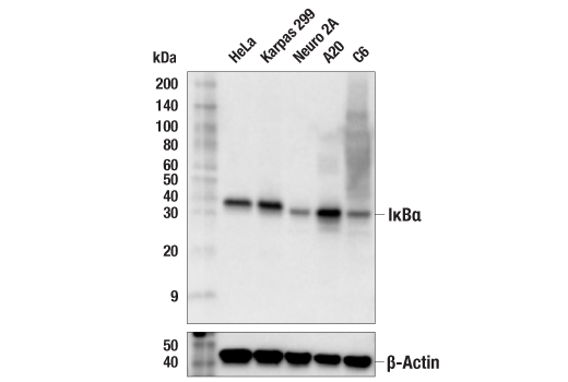
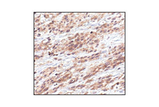
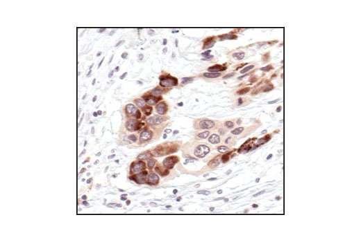
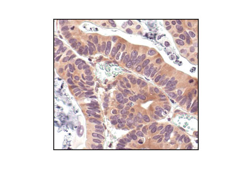
Specificity / Sensitivity

IκBα (L35A5) Mouse mAb (Amino-terminal Antigen) #4814 detects endogenous levels of total IκBα protein. Phospho-IκBα (Ser32/36) (5A5) Mouse mAb #9246 detects endogenous levels of IκBα only when phosphorylated at Ser32/36. Neither antibody cross-reacts with other IκB family members at physiological levels.

Source / Purification

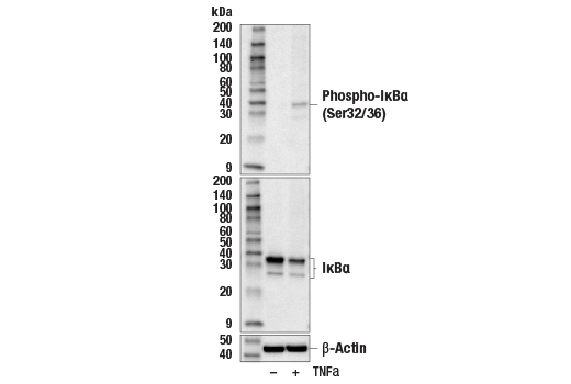
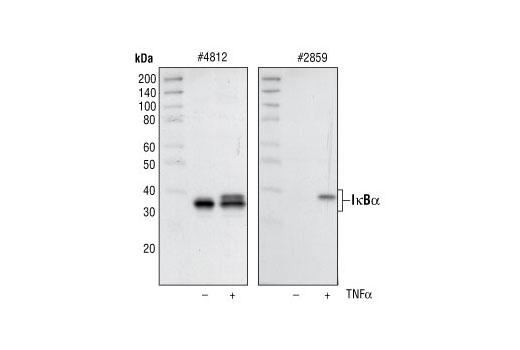
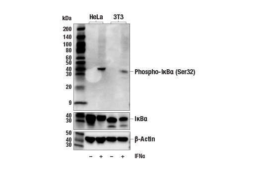
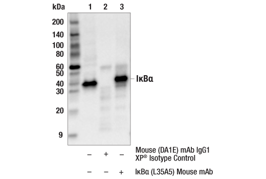
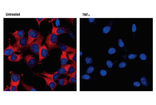
IκBα (L35A5) Mouse mAb (Amino-terminal Antigen) #4814 is produced by immunizing mice with a GST-IκBα fusion protein corresponding the amino-terminus of human IκBα. Phospho-IκBα (Ser32/36) (5A5) Mouse mAb #9246 is produced by immunizing mice with a synthetic phosphopeptide corresponding to residues surrounding Ser32/36 of human IκBα. Total cell extracts from HeLa cells prepared without treatment serve as a negative control. Supplied in SDS Sample Buffer. Total cell extracts from HeLa cells prepared with TNF-α treatment (#2169, 20 ng/ml for 5 minutes) serve as a positive control. Supplied in SDS Sample Buffer.
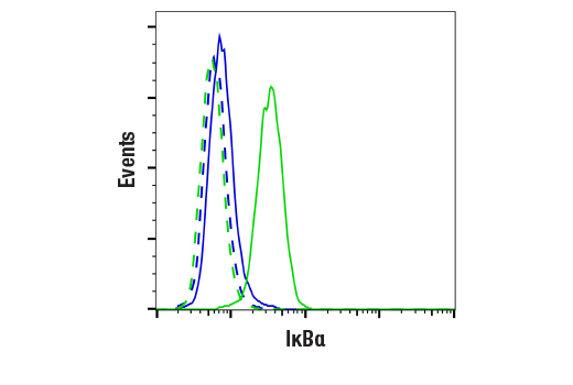
Product Description

The PhosphoPlus® IκBα (Ser32/36) Antibody Kit provides reagents and protocols for the rapid analysis of IκBα phosphorylation at Ser32/36. The kit contains a total IκBα antibody, a phospho-specific antibody, and positive/negative control whole cell lysates, along with secondary antibodies and reagents for Western blotting.

Background

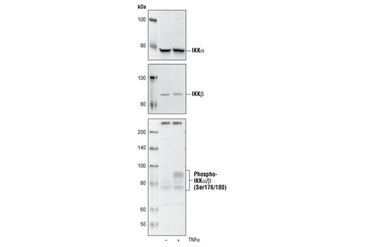
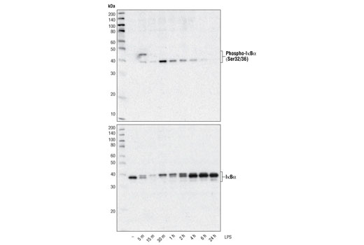
The NF-κB/Rel transcription factors are present in the cytosol in an inactive state complexed with the inhibitory IκB proteins (1-3). Activation occurs via phosphorylation of IκBα at Ser32 and Ser36 followed by proteasome-mediated degradation that results in the release and nuclear translocation of active NF-κB (3-7). IκBα phosphorylation and resulting Rel-dependent transcription are activated by a highly diverse group of extracellular signals including inflammatory cytokines, growth factors, and chemokines. Kinases that phosphorylate IκB at these activating sites have been identified (8).

  1. Baeuerle, P.A. and Baltimore, D. (1988) Science 242, 540-6.
  2. Beg, A.A. and Baldwin, A.S. (1993) Genes Dev 7, 2064-70.
  3. Finco, T.S. et al. (1994) Proc Natl Acad Sci USA 91, 11884-8.
  4. Brown, K. et al. (1995) Science 267, 1485-8.
  5. Brockman, J.A. et al. (1995) Mol Cell Biol 15, 2809-18.
  6. Traenckner, E.B. et al. (1995) EMBO J 14, 2876-83.
  7. Chen, Z.J. et al. (1996) Cell 84, 853-62.
  8. Karin, M. and Ben-Neriah, Y. (2000) Annu Rev Immunol 18, 621-63.

Species Reactivity

Species reactivity is determined by testing in at least one approved application (e.g., western blot).

Cross-Reactivity Key

H: human M: mouse R: rat Hm: hamster Mk: monkey Vir: virus Mi: mink C: chicken Dm: D. melanogaster X: Xenopus Z: zebrafish B: bovine Dg: dog Pg: pig Sc: S. cerevisiae Ce: C. elegans Hr: horse GP: Guinea Pig Rab: rabbit All: all species expected

Trademarks and Patents

Cell Signaling Technology is a trademark of Cell Signaling Technology, Inc.
PhosphoPlus is a registered trademark of Cell Signaling Technology, Inc.
All other trademarks are the property of their respective owners. Visit cellsignal.com/trademarks for more information.

使用に関する制限

法的な権限を与えられたCSTの担当者が署名した書面によって別途明示的に合意された場合を除き、 CST、その関連会社または代理店が提供する製品には以下の条件が適用されます。お客様が定める条件でここに定められた条件に含まれるものを超えるもの、 または、ここに定められた条件と異なるものは、法的な権限を与えられたCSTの担当者が別途書面にて受諾した場合を除き、拒絶され、 いかなる効力も効果も有しません。

研究専用 (For Research Use Only) またはこれに類似する表示がされた製品は、 いかなる目的についても FDA または外国もしくは国内のその他の規制機関により承認、認可または許可を受けていません。 お客様は製品を診断もしくは治療目的で使用してはならず、また、製品に表示された内容に違反する方法で使用してはなりません。 CST が販売または使用許諾する製品は、エンドユーザーであるお客様に対し、使途を研究および開発のみに限定して提供されるものです。 診断、予防もしくは治療目的で製品を使用することまたは製品を再販売 (単独であるか他の製品等の一部であるかを問いません) もしくはその他の商業的利用の目的で購入することについては、CST から別途許諾を得る必要があります。 お客様は以下の事項を遵守しなければなりません。(a) CST の製品 (単独であるか他の資材と一緒であるかを問いません) を販売、使用許諾、貸与、寄付もしくはその他の態様で第三者に譲渡したり使用させたりしてはなりません。また、商用の製品を製造するために CST の製品を使用してはなりません。(b) 複製、改変、リバースエンジニアリング、逆コンパイル、 分解または他の方法により製品の構造または技術を解明しようとしてはなりません。また、 CST の製品またはサービスと競合する製品またはサービスを開発する目的で CST の製品を使用してはなりません。(c) CST の製品の商標、商号、ロゴ、特許または著作権に関する通知または表示を除去したり改変したりしてはなりません。(d) CST の製品をCST 製品販売条件(CST’s Product Terms of Sale) および該当する書面のみに従って使用しなければなりません。(e) CST の製品に関連してお客様が使用する第三者の製品またはサービスに関する使用許諾条件、 サービス提供条件またはこれに類する合意事項を遵守しなければなりません。

Revision 1
#9240

PhosphoPlus® IκBα (Ser32/36) Antibody Kit

PhosphoPlus® IκBα (Ser32/36) Antibody Kit: Image 1 Expand Image
Western blot analysis of extracts from various cell lines using IκBα (L35A5) Mouse mAb (Amino-terminal Antigen) #4814 (upper) or β-Actin (D6A8) Rabbit mAb #8457 (lower).
PhosphoPlus® IκBα (Ser32/36) Antibody Kit: Image 2 Expand Image
After the primary antibody is bound to the target protein, a complex with HRP-linked secondary antibody is formed. The LumiGLO® is added and emits light during enzyme catalyzed decomposition.
PhosphoPlus® IκBα (Ser32/36) Antibody Kit: Image 3 Expand Image
After the primary antibody is bound to the target protein, a complex with HRP-linked secondary antibody is formed. The LumiGLO* is added and emits light during enzyme catalyzed decomposition.
PhosphoPlus® IκBα (Ser32/36) Antibody Kit: Image 4 Expand Image
Western blot analysis of NF-κB Control Cell Extracts #9243, using IKKα Antibody #2682 (upper), IKKβ (2C8) Rabbit mAb #2370 (middle) and Phospho-IKKα/β (Ser176/180) (16A6) Rabbit mAb #2697 (lower).
PhosphoPlus® IκBα (Ser32/36) Antibody Kit: Image 5 Expand Image
Western blot analysis of extracts from NIH/3T3 cells, untreated or TNF-α-treated (#8902, 20 ng/ml) for 5 minutes, using Phospho-IκBα (Ser32/36) (5A5) Mouse mAb #9246 (upper) or IκBα (L35A5) Mouse mAb (Amino-terminal Antigen) #4814, and β-Actin (D6A8) Rabbit mAb #8457 (lower).
PhosphoPlus® IκBα (Ser32/36) Antibody Kit: Image 6 Expand Image
Western blot analysis of extracts from THP-1 cells, differentiated with TPA (#9905, 80 nM for 24h) and treated with 1 μg/ml LPS for the indicated times, using Phospho-IκBα (Ser32/36) (5A5) Mouse mAb #9246 (upper) and IκBα (L35A5) Mouse mAb (Amino-terminal Antigen) (lower).
PhosphoPlus® IκBα (Ser32/36) Antibody Kit: Image 7 Expand Image
Western blot analysis of NF-κB Control Cell Extracts #9243, using IκBα (44D4) Rabbit mAb #4812 (left) and Phospho-IκBα (Ser32) (14D4) Rabbit mAb #2859 (right).
PhosphoPlus® IκBα (Ser32/36) Antibody Kit: Image 8 Expand Image
Western blot analysis of extracts from various cell lines, untreated or treated with IFNa (#36000, 20 ng/mL, 5 min); using Phospho-IκBα (Ser32/36) (5A5) Mouse mAb #9246 (upper), IκBα (L35A5) Mouse mAb (Amino-terminal Antigen) #4814 (middle), and β-Actin (D6A8) Rabbit mAb #8457 (lower).
PhosphoPlus® IκBα (Ser32/36) Antibody Kit: Image 9 Expand Image
Immunoprecipitation of IkBa from HeLa cell extracts. Lane 1 is 10% input, lane 2 is precipitated with Mouse (G3A1) mAb IgG1 Isotype Control #5415, and lane 3 is IκBα (L35A5) Mouse mAb (Amino-terminal Antigen), #4814. Western blot was performed using IκBα Antibody #9242.
PhosphoPlus® IκBα (Ser32/36) Antibody Kit: Image 10 Expand Image
Western blot analysis of extracts from THP-1 cells, differentiated with TPA (#9905, 80 nM for 24 h) and treated with 1 μg/ml LPS for the indicated times, using Phospho-IκBα (Ser32/36) (5A5) Mouse mAb (upper) and IκBα (L35A5) Mouse mAb (Amino-terminal Antigen) #4814 (lower).
PhosphoPlus® IκBα (Ser32/36) Antibody Kit: Image 11 Expand Image
Immunohistochemical analysis of paraffin-embedded human leiomyoma, using IκBα (L35A5) Mouse mAb (Amino-terminal Antigen).
PhosphoPlus® IκBα (Ser32/36) Antibody Kit: Image 12 Expand Image
Immunohistochemical analysis of paraffin-embedded human lung carcinoma, using IκBα (L35A5) Mouse mAb (Amino-terminal Antigen).
PhosphoPlus® IκBα (Ser32/36) Antibody Kit: Image 13 Expand Image
Immunohistochemical analysis of paraffin-embedded human renal adenocarcinoma, using IκBα (L35A5) Mouse mAb (Amino-terminal Antigen).
PhosphoPlus® IκBα (Ser32/36) Antibody Kit: Image 14 Expand Image
Confocal immunofluorescent analysis of HeLa cells, untreated (left), or TNF-α-treated (#8902, 10 ng/ml for 15 min, right) using IκBα (L35A5) Mouse mAb (Amino-terminal Antigen) (red). Blue pseudocolor = DRAQ5® #4084 (fluorescent DNA dye).
PhosphoPlus® IκBα (Ser32/36) Antibody Kit: Image 15 Expand Image
Flow cytometric analysis of NIH/3T3 cells, treated with TNF-α (#8902, 10 ng/ml for 5 min; blue, negative) or untreated (green, positive) using IκBα (L35A5) Mouse mAb (Amino-terminal Antigen) (solid lines) or concentration-matched Mouse (G3A1) mAb IgG1 Isotype Control #5415 (dashed lines). Anti-mouse IgG (H+L), F(ab')2 Fragment (Alexa Fluor® 488 Conjugate) #4408 was used as a secondary antibody.