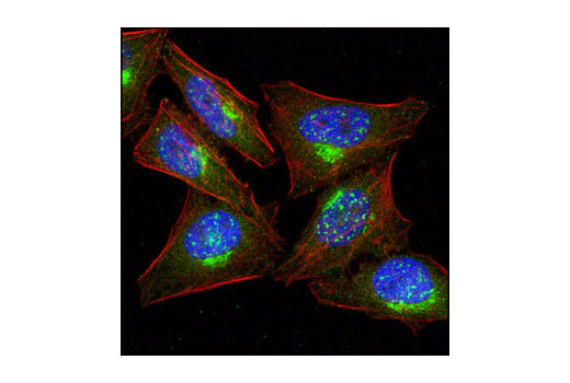
WB, IP, IF-IC, FC-FP
H M R
Endogenous
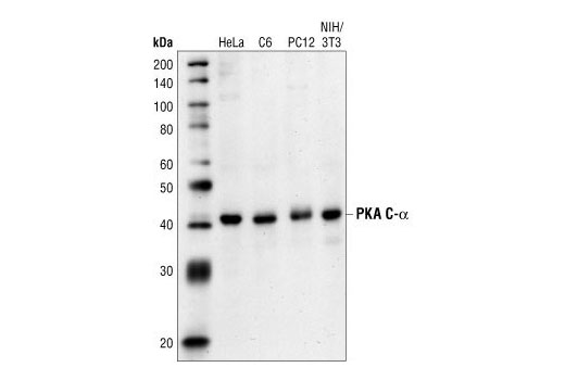
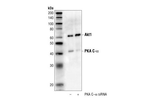
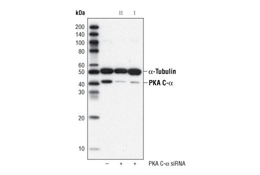
42
Rabbit
#P17612
5566
Product Information
Product Usage Information
| Application | Dilution |
|---|---|
| Western Blotting | 1:1000 |
| Immunoprecipitation | 1:50 |
| Immunofluorescence (Immunocytochemistry) | 1:50 - 1:100 |
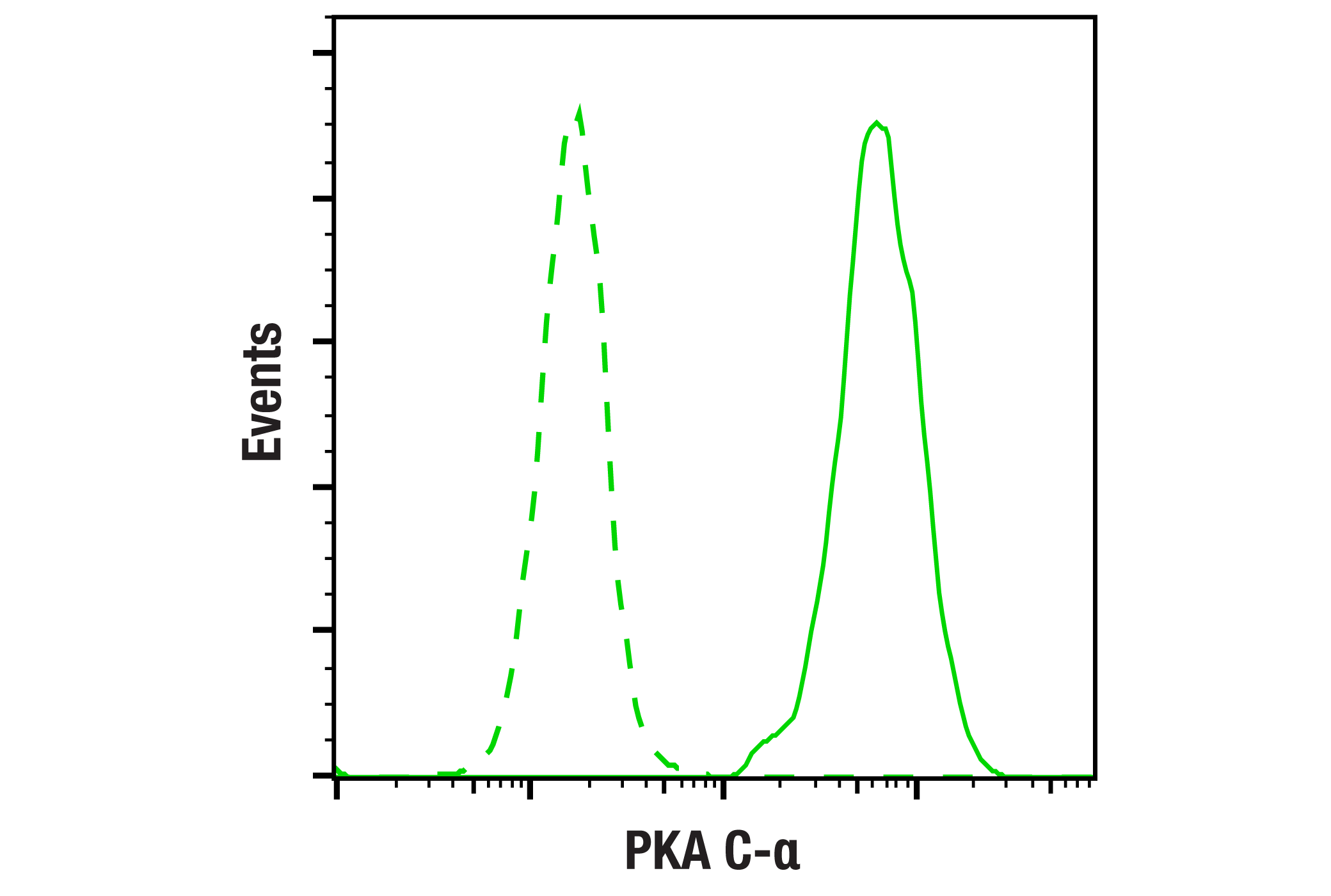
| Flow Cytometry (Fixed/Permeabilized) | 1:50 - 1:200 |
Storage
Specificity / Sensitivity
Species Reactivity:
Human, Mouse, Rat
Source / Purification
Polyclonal antibodies are produced by immunizing animals with a synthetic peptide corresponding to the carboxy terminal sequence of human PKA C-α. Antibodies are purified by protein A and peptide affinity chromatography.
Background
The second messenger cyclic AMP (cAMP) activates cAMP-dependent protein kinase (PKA or cAPK) in mammalian cells and controls many cellular mechanisms such as gene transcription, ion transport, and protein phosphorylation (1). Inactive PKA is a heterotetramer composed of a regulatory subunit (R) dimer and a catalytic subunit (C) dimer. In this inactive state, the pseudosubstrate sequences on the R subunits block the active sites on the C subunits. Three C subunit isoforms (C-α, C-β, and C-γ) and two families of regulatory subunits (RI and RII) with distinct cAMP binding properties have been identified. The two R families exist in two isoforms, α and β (RI-α, RI-β, RII-α, and RII-β). Upon binding of cAMP to the R subunits, the autoinhibitory contact is eased and active monomeric C subunits are released. PKA shares substrate specificity with Akt (PKB) and PKC, which are characterized by an arginine at position -3 relative to the phosphorylated serine or threonine residue (2). Substrates that present this consensus sequence and have been shown to be phosphorylated by PKA are Bad (Ser155), CREB (Ser133), and GSK-3 (GSK-3α Ser21 and GSK-3β Ser9) (3-5). In addition, combined knock-down of PKA C-α and -β blocks cAMP-mediated phosphorylation of Raf (Ser43 and Ser259) (6). Autophosphorylation and phosphorylation by PDK-1 are two known mechanisms responsible for phosphorylation of the C subunit at Thr197 (7).
- Montminy, M. (1997) Annu. Rev. Biochem. 66, 807-822.
- Dell'Acqua, M.L. and Scott, J.D. (1997) J. Biol. Chem. 272, 12881-12884.
- Tan, Y. et al. (2000) J. Biol. Chem. 275, 25865-25869.
- Gonzalez, G.A. and Montminy, M.R. (1989) Cell 59, 675-680.
- Fang, X. et al. (2000) Proc. Natl. Acad. Sci. USA 97, 11960-11965.
- Dumaz, N. and Marais, R. (2003) J. Biol. Chem. 278, 29819 -29823.
- Moore, M.J. et al. (2002) J. Biol. Chem. 277, 47878-47884.
Species Reactivity
Species reactivity is determined by testing in at least one approved application (e.g., western blot).
Western Blot Buffer
IMPORTANT: For western blots, incubate membrane with diluted primary antibody in 5% w/v BSA, 1X TBS, 0.1% Tween® 20 at 4°C with gentle shaking, overnight.
Applications Key
WB: Western Blotting IP: Immunoprecipitation IF-IC: Immunofluorescence (Immunocytochemistry) FC-FP: Flow Cytometry (Fixed/Permeabilized)
Cross-Reactivity Key
H: human M: mouse R: rat Hm: hamster Mk: monkey Vir: virus Mi: mink C: chicken Dm: D. melanogaster X: Xenopus Z: zebrafish B: bovine Dg: dog Pg: pig Sc: S. cerevisiae Ce: C. elegans Hr: horse GP: Guinea Pig Rab: rabbit All: all species expected
Trademarks and Patents
使用に関する制限
法的な権限を与えられたCSTの担当者が署名した書面によって別途明示的に合意された場合を除き、 CST、その関連会社または代理店が提供する製品には以下の条件が適用されます。お客様が定める条件でここに定められた条件に含まれるものを超えるもの、 または、ここに定められた条件と異なるものは、法的な権限を与えられたCSTの担当者が別途書面にて受諾した場合を除き、拒絶され、 いかなる効力も効果も有しません。
研究専用 (For Research Use Only) またはこれに類似する表示がされた製品は、 いかなる目的についても FDA または外国もしくは国内のその他の規制機関により承認、認可または許可を受けていません。 お客様は製品を診断もしくは治療目的で使用してはならず、また、製品に表示された内容に違反する方法で使用してはなりません。 CST が販売または使用許諾する製品は、エンドユーザーであるお客様に対し、使途を研究および開発のみに限定して提供されるものです。 診断、予防もしくは治療目的で製品を使用することまたは製品を再販売 (単独であるか他の製品等の一部であるかを問いません) もしくはその他の商業的利用の目的で購入することについては、CST から別途許諾を得る必要があります。 お客様は以下の事項を遵守しなければなりません。(a) CST の製品 (単独であるか他の資材と一緒であるかを問いません) を販売、使用許諾、貸与、寄付もしくはその他の態様で第三者に譲渡したり使用させたりしてはなりません。また、商用の製品を製造するために CST の製品を使用してはなりません。(b) 複製、改変、リバースエンジニアリング、逆コンパイル、 分解または他の方法により製品の構造または技術を解明しようとしてはなりません。また、 CST の製品またはサービスと競合する製品またはサービスを開発する目的で CST の製品を使用してはなりません。(c) CST の製品の商標、商号、ロゴ、特許または著作権に関する通知または表示を除去したり改変したりしてはなりません。(d) CST の製品をCST 製品販売条件(CST’s Product Terms of Sale) および該当する書面のみに従って使用しなければなりません。(e) CST の製品に関連してお客様が使用する第三者の製品またはサービスに関する使用許諾条件、 サービス提供条件またはこれに類する合意事項を遵守しなければなりません。