WB, IP, IHC-P, IF-IC, FC-FP
H Mk
Endogenous
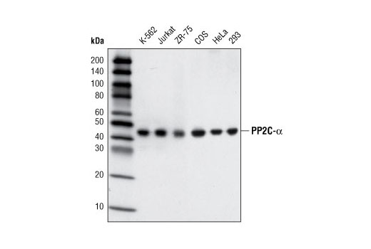
43
Rabbit IgG
#P35813
5494
Product Information
Product Usage Information
| Application | Dilution |
|---|---|
| Western Blotting | 1:1000 |
| Immunoprecipitation | 1:100 |
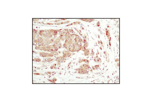
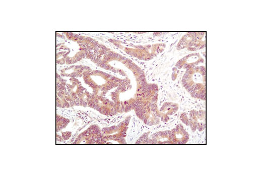
| Immunohistochemistry (Paraffin) | 1:200 |
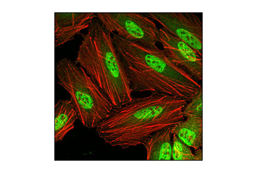
| Immunofluorescence (Immunocytochemistry) | 1:400 |
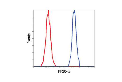
| Flow Cytometry (Fixed/Permeabilized) | 1:100 |
Storage
Specificity / Sensitivity
Species Reactivity:
Human, Monkey
Source / Purification
Monoclonal antibody is produced by immunizing animals with a synthetic peptide corresponding to residues surrounding Ser375 of human PP2C-α.
Background
The α isoform of protein phosphatase 2C (PP2C-α) is the catalytic subunit of a widely expressed serine/threonine phosphatase involved in regulation of the cell stress response (1,2). Also known as magnesium-dependent protein phosphatase (PPM1A), this monomeric phosphatase is a member of a conserved group of proteins that acts on many different substrates in numerous pathways. PP2C-α inhibits p38 MAPK and SAPK/JNK pathways activated in response to cell stress as seen in both in vivo and in vitro studies. Specifically, PP2C-α removes phosphates from MKK3 and MKK7, reducing activity of both proteins and inhibiting activation of the downstream kinases JNK and p38 MAPK, respectively (3). Another PP2C-α substrate is IKKβ, the critical regulator of NF-κB signaling. Dephosphorylation of IKKβ at Ser177/181 by PPM1A and PPM1B results in inactivation of IKKβ and inhibition of NF-κB signaling (4). PP2C-α is one of the phosphatases responsible for removing phosphate residues from cyclin dependent protein kinases. In a study using HeLa cell extracts, PP2C-α dephospohrylates CDK2 and CDK6, with a preference toward interacting with CDK2 phosphorylated at Thr160, a residue found in the activating T-loop of the kinase. Removal of phosphates from this site is thought to inactivate cyclin-associated kinases (5). PP2C-α induces cell cycle arrest and apoptosis, likely through activation of p53 though other pathways may also contribute to PP2C-α mediated cell death (6). Additional PP2C-α substrates include the Wnt signaling pathway protein axin (7) and CFTR, a chloride channel protein implicated in cystic fibrosis (8).
- Marley, A.E. et al. (1998) FEBS Lett 431, 121-4.
- Stern, A. et al. (2007) J Mol Evol 64, 61-70.
- Takekawa, M. et al. (1998) EMBO J 17, 4744-52.
- Sun, W. et al. (2009) Cell Signal 21, 95-102.
- Cheng, A. et al. (2000) J Biol Chem 275, 34744-9.
- Ofek, P. et al. (2003) J Biol Chem 278, 14299-305.
- Strovel, E.T. et al. (2000) J Biol Chem 275, 2399-403.
- Travis, S.M. et al. (1997) Proc Natl Acad Sci USA 94, 11055-60.
Species Reactivity
Species reactivity is determined by testing in at least one approved application (e.g., western blot).
Western Blot Buffer
IMPORTANT: For western blots, incubate membrane with diluted primary antibody in 5% w/v BSA, 1X TBS, 0.1% Tween® 20 at 4°C with gentle shaking, overnight.
Applications Key
WB: Western Blotting IP: Immunoprecipitation IHC-P: Immunohistochemistry (Paraffin) IF-IC: Immunofluorescence (Immunocytochemistry) FC-FP: Flow Cytometry (Fixed/Permeabilized)
Cross-Reactivity Key
H: human M: mouse R: rat Hm: hamster Mk: monkey Vir: virus Mi: mink C: chicken Dm: D. melanogaster X: Xenopus Z: zebrafish B: bovine Dg: dog Pg: pig Sc: S. cerevisiae Ce: C. elegans Hr: horse GP: Guinea Pig Rab: rabbit All: all species expected
Trademarks and Patents
使用に関する制限
法的な権限を与えられたCSTの担当者が署名した書面によって別途明示的に合意された場合を除き、 CST、その関連会社または代理店が提供する製品には以下の条件が適用されます。お客様が定める条件でここに定められた条件に含まれるものを超えるもの、 または、ここに定められた条件と異なるものは、法的な権限を与えられたCSTの担当者が別途書面にて受諾した場合を除き、拒絶され、 いかなる効力も効果も有しません。
研究専用 (For Research Use Only) またはこれに類似する表示がされた製品は、 いかなる目的についても FDA または外国もしくは国内のその他の規制機関により承認、認可または許可を受けていません。 お客様は製品を診断もしくは治療目的で使用してはならず、また、製品に表示された内容に違反する方法で使用してはなりません。 CST が販売または使用許諾する製品は、エンドユーザーであるお客様に対し、使途を研究および開発のみに限定して提供されるものです。 診断、予防もしくは治療目的で製品を使用することまたは製品を再販売 (単独であるか他の製品等の一部であるかを問いません) もしくはその他の商業的利用の目的で購入することについては、CST から別途許諾を得る必要があります。 お客様は以下の事項を遵守しなければなりません。(a) CST の製品 (単独であるか他の資材と一緒であるかを問いません) を販売、使用許諾、貸与、寄付もしくはその他の態様で第三者に譲渡したり使用させたりしてはなりません。また、商用の製品を製造するために CST の製品を使用してはなりません。(b) 複製、改変、リバースエンジニアリング、逆コンパイル、 分解または他の方法により製品の構造または技術を解明しようとしてはなりません。また、 CST の製品またはサービスと競合する製品またはサービスを開発する目的で CST の製品を使用してはなりません。(c) CST の製品の商標、商号、ロゴ、特許または著作権に関する通知または表示を除去したり改変したりしてはなりません。(d) CST の製品をCST 製品販売条件(CST’s Product Terms of Sale) および該当する書面のみに従って使用しなければなりません。(e) CST の製品に関連してお客様が使用する第三者の製品またはサービスに関する使用許諾条件、 サービス提供条件またはこれに類する合意事項を遵守しなければなりません。