| Product Includes | Product # | Quantity | Mol. Wt | Isotype/Source |
|---|---|---|---|---|
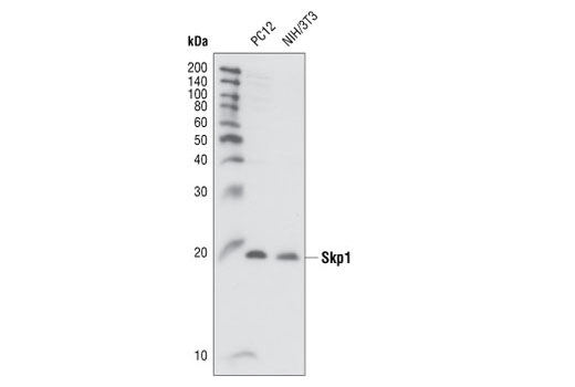
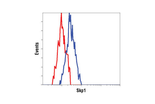
| Skp1 Antibody | 2156 | 20 µl | 19 kDa | Rabbit |
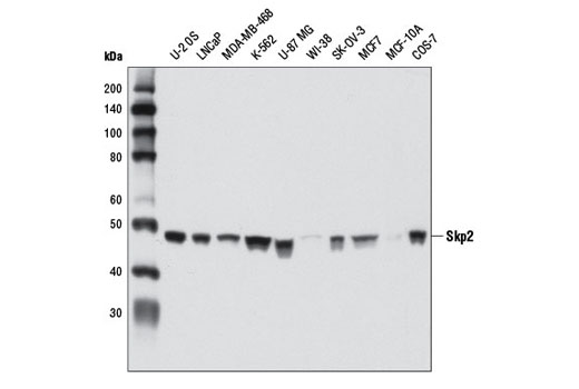
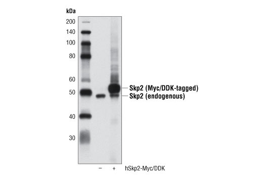
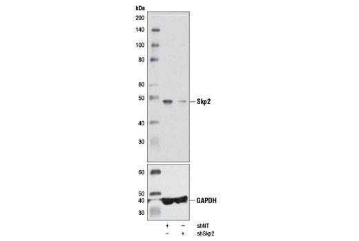
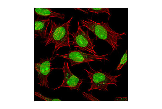
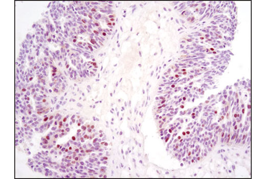
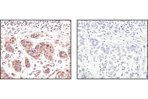
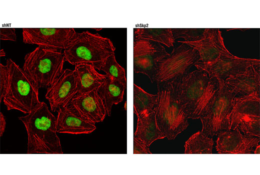
| Skp2 (D3G5) XP® Rabbit mAb | 2652 | 20 µl | 48 kDa | Rabbit IgG |
| ISG15 (22D2) Rabbit mAb | 2758 | 20 µl | 15 kDa | Rabbit IgG |
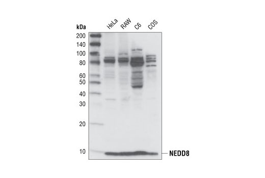
| NEDD8 (19E3) Rabbit mAb | 2754 | 20 µl | 9 kDa | Rabbit IgG |
| Ubiquitin (P4D1) Mouse mAb | 3936 | 20 µl | Mouse IgG1 | |
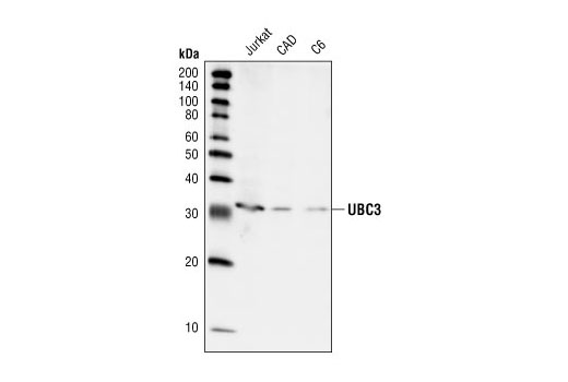
| UBC3 Antibody | 4997 | 20 µl | 32 kDa | Rabbit |
| SUMO-1 Antibody | 4930 | 20 µl | Rabbit | |
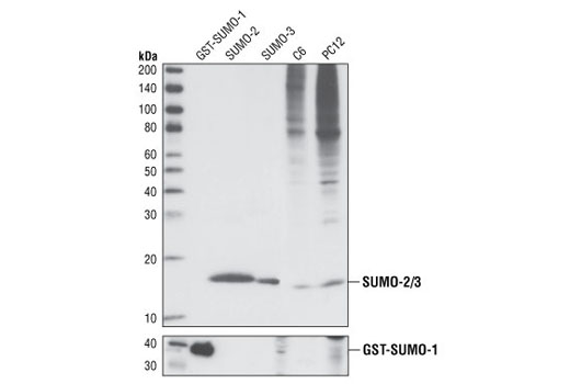
| SUMO-2/3 (18H8) Rabbit mAb | 4971 | 20 µl | Rabbit IgG | |
| Anti-rabbit IgG, HRP-linked Antibody | 7074 | 100 µl | Goat | |
| Anti-mouse IgG, HRP-linked Antibody | 7076 | 100 µl | Horse |
Please visit cellsignal.com for individual component applications, species cross-reactivity, dilutions, protocols, and additional product information.
Description
This sampler kit provides an economical means to investigate protein folding and stability. The kit contains primary and secondary antibodies to perform two Western blots with each antibody.
Storage
Background
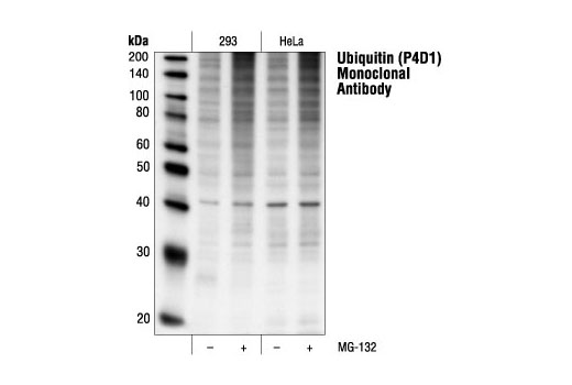
The small regulatory protein ubiquitin is often covalently linked to many cellular proteins, labeling these targeted proteins for proteasome-mediated degradation. Ubiquitin is first activated by forming a thiolester complex with the E1 activation component. Activated ubiquitin is subsequently transferred to the ubiquitin-carrier protein E2, and then to an E3 ubiquitin ligase for final delivery to the ε-NH2 of the target protein lysine residue (1). The ubiquitin-proteasome pathway has been implicated in a wide range of both normal biological processes and diseases (2,3).
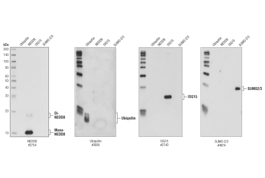
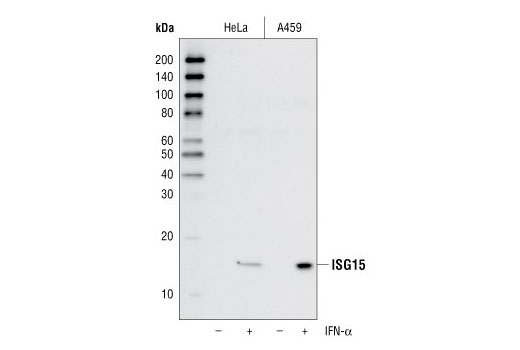
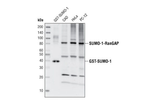
The ubiquitin-like protein family contains three small ubiquitin-related modifier proteins (SUMO-1, -2 and -3), neural precursor cell-expressed developmentally down-regulated protein 8 (NEDD8) and interferon-stimulated 15 kDa protein (ISG15) (4-6). Their covalent attachment to target proteins is a reversible, multi-step process that is analogous to protein ubiquitination. Mature molecules are linked to the activating enzyme E1, conjugated to E2 and ligated to the target proteins by E3 (7-10). Ubiquitin is the predominant regulator for the degradation of a wide range of target proteins (8) while SUMO, NEDD8 and ISG15 modify a limited set of substrates to regulate various other biological processes (4, 11-18).
During ubiquitination, the combinatorial interaction of different E2 and E3 proteins produces variable substrate specificity (4). UBC3 and UBC3B are E2 ubiquitin-carrier proteins (19, 20). The SCF (Skp1/CUL1/F-box) E3 ubiquitin ligase protein complex is composed of three protein components, including the S phase kinase associated protein 1 (Skp1), Cullin homolog 1 (CUL1) and the Skp2 F-box protein (21-23).
- Ciechanover, A. (1998) EMBO J. 17, 7151-7160.
- Bernardi, R. et al. (2000) Oncogene 19, 2447-2454.
- Jesenberger, V. and Jentsch, S. (2002) Nat. Rev. Mol. Cell Biol. 3, 112-121.
- Schwartz, D.C. and Hochstrasser, M. (2003) Trends Biochem. Sci. 28, 321-328.
- Chiba, T. and Tanaka, K. (2004) Curr. Protein Pept. Sci. 5, 177-184.
- Ritchie, K.J. and Zhang, D.E. (2004) Semin. Cell Dev. Biol. 15, 237-246.
- Kim, K.I. et al. (2002) J. Cell Physiol. 191, 257-268.
- Osaka, F. et al. (1998) Genes Dev. 12, 2263-2268.
- Loeb, K.R. and Haas, A.L. (1992) J. Biol. Chem. 267, 7806-7813.
- Zhao, C. et al. (2005) Proc. Natl. Acad. Sci. USA 102, 10200-10205.
- Matunis, M.J. et al. (1996) J. Cell Biol. 135, 1457-1470.
- Duprez, E. et al. (1999) J. Cell Sci. 112 ( Pt 3), 381-393.
- Gostissa, M. et al. (1999) EMBO J. 18, 6462-6471.
- Rodriguez, M.S. et al. (1999) EMBO J. 18, 6455-6461.
- Desterro, J.M. et al. (1998) Mol. Cell 2, 233-239.
- Stickle, N.H. et al. (2004) Mol. Cell. Biol. 24, 3251-3261.
- Xirodimas, D.P. et al. (2004) Cell 118, 83-97.
- Hamerman, J.A. et al. (2002) J. Immunol. 168, 2415-2423.
- Semplici, F. et al. (2002) Oncogene 21, 3978-3987.
- Pagano, M. et al. (1995) Science 269, 682-685.
- Yu, Z.K. et al. (1998) Proc. Natl. Acad. Sci. USA 95, 11324-11329.
- Pagano, M. (2004) Mol. Cell 14, 414-416.
- Reed, S.I. (2003) Nat. Rev. Mol. Cell Biol. 4, 855-864.
Background References
Trademarks and Patents
使用に関する制限
法的な権限を与えられたCSTの担当者が署名した書面によって別途明示的に合意された場合を除き、 CST、その関連会社または代理店が提供する製品には以下の条件が適用されます。お客様が定める条件でここに定められた条件に含まれるものを超えるもの、 または、ここに定められた条件と異なるものは、法的な権限を与えられたCSTの担当者が別途書面にて受諾した場合を除き、拒絶され、 いかなる効力も効果も有しません。
研究専用 (For Research Use Only) またはこれに類似する表示がされた製品は、 いかなる目的についても FDA または外国もしくは国内のその他の規制機関により承認、認可または許可を受けていません。 お客様は製品を診断もしくは治療目的で使用してはならず、また、製品に表示された内容に違反する方法で使用してはなりません。 CST が販売または使用許諾する製品は、エンドユーザーであるお客様に対し、使途を研究および開発のみに限定して提供されるものです。 診断、予防もしくは治療目的で製品を使用することまたは製品を再販売 (単独であるか他の製品等の一部であるかを問いません) もしくはその他の商業的利用の目的で購入することについては、CST から別途許諾を得る必要があります。 お客様は以下の事項を遵守しなければなりません。(a) CST の製品 (単独であるか他の資材と一緒であるかを問いません) を販売、使用許諾、貸与、寄付もしくはその他の態様で第三者に譲渡したり使用させたりしてはなりません。また、商用の製品を製造するために CST の製品を使用してはなりません。(b) 複製、改変、リバースエンジニアリング、逆コンパイル、 分解または他の方法により製品の構造または技術を解明しようとしてはなりません。また、 CST の製品またはサービスと競合する製品またはサービスを開発する目的で CST の製品を使用してはなりません。(c) CST の製品の商標、商号、ロゴ、特許または著作権に関する通知または表示を除去したり改変したりしてはなりません。(d) CST の製品をCST 製品販売条件(CST’s Product Terms of Sale) および該当する書面のみに従って使用しなければなりません。(e) CST の製品に関連してお客様が使用する第三者の製品またはサービスに関する使用許諾条件、 サービス提供条件またはこれに類する合意事項を遵守しなければなりません。