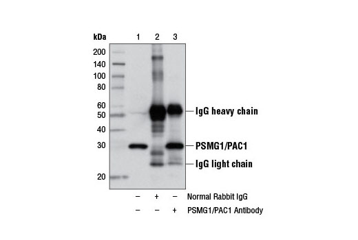
WB, IP
H Mk
Endogenous
30
Rabbit
#O95456
8624
Product Information
Product Usage Information
| Application | Dilution |
|---|---|
| Western Blotting | 1:1000 |
| Immunoprecipitation | 1:200 |
Storage
Specificity / Sensitivity
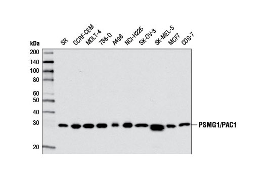
Species Reactivity:
Human, Monkey
Source / Purification
Polyclonal antibodies are produced by immunizing animals with a synthetic peptide corresponding to residues near the amino terminus of human PSMG1 (PAC1) protein. Antibodies are purified by protein A and peptide affinity chromatography.
Background
The 26S proteasome is a highly abundant proteolytic complex involved in the degradation of ubiquitinated substrate proteins. It consists largely of two sub-complexes, the 20S catalytic core particle (CP) and the 19S/PA700 regulatory particle (RP) that can cap either end of the CP. The CP consists of two stacked heteroheptameric β-rings (β1-7) that contain three catalytic β-subunits and are flanked on either side by two heteroheptameric α-rings (α1-7). The RP includes a base and a lid, each having multiple subunits. The base, in part, is composed of a heterohexameric ring of ATPase subunits belonging to the AAA (ATPases Associated with diverse cellular Activities) family. The ATPase subunits function to unfold the substrate and open the gate formed by the α-subunits, thus exposing the unfolded substrate to the catalytic β-subunits. The lid consists of ubiquitin receptors and DUBs that function in recruitment of ubiquitinated substrates and modification of ubiquitin chain topology (1,2). Other modulators of proteasome activity, such as PA28/11S REG, can also bind to the end of the 20S CP and activate it (1,2).
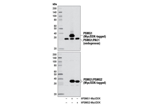
Proteasome assembly chaperone 1 (PSMG1, PAC1) is an evolutionarily conserved, ubiquitously expressed chaperone protein that promotes proper biogenesis of the α-ring of the 20S CP of the eukaryotic proteasome (3,4). PSMG1 (PAC1) functions in a heterodimeric complex with PSMG2 (PAC2) and was originally identified as a proteasome subunit binding partner (4). Intact PSMG1-PSMG2 heterodimers both promote heteroheptameric α-ring assembly and/or stability and prevent accumulation of non-productive α-ring dimers (4). Research studies targeting the disruption of the murine Psmg1 locus have substantiated the importance of proteasome chaperones in contributing to normal proteasome maturation and cellular homeostasis by demonstrating that loss of Psmg1/Pac1 function leads to embryonic lethality (5).
Species Reactivity
Species reactivity is determined by testing in at least one approved application (e.g., western blot).
Western Blot Buffer
IMPORTANT: For western blots, incubate membrane with diluted primary antibody in 5% w/v nonfat dry milk, 1X TBS, 0.1% Tween® 20 at 4°C with gentle shaking, overnight.
Applications Key
WB: Western Blotting IP: Immunoprecipitation
Cross-Reactivity Key
H: human M: mouse R: rat Hm: hamster Mk: monkey Vir: virus Mi: mink C: chicken Dm: D. melanogaster X: Xenopus Z: zebrafish B: bovine Dg: dog Pg: pig Sc: S. cerevisiae Ce: C. elegans Hr: horse GP: Guinea Pig Rab: rabbit All: all species expected
Trademarks and Patents
使用に関する制限
法的な権限を与えられたCSTの担当者が署名した書面によって別途明示的に合意された場合を除き、 CST、その関連会社または代理店が提供する製品には以下の条件が適用されます。お客様が定める条件でここに定められた条件に含まれるものを超えるもの、 または、ここに定められた条件と異なるものは、法的な権限を与えられたCSTの担当者が別途書面にて受諾した場合を除き、拒絶され、 いかなる効力も効果も有しません。
研究専用 (For Research Use Only) またはこれに類似する表示がされた製品は、 いかなる目的についても FDA または外国もしくは国内のその他の規制機関により承認、認可または許可を受けていません。 お客様は製品を診断もしくは治療目的で使用してはならず、また、製品に表示された内容に違反する方法で使用してはなりません。 CST が販売または使用許諾する製品は、エンドユーザーであるお客様に対し、使途を研究および開発のみに限定して提供されるものです。 診断、予防もしくは治療目的で製品を使用することまたは製品を再販売 (単独であるか他の製品等の一部であるかを問いません) もしくはその他の商業的利用の目的で購入することについては、CST から別途許諾を得る必要があります。 お客様は以下の事項を遵守しなければなりません。(a) CST の製品 (単独であるか他の資材と一緒であるかを問いません) を販売、使用許諾、貸与、寄付もしくはその他の態様で第三者に譲渡したり使用させたりしてはなりません。また、商用の製品を製造するために CST の製品を使用してはなりません。(b) 複製、改変、リバースエンジニアリング、逆コンパイル、 分解または他の方法により製品の構造または技術を解明しようとしてはなりません。また、 CST の製品またはサービスと競合する製品またはサービスを開発する目的で CST の製品を使用してはなりません。(c) CST の製品の商標、商号、ロゴ、特許または著作権に関する通知または表示を除去したり改変したりしてはなりません。(d) CST の製品をCST 製品販売条件(CST’s Product Terms of Sale) および該当する書面のみに従って使用しなければなりません。(e) CST の製品に関連してお客様が使用する第三者の製品またはサービスに関する使用許諾条件、 サービス提供条件またはこれに類する合意事項を遵守しなければなりません。